ATRX/DAXX and ALT: History
Please note this is an old version of this entry, which may differ significantly from the current revision.
Contributor: ,

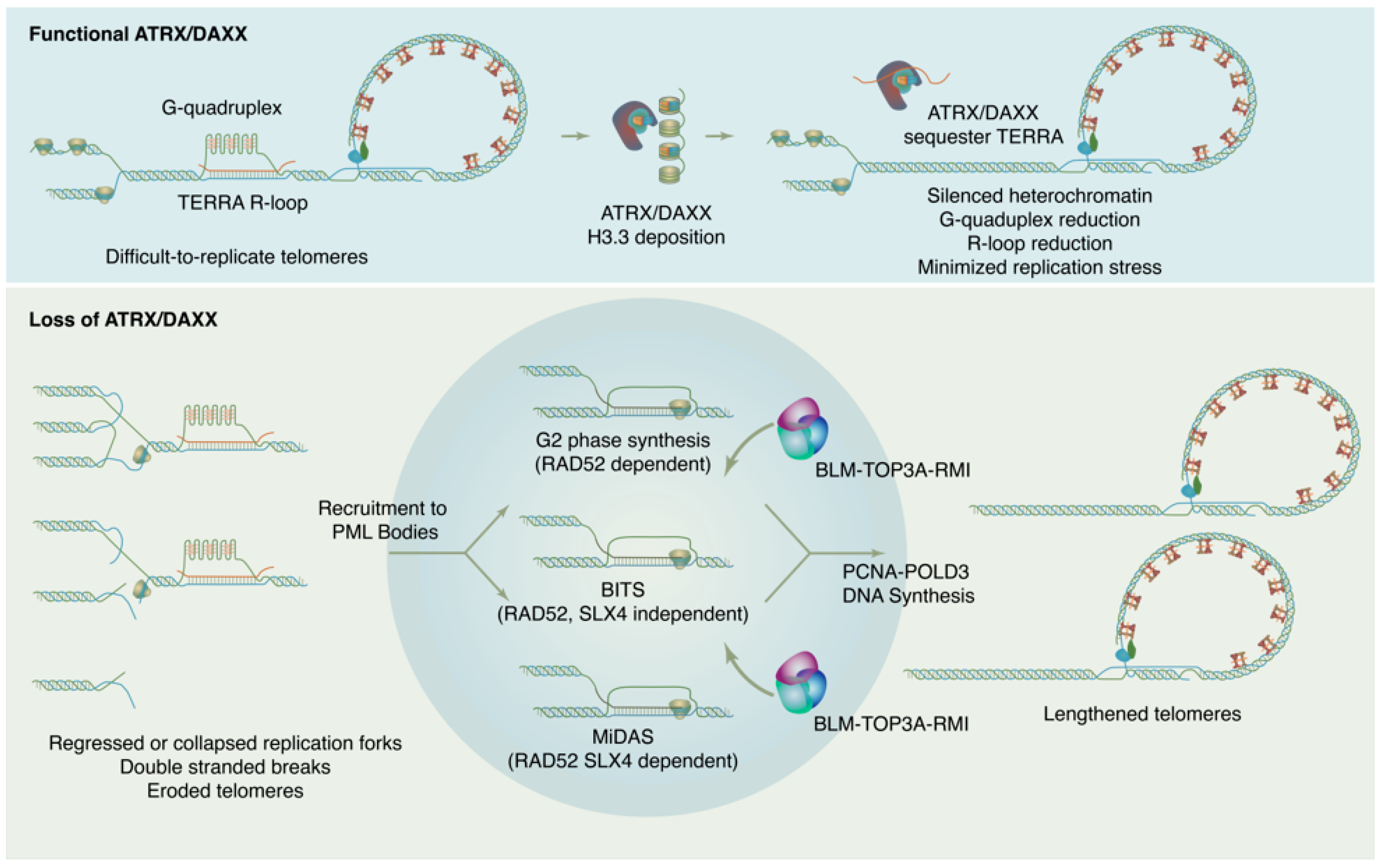
ATRX is named for its causal role in ATR-X syndrome (α-thalassemia with mental impairment, X-linked), an X-linked disorder characterized by developmental delays, urogenital abnormalities, distinctive craniofacial features, and α-thalassemia caused by insufficient α-globin expression. Because of the central role of decreased α-globin mRNA expression in the ATR-X phenotype, research on ATRX initially focused on its potential as a transcriptional regulator. In fact, ATRX in concert with DAXX play wide-ranging roles in maintaining chromatin and reckoning with problematic DNA repeat sequences, with downstream effects on gene expression that have critical impacts in development. Proliferating cells must enact a telomere maintenance mechanism to ensure genomic stability. In a subset of tumors, telomeres are maintained not by telomerase, but through a homologous recombination-based mechanism termed Alternative Lengthening of Telomeres or ALT. The ALT process is linked to mutations in the ATRX/DAXX/H3.3 histone chaperone complex. This complex is responsible for depositing non-replicative histone variant H3.3 at pericentric and telomeric heterochromatin but has also been found to have roles in ameliorating replication in repeat sequences and in promoting DNA repair.

  • ATRX
  • DAXX
  • ALT

1. Introduction

To counteract the erosion of DNA ends due to the end-replication problem, organisms with linear chromosomes must enact a program of telomere maintenance. In mammalian cells, telomeres are lengthened through the action of the ribonucleoprotein telomerase, which synthesizes new DNA repeats from an RNA template. Telomerase is not expressed in most somatic cells, however, so in the process of immortalization cancer cells must acquire a telomere maintenance program. Most tumors reactivate the expression of telomerase, but 10–15% of tumors lengthen telomeres through a DNA-templated process known as Alternative Lengthening of Telomeres or ALT [1][2][3][4].
ALT is strongly correlated with loss of ATRX or DAXX [5][6][7], which together form a complex that deposits non-canonical histone variant H3.3 at pericentromeric and telomeric heterochromatin [8][9][10]. Though ALT-related ATRX or DAXX mutations are often truncating nonsense mutations or deletions, protein loss has been observed without obvious coding sequence changes, implying loss of expression due to alterations in promoters or splicing, or due to epigenetic changes [6]. Notably, ATRX and DAXX loss are mutually exclusive. ALT is so highly correlated with loss of ATRX or DAXX that mutations leading to truncation of these proteins have been considered synonymous with ALT status, though using this metric as a reliable diagnostic of ALT has also been controversial [11][12][13]. ATRX/DAXX mutations associated with ALT telomere maintenance are most frequently observed in liposarcomas, adult gliomas, pancreatic neuro-endocrine tumors, and osteosarcomas [11]. ALT is rarely observed in carcinomas, or tumors arising from highly proliferative tissue, perhaps due to their predisposition to reactivate telomerase. Many ALT-prone tumors have poor prognoses, so a better understanding of the role of ATRX and DAXX mutations in ALT and in tumorigenesis generally is desirable to open the door to new targeted therapies.

2. ATRX/DAXX and the Suppression of ALT

ALT leverages recombination machinery to synthesize new telomere sequence. At the time of the initial discovery of ALT in mammalian cells the possibility of telomere maintenance without telomerase was already well established in other organisms, including yeast and Drosophila [14][15][16]. In budding yeast, it was recognized that multiple recombination pathways could compensate for a lack of telomerase activity [15]. Thus, the notion that human telomeres could be lengthened via a recombination mechanism was not difficult to imagine, and indeed it was soon found that in ALT cells lines telomere sequence was copied to other telomeres, implicating a recombination mechanism [17].
In surveys of cell lines and tumors without telomerase activity, it was observed that loss of ATRX/DAXX was a consistent feature [5][6][7]. Recent large-scale sequencing studies demonstrated that across tumor types truncation of ATRX or DAXX correlates strongly with telomere variant repeats and telomere insertions into non-telomeric regions with concurrent copy number loss at the insertion site [11]. These sequencing results highlight the risk of ALT in driving genome instability and tumorigenesis.
ALT is more common in tumors of mesenchymal origin, including osteosarcoma [5]. While ALT correlates strongly with ATRX mutations in osteosarcoma, DAXX mutations are frequently observed in pancreatic neuroendocrine tumors (PanNETs) and correlate with poorer prognosis [11][18][19][20]. Patients with multiple endocrine neoplasia-1 (MEN-1) syndrome have a predisposition to PanNETs. In these patients ATRX/DAXX was found to be intact in microadenomas but had been lost in some larger PanNETs with concurrent acquisition of ALT, implying that the acquisition of a telomere maintenance program is a late even in PanNET tumorigenesis [21]. In pediatric gliomas, mutations in H3.3 genes are also observed in addition to ATRX mutations, with strong correlation to the ALT phenotype [22].
ALT refers to a set of related mechanisms that leverage the homologous recombination machinery to achieve maintenance of telomere length in the absence of telomerase. Hallmarks of ALT telomere maintenance include long and heterogeneous telomeres [4], extra-chromosomal telomere repeat DNA (ECTRs) [23], clustering of telomere repeats at PML nuclear bodies forming ALT-associated PML Bodies (APBs) [24][25], and elevated levels of telomere sister chromatid exchange [26]. Together, these symptoms of ALT paint a picture of telomeres that have broken free of regulation. They can also serve as markers to assay for ALT activity.
ECTRs include both linear and circular DNA species and may have multiple origins. ECTRs are known to be the result of telomere trimming by XRCC3 and Nbs1 [27], but may also form from internal DNA loops (i-loops) in ALT cells [28]. Of the circular ECTRs, there exist T-circles that are double-stranded, and C-circles that are predominantly single-stranded. These C-circles can be specifically amplified through rolling-circle amplification, self-primed from a double-stranded region [29]. Detection of C-circles using this assay is a common metric of ALT activity. Notably, ECTRs can also be found in the cytoplasm in ALT cell lines, which would be expected to trigger the cGAS-STING DNA sensing pathway to trigger production of interferon b and the type I interferon response. It was found that STING expression was lost in ALT cells, but even when STING was restored to the ATRXnull ALT cell line U2OS it was necessary to also restore ATRX to recover functional DNA sensing [30]. This implies that a role in DNA sensing represents yet another function for the ATRX/DAXX complex.
The presence of APBs is another key indicator of ALT activity. In ALT, telomeres cluster at PML bodies forming large, intense foci [24]. APBs are the location of new DNA synthesis in ALT, and ALT does not proceed in the absence of PML protein [31][32]. As previously mentioned, PML bodies are organized through SUMO-SIM interactions, and APBs are no exception to that rule. In ALT, the SMC5/6 complex SUMO ligase MMS21, as well as the SUMO ligase PIAS4, sumoylate telomere binding proteins including TRF1 and TRF2 [33][34]. This sumoylation is required for recruitment of telomeres to APBs [33]. Curiously, synthetic APB-like condensates have been induced to form in cells to test the notion that it is the liquid properties of APBs rather than the specific proteins that drive clustering [35]. Indeed, it was possibly to induce the clustering of telomeres with PML bodies using a SIM-coupled dimerization-induction system, but these clusters were not functional for new telomere synthesis. When telomeres were tethered with PML in an ALT cell line, however, new DNA synthesis was generated at the synthetic APB [34]. Thus, the ALT mechanism requires correct signaling to engage the DNA repair machinery, not mere telomere proximity to PML.
If proximity is insufficient, how is ALT triggered de novo? Despite the strong correlation of ATRX/DAXX loss to ALT, ablation of ATRX is of itself insufficient to trigger the ALT phenotype [36][37][38], though upon crisis cells lacking ATRX are predisposed to activate ALT instead of telomerase [38]. One successful strategy for activating ALT de novo in telomerase-positive fibrosarcoma cells involved shRNA knockdown of both ATRX and DAXX plus infliction of constitutive telomere damage. This was accomplished using inducible overexpression of a TPP1 ∆OBRD fold construct to induce telomere-specific DNA damage and inhibit telomerase activity [39]. In this context, C-circle production was triggered, cells formed APBs, and telomere length was maintained, indicating adoption of ALT telomere maintenance.
ALT has also been activated through depletion of the histone H3/H4 chaperone ASF1 in human primary fibroblasts and immortalized cell lines [40]. ASF1 is an H3/H4 chaperone that delivers both canonical replication-coupled H3/H4 heterodimers to the replication fork as well as providing H3.3/H4 heterodimers to ATRX/DAXX and HIRA. The co-depletion of ASF1a and ASF1b induces C-circle production, APBs, and telomere length maintenance in absence of telomerase. This induction of ALT is relatively rapid, occurring withing 72 h of ASF1 depletion, and persists even after ASF1 protein levels rebound. These findings are phenocopied by loss of TLK (tousled-like kinase) activity. The TLKs (TLK1 and TLK2) are Ser-Thr kinases that regulate the activity of ASF1A and ASF1B. Depletion of TLKs results in replication stress and impaired de novo nucleosome assembly [41]. In the ALT+ U2OS osteosarcoma cell line, depletion of TLKs results in increases in markers of ALT activity, while in telomerase-expressing HeLa cells APBs and C-circles were observed when TLK1 was knocked out. Taken together, these experiments strongly suggest that provisioning of H3.3 at the replication fork is necessary for ALT suppression. Importantly, it was recently observed that HIRA can compensate for H3.3 deposition at telomeres in ALT cells, and loss of HIRA is synthetic lethal with ALT [42]. Thus, ALT seems tied to reduction of telomeric H3.3, but total elimination of H3.3 at telomeres is not tolerated.
How is ALT DNA synthesis accomplished? Though details of the ALT mechanism continue to be worked out, current evidence indicates that new DNA synthesis at ALT telomeres may proceed through at least three distinct RAD51 independent pathways (Figure 1). These pathways differ in their details but may all be considered variations of a break-induced replication (BIR) mechanism [43][44]. BIR is a repair pathway for single-ended DNA breaks that results in conservative synthesis of new DNA. Single-ended breaks can occur due to the collapse of a replication fork or can be mimicked by an eroded telomere. As in other modes of homologous recombination, BIR begins with resection at the break site to produce a single-stranded filament to engage in a homology search. From there, the separate sub-pathways diverge. In a model system with experimentally-generated DSBs at telomeres, breaks are repaired through a RAD52, SLX4 independent mechanism termed Break Induced Telomere Synthesis or BITS [43], which can extend into mitosis. RAD52 and SLX4 are both required for telomeric mitotic DNA synthesis (MiDAS), a DNA repair pathway used at DNA common fragile sites and observed at telomeres in both ALT and telomerase positive cells [44][45]. As MiDAS is relatively infrequent, most ALT telomere synthesis spontaneously arises in the G2 phase and utilizes a RAD52 dependent, SLX4 independent pathway [46]. After homology search and strand invasion, the ALT mechanism absolutely requires the action of the BLM–TOP3A–RMI (BTR) dissolvase complex to promote long-track DNA synthesis by PCNA-POLD3, then dissolve the resulting recombination intermediate without crossover [47][48].
Figure 1. ATRX/DAXX suppresses ALT by maintaining chromatin states and reducing replication stress at telomeres. In absence of ATRX/DAXX, chromatin loses silencing, TERRA transcription increases, G4s and R-loops increase, and replication stress results. This replication ultimately feeds forward into self-perpetuating ALT.
ALT telomeres have reduced chromatin compaction, consistent with the role of ATRX/DAXX in H3.3 deposition. In addition to increased nuclease sensitivity, ALT telomeres are low in H3K9me3 and express increased levels of TERRA [6][49][50][51]. TERRA expression appears to be a critical driver of ALT in multiple ways. TERRA transcription destabilizes chromosomes and increases their replication stress. Inhibition of TERRA expression reduces ALT activity [52]. In ALT, TERRA recruits the DNA repair factor RAD51AP1, which is thought to promote formation of recombination intermediates that are important for ALT [53]. One model suggests that RAD52 and RAD51AP1 play distinct roles at telomeres, where RAD52 promotes telomeric D-loops and RAD51AP1 enables formation of R-loops with TERRA. These R-loops represent a sort of HR intermediate that can be “swapped out” in a RAD52 independent manner for an invading D-loop to enable BIR synthesis [54].
In ALT cell lines that lack fully functional ATRX or DAXX, repletion of the dysfunctional protein is sufficient to suppress ALT. When ATRX was restored to U2OS cells, C-circles were reduced, APBs diminished, and telomeres eroded, indicating that ALT was no longer active in these cells. Histone H3.3 occupancy was increased at telomeres and replication stress was alleviated. These effects were mitigated by the addition of a G4 stabilizing drug, indicating that the effect of ATRX was at least in part related to its role in reducing replication stress due to G4s [55]. In other work, wild-type DAXX was restored to the ALT + G292 osteosarcoma cell line, in which ATRX is wild type but DAXX has undergone a fusion event with the non-canonical kinesin KIFC3. In this cell line DAXX has lost the C-terminal SIM motif that targets it to PML bodies, resulting in mislocalization of DAXX and ATRX. Restoration of wild-type DAXX in G292 localizes ATRX and abrogates ALT. Notably, the endogenous DAXX in G292 is nearly full-length except for the localization motif, and it binds both ATRX and H3.3 competently, indicating that presence of ATRX/DAXX not only in the nucleus but specifically at PML bodies is essential for ALT suppression [56][57].
Why does the loss of ATRX/DAXX lead to ALT in cancer cells, but genetic knock-out of ATRX/DAXX does not? ALT appears to rely on a certain level of replication stress at telomeres to perpetuate itself. There seems to be a “Goldilocks zone” of replication stress that can maintain ALT without leading to cell toxicity. If too much replication stress occurs in ALT cells, as in the case of FANCM depletion, for example [58][59], ALT activity is increased to the point of toxicity and cells cannot survive. Remarkably, it was recently observed that BIR itself produces replication stress, creating a self-reinforcing cycle of DNA damage to perpetuate ALT telomere maintenance [34]. Thus, the normal activities of ATRX/DAXX at telomeres maintain heterochromatin silencing, leading to reduction in TERRA transcription, reduced G4s and R-loops, and less replication stress (Figure 1). In the absence of ATRX/DAXX, the levels of replication stress may increase, but not enough for the ALT mechanism to be self-sustaining. Only through additional insults does the level of replication stress attain the vicious cycle necessary to perpetuate ALT.

This entry is adapted from the peer-reviewed paper 10.3390/genes14040790

References

  1. Kim, N.W.; Piatyszek, M.A.; Prowse, K.R.; Harley, C.B.; West, M.D.; Ho, P.L.; Coviello, G.M.; Wright, W.E.; Weinrich, S.L.; Shay, J.W. Specific association of human telomerase activity with immortal cells and cancer. Science 1994, 266, 2011–2015.
  2. Shay, J.W.; Bacchetti, S. A survey of telomerase activity in human cancer. Eur. J. Cancer 1997, 33, 787–791.
  3. Bryan, T.M.; Englezou, A.; Dalla-Pozza, L.; Dunham, M.A.; Reddel, R.R. Evidence for an alternative mechanism for maintaining telomere length in human tumors and tumor-derived cell lines. Nat. Med 1997, 3, 1271–1274.
  4. Bryan, T.M.; Englezou, A.; Gupta, J.; Bacchetti, S.; Reddel, R.R. Telomere elongation in immortal human cells without detectable telomerase activity. EMBO J. 1995, 14, 4240–4248.
  5. Heaphy, C.M.; de Wilde, R.F.; Jiao, Y.; Klein, A.P.; Edil, B.H.; Shi, C.; Bettegowda, C.; Rodriguez, F.J.; Eberhart, C.G.; Hebbar, S.; et al. Altered telomeres in tumors with ATRX and DAXX mutations. Science 2011, 333, 425.
  6. Lovejoy, C.A.; Li, W.; Reisenweber, S.; Thongthip, S.; Bruno, J.; de Lange, T.; De, S.; Petrini, J.H.J.; Sung, P.A.; Jasin, M.; et al. Loss of ATRX, genome instability, and an altered DNA damage response are hallmarks of the alternative lengthening of telomeres pathway. PLoS Genet. 2012, 8, e1002772.
  7. Bower, K.; Napier, C.E.; Cole, S.L.; Dagg, R.A.; Lau, L.M.; Duncan, E.L.; Moy, E.L.; Reddel, R.R. Loss of wild-type ATRX expression in somatic cell hybrids segregates with activation of Alternative Lengthening of Telomeres. PLoS ONE 2012, 7, e50062.
  8. Goldberg, A.D.; Banaszynski, L.A.; Noh, K.M.; Lewis, P.W.; Elsaesser, S.J.; Stadler, S.; Dewell, S.; Law, M.; Guo, X.; Li, X.; et al. Distinct factors control histone variant H3.3 localization at specific genomic regions. Cell 2010, 140, 678–691.
  9. Lewis, P.W.; Elsaesser, S.J.; Noh, K.M.; Stadler, S.C.; Allis, C.D. Daxx is an H3.3-specific histone chaperone and cooperates with ATRX in replication-independent chromatin assembly at telomeres. Proc. Natl. Acad. Sci. USA 2010, 107, 14075–14080.
  10. Drane, P.; Ouararhni, K.; Depaux, A.; Shuaib, M.; Hamiche, A. The death-associated protein DAXX is a novel histone chaperone involved in the replication-independent deposition of H3.3. Genes Dev. 2010, 24, 1253–1265.
  11. Sieverling, L.; Hong, C.; Koser, S.D.; Ginsbach, P.; Kleinheinz, K.; Hutter, B.; Braun, D.M.; Cortes-Ciriano, I.; Xi, R.; Kabbe, R.; et al. Genomic footprints of activated telomere maintenance mechanisms in cancer. Nat. Commun. 2020, 11, 733.
  12. de Nonneville, A.; Reddel, R.R. Alternative lengthening of telomeres is not synonymous with mutations in ATRX/DAXX. Nat. Commun. 2021, 12, 1552.
  13. Feuerbach, L. Formal reply to “Alternative lengthening of telomeres is not synonymous with mutations in ATRX/DAXX”. Nat. Commun. 2021, 12, 1551.
  14. Wang, S.S.; Zakian, V.A. Telomere-telomere recombination provides an express pathway for telomere acquisition. Nature 1990, 345, 456–458.
  15. Lundblad, V.; Blackburn, E.H. An alternative pathway for yeast telomere maintenance rescues est1- senescence. Cell 1993, 73, 347–360.
  16. Biessmann, H.; Mason, J.M.; Ferry, K.; d’Hulst, M.; Valgeirsdottir, K.; Traverse, K.L.; Pardue, M.L. Addition of telomere-associated HeT DNA sequences “heals” broken chromosome ends in Drosophila. Cell 1990, 61, 663–673.
  17. Dunham, M.A.; Neumann, A.A.; Fasching, C.L.; Reddel, R.R. Telomere maintenance by recombination in human cells. Nat. Genet. 2000, 26, 447–450.
  18. Cives, M.; Partelli, S.; Palmirotta, R.; Lovero, D.; Mandriani, B.; Quaresmini, D.; Pelle, E.; Andreasi, V.; Castelli, P.; Strosberg, J.; et al. DAXX mutations as potential genomic markers of malignant evolution in small nonfunctioning pancreatic neuroendocrine tumors. Sci. Rep. 2019, 9, 18614.
  19. Singhi, A.D.; Liu, T.C.; Roncaioli, J.L.; Cao, D.; Zeh, H.J.; Zureikat, A.H.; Tsung, A.; Marsh, J.W.; Lee, K.K.; Hogg, M.E.; et al. Alternative Lengthening of Telomeres and Loss of DAXX/ATRX Expression Predicts Metastatic Disease and Poor Survival in Patients with Pancreatic Neuroendocrine Tumors. Clin. Cancer Res. 2017, 23, 600–609.
  20. Jiao, Y.; Shi, C.; Edil, B.H.; de Wilde, R.F.; Klimstra, D.S.; Maitra, A.; Schulick, R.D.; Tang, L.H.; Wolfgang, C.L.; Choti, M.A.; et al. DAXX/ATRX, MEN1, and mTOR pathway genes are frequently altered in pancreatic neuroendocrine tumors. Science 2011, 331, 1199–1203.
  21. de Wilde, R.F.; Heaphy, C.M.; Maitra, A.; Meeker, A.K.; Edil, B.H.; Wolfgang, C.L.; Ellison, T.A.; Schulick, R.D.; Molenaar, I.Q.; Valk, G.D.; et al. Loss of ATRX or DAXX expression and concomitant acquisition of the alternative lengthening of telomeres phenotype are late events in a small subset of MEN-1 syndrome pancreatic neuroendocrine tumors. Mod. Pathol. 2012, 25, 1033–1039.
  22. Minasi, S.; Baldi, C.; Gianno, F.; Antonelli, M.; Buccoliero, A.M.; Pietsch, T.; Massimino, M.; Buttarelli, F.R. Alternative lengthening of telomeres in molecular subgroups of paediatric high-grade glioma. Child’s Nerv. Syst. 2021, 37, 809–818.
  23. Ogino, H.; Nakabayashi, K.; Suzuki, M.; Takahashi, E.; Fujii, M.; Suzuki, T.; Ayusawa, D. Release of telomeric DNA from chromosomes in immortal human cells lacking telomerase activity. Biochem. Biophys. Res. Commun. 1998, 248, 223–227.
  24. Yeager, T.R.; Neumann, A.A.; Englezou, A.; Huschtscha, L.I.; Noble, J.R.; Reddel, R.R. Telomerase-negative immortalized human cells contain a novel type of promyelocytic leukemia (PML) body. Cancer Res. 1999, 59, 4175–4179.
  25. Draskovic, I.; Arnoult, N.; Steiner, V.; Bacchetti, S.; Lomonte, P.; Londono-Vallejo, A. Probing PML body function in ALT cells reveals spatiotemporal requirements for telomere recombination. Proc. Natl. Acad. Sci. USA 2009, 106, 15726–15731.
  26. Murnane, J.P.; Sabatier, L.; Marder, B.A.; Morgan, W.F. Telomere dynamics in an immortal human cell line. EMBO J. 1994, 13, 4953–4962.
  27. Rivera, T.; Haggblom, C.; Cosconati, S.; Karlseder, J. A balance between elongation and trimming regulates telomere stability in stem cells. Nat. Struct. Mol. Biol. 2017, 24, 30–39.
  28. Mazzucco, G.; Huda, A.; Galli, M.; Piccini, D.; Giannattasio, M.; Pessina, F.; Doksani, Y. Telomere damage induces internal loops that generate telomeric circles. Nat. Commun. 2020, 11, 5297.
  29. Henson, J.D.; Lau, L.M.; Koch, S.; Martin La Rotta, N.; Dagg, R.A.; Reddel, R.R. The C-Circle Assay for alternative-lengthening-of-telomeres activity. Methods 2016, 114, 74–84.
  30. Chen, Y.A.; Shen, Y.L.; Hsia, H.Y.; Tiang, Y.P.; Sung, T.L.; Chen, L.Y. Extrachromosomal telomere repeat DNA is linked to ALT development via cGAS-STING DNA sensing pathway. Nat. Struct. Mol. Biol. 2017, 24, 1124–1131.
  31. Zhang, J.M.; Yadav, T.; Ouyang, J.; Lan, L.; Zou, L. Alternative Lengthening of Telomeres through Two Distinct Break-Induced Replication Pathways. Cell Rep. 2019, 26, 955–968.e953.
  32. Loe, T.K.; Li, J.S.Z.; Zhang, Y.; Azeroglu, B.; Boddy, M.N.; Denchi, E.L. Telomere length heterogeneity in ALT cells is maintained by PML-dependent localization of the BTR complex to telomeres. Genes Dev. 2020, 34, 650–662.
  33. Potts, P.R.; Yu, H. The SMC5/6 complex maintains telomere length in ALT cancer cells through SUMOylation of telomere-binding proteins. Nat. Struct. Mol. Biol. 2007, 14, 581–590.
  34. Zhang, J.M.; Genois, M.M.; Ouyang, J.; Lan, L.; Zou, L. Alternative lengthening of telomeres is a self-perpetuating process in ALT-associated PML bodies. Mol. Cell 2021, 81, 1027–1042.e1024.
  35. Zhang, H.; Zhao, R.; Tones, J.; Liu, M.; Dilley, R.L.; Chenoweth, D.M.; Greenberg, R.A.; Lampson, M.A. Nuclear body phase separation drives telomere clustering in ALT cancer cells. Mol. Biol. Cell 2020, 31, 2048–2056.
  36. Clynes, D.; Jelinska, C.; Xella, B.; Ayyub, H.; Taylor, S.; Mitson, M.; Bachrati, C.Z.; Higgs, D.R.; Gibbons, R.J. ATRX dysfunction induces replication defects in primary mouse cells. PloS ONE 2014, 9, e92915.
  37. Eid, R.; Demattei, M.-V.; Episkopou, H.; Augé-Gouillou, C.; Decottignies, A.; Grandin, N.; Charbonneau, M. Genetic inactivation of ATRX leads to a decrease in the amount of telomeric cohesin and level of telomere transcription in human glioma cells. Mol. Cell Biol. 2015, 35, 2818–2830.
  38. Napier, C.E.; Huschtscha, L.I.; Harvey, A.; Bower, K.; Noble, J.R.; Hendrickson, E.A.; Reddel, R.R. ATRX represses alternative lengthening of telomeres. Oncotarget 2015, 6, 16543–16558.
  39. Hu, Y.; Shi, G.; Zhang, L.; Li, F.; Jiang, Y.; Jiang, S.; Ma, W.; Zhao, Y.; Songyang, Z.; Huang, J. Switch telomerase to ALT mechanism by inducing telomeric DNA damages and dysfunction of ATRX and DAXX. Sci. Rep. 2016, 6, 32280.
  40. O’Sullivan, R.J.; Arnoult, N.; Lackner, D.H.; Oganesian, L.; Haggblom, C.; Corpet, A.; Almouzni, G.; Karlseder, J. Rapid induction of alternative lengthening of telomeres by depletion of the histone chaperone ASF1. Nat. Struct. Mol. Biol. 2014, 21, 167–174.
  41. Lee, S.B.; Segura-Bayona, S.; Villamor-Paya, M.; Saredi, G.; Todd, M.A.M.; Attolini, C.S.; Chang, T.Y.; Stracker, T.H.; Groth, A. Tousled-like kinases stabilize replication forks and show synthetic lethality with checkpoint and PARP inhibitors. Sci. Adv. 2018, 4, eaat4985.
  42. Hoang, S.M.; Kaminski, N.; Bhargava, R.; Barroso-Gonzalez, J.; Lynskey, M.L.; Garcia-Exposito, L.; Roncaioli, J.L.; Wondisford, A.R.; Wallace, C.T.; Watkins, S.C.; et al. Regulation of ALT-associated homology-directed repair by polyADP-ribosylation. Nat. Struct. Mol. Biol. 2020, 27, 1152–1164.
  43. Dilley, R.L.; Verma, P.; Cho, N.W.; Winters, H.D.; Wondisford, A.R.; Greenberg, R.A. Break-induced telomere synthesis underlies alternative telomere maintenance. Nature 2016, 539, 54–58.
  44. Min, J.; Wright, W.E.; Shay, J.W. Alternative Lengthening of Telomeres Mediated by Mitotic DNA Synthesis Engages Break-Induced Replication Processes. Mol. Cell Biol 2017, 37, e00226-17.
  45. Ozer, O.; Bhowmick, R.; Liu, Y.; Hickson, I.D. Human cancer cells utilize mitotic DNA synthesis to resist replication stress at telomeres regardless of their telomere maintenance mechanism. Oncotarget 2018, 9, 15836–15846.
  46. Verma, P.; Dilley, R.L.; Zhang, T.; Gyparaki, M.T.; Li, Y.; Greenberg, R.A. RAD52 and SLX4 act nonepistatically to ensure telomere stability during alternative telomere lengthening. Genes Dev. 2019, 33, 221–235.
  47. Sobinoff, A.P.; Allen, J.A.; Neumann, A.A.; Yang, S.F.; Walsh, M.E.; Henson, J.D.; Reddel, R.R.; Pickett, H.A. BLM and SLX4 play opposing roles in recombination-dependent replication at human telomeres. EMBO J. 2017, 36, 2907–2919.
  48. Min, J.; Wright, W.E.; Shay, J.W. Clustered telomeres in phase-separated nuclear condensates engage mitotic DNA synthesis through BLM and RAD52. Genes Dev. 2019, 33, 814–827.
  49. Azzalin, C.M.; Reichenbach, P.; Khoriauli, L.; Giulotto, E.; Lingner, J. Telomeric repeat containing RNA and RNA surveillance factors at mammalian chromosome ends. Science 2007, 318, 798–801.
  50. Schoeftner, S.; Blasco, M.A. Developmentally regulated transcription of mammalian telomeres by DNA-dependent RNA polymerase II. Nat. Cell Biol. 2008, 10, 228–236.
  51. Episkopou, H.; Draskovic, I.; Van Beneden, A.; Tilman, G.; Mattiussi, M.; Gobin, M.; Arnoult, N.; Londoño-Vallejo, A.; Decottignies, A. Alternative Lengthening of Telomeres is characterized by reduced compaction of telomeric chromatin. Nucleic Acids Res. 2014, 42, 4391–4405.
  52. Silva, B.; Arora, R.; Bione, S.; Azzalin, C.M. TERRA transcription destabilizes telomere integrity to initiate break-induced replication in human ALT cells. Nat. Commun. 2021, 12, 3760.
  53. Kaminski, N.; Wondisford, A.R.; Kwon, Y.; Lynskey, M.L.; Bhargava, R.; Barroso-Gonzalez, J.; Garcia-Exposito, L.; He, B.; Xu, M.; Mellacheruvu, D.; et al. RAD51AP1 regulates ALT-HDR through chromatin-directed homeostasis of TERRA. Mol. Cell 2022, 82, 4001–4017.e4007.
  54. Yadav, T.; Zhang, J.M.; Ouyang, J.; Leung, W.; Simoneau, A.; Zou, L. TERRA and RAD51AP1 promote alternative lengthening of telomeres through an R- to D-loop switch. Mol. Cell 2022, 82, 3985–4000.e3984.
  55. Clynes, D.; Jelinska, C.; Xella, B.; Ayyub, H.; Scott, C.; Mitson, M.; Taylor, S.; Higgs, D.R.; Gibbons, R.J. Suppression of the alternative lengthening of telomere pathway by the chromatin remodelling factor ATRX. Nat. Commun. 2015, 6, 7538.
  56. Mason-Osann, E.; Dai, A.; Floro, J.; Lock, Y.J.; Reiss, M.; Gali, H.; Matschulat, A.; Labadorf, A.; Flynn, R.L. Identification of a novel gene fusion in ALT positive osteosarcoma. Oncotarget 2018, 9, 32868–32880.
  57. Yost, K.E.; Clatterbuck Soper, S.F.; Walker, R.L.; Pineda, M.A.; Zhu, Y.J.; Ester, C.D.; Showman, S.; Roschke, A.V.; Waterfall, J.J.; Meltzer, P.S. Rapid and reversible suppression of ALT by DAXX in osteosarcoma cells. Sci. Rep. 2019, 9, 4544.
  58. Lu, R.; O’Rourke, J.J.; Sobinoff, A.P.; Allen, J.A.M.; Nelson, C.B.; Tomlinson, C.G.; Lee, M.; Reddel, R.R.; Deans, A.J.; Pickett, H.A. The FANCM-BLM-TOP3A-RMI complex suppresses alternative lengthening of telomeres (ALT). Nat. Commun. 2019, 10, 2252.
  59. Pan, X.; Drosopoulos, W.C.; Sethi, L.; Madireddy, A.; Schildkraut, C.L.; Zhang, D. FANCM, BRCA1, and BLM cooperatively resolve the replication stress at the ALT telomeres. Proc. Natl. Acad. Sci. USA 2017, 114, E5940–E5949.
More
This entry is offline, you can click here to edit this entry!
Video Production Service