Material Design in Implantable Biosensors: History
Please note this is an old version of this entry, which may differ significantly from the current revision.

The impressive development in biosensing devices has led to the creation of man-made implantable devices that are temporarily or permanently introduced into the human body, and thus, diminishing the pain and discomfort of the person. Biosensors in the in vivo environment are required to have specific features, including biocompatibility (minimal immune response or biofouling), biodegradability, reliability, high accuracy, and miniaturization (flexible, stretchable, lightweight, and ultra-thin). However, the performance of implantable biosensors is limited by body responses and insufficient power supplies (due to minimized batteries/electronics and data transmission without wires).

  • implantable biosensors
  • diagnostics
  • therapy
  • polymers
  • nanomaterials

1. Introduction

One of the biggest concerns of human beings in all eras has been the diagnosis and treatment of different disorders and ultimately advancing quality of life. The remarkable progress in various scientific fields (physics, electronics, mechanics, chemistry, biochemistry, computer, and medicine) has led to the creation of new diagnostic and therapeutic methods and devices by employing the already-existing techniques and tools at hand. Biosensors have been one of the most successful outputs of supplementary science, especially after the first invention of a glucose biosensor in 1962 [1]. They found their proper place in various fields, including biomedicine, bioprocessing, homeland security, food safety, agriculture, environmental, and industrial monitoring. What is known today as pervasive computing (people interaction environment with various companions, embedded, and invisible computers) could bring the application and performance of biosensors to a higher level. Pervasive computing technology provides the automatic environmental reaction to the user’s computing needs without spending time and energy. The implantation of micro-sensors in the human body is an essential part of pervasive computing. This technology’s application undeniably provides high-performance, proportionally inexpensive, and people-centered solutions for health care and monitoring. Such a system, centralizing each patient individually, reduces the burden on society’s health systems, expended time and costs, and the risk of incorrect diagnosis and treatment while requiring low sample volumes and fewer testing reagents.

2. Implantable Biosensors and Their Design

Implantable devices were introduced in the early 20th century and with technological developments, the concept of implantable biosensors has seen many breakthroughs. Implantable biosensors consist of (i) a wireless sensing network inside the human body and (ii) some external entities outside of the body that are responsible for data collection and dissemination, respectively [2][3]. For instance, implantable biosensors in the subcutaneous skin layer, nasal area, and tongue can recognize toxins in ingested food and inhaled air. Depending on the biosensor infrastructures, these devices can take corrective action after toxin detection or inform the host about it. The retina implantable biosensors are another success, where the biosensor collects light signals from outside and stimulates the optical cells to give partial vision [4]. Since the invention of implantable biosensors, many of them have been approved by the FDA and reached industrialization, such as cochlear, heart pacemakers, and vagus nerve stimulators [5][6][7][8]. Despite all the positive impacts of implantable devices on medical care and treatment, their side effects and high cost are still thought-provoking [9]. Changing materials and manipulating devices’ architectures are among the primary strategies selected to reduce or eliminate such consequences [10].
The materials used in implantable biosensors must have in vivo biocompatibility, mechanical suitability, flexibility, and biodegradability. The in vivo biocompatibility experimentation is hardly recommended due to the different body responses to foreign materials [5][6][7][8]. Introducing foreign materials into the human body causes some biological reactions (i.e., host response) that lead to tissue or organ malfunction. Other than biocompatibility, the flexibility of the materials is an important criterion due to the soft nature of tissues and organs and the ability of the device to adhere to its allocated and targeted region [11][12]. Moreover, the morphology and size of the device are important parameters. The chance of biological rejection is high for bulky devices [11]. Although implantable devices with good biocompatibility and soft materials are suitable for clinical application, the long-term presence of foreign substances in the body can increase the risk of malfunction, inflammation, and deformity of tissues. 
Other than the host response of the human body to implantable biosensors, the chances of devices breaking down and malfunctioning in the body after a long time are high [13]. The most common reason is the repeated motion and size change of organs and tissues. To overcome this lack, self-healable materials with the ability to create energy dissipation mechanism-based reversible chemical bands and adaptable geometry are worthy of being utilized [14][15]. Another criterion to mention in the design of implantable sensors is their power suppliers, which often need to be out-of-body sources, that cause some drawbacks, including implantation complexity, discomfort of subjects, and infection risks [2][16]
The present solutions are designed around using two types of wireless communication tools: (i) passive devices (based on electromagnetic transmitters or readers) and (ii) chip-integrated devices (based on the measurement of electrical signals sent from the chip) [16][17]. However, the choice of devices should be meticulously selected depending on the intended use. The passive component-based devices are more favored due to the flexibility, self-healing ability, biocompatibility, and biodegradability of the applied substances and their lack of need for a hard and voluminous chip nor an inner power source (Figure 1) [2][3]. However, passive sensors have limited temporal resolution and poor performance when measuring multiple or complex parameters, limiting their use as implantable sensors in many medical applications. They also suffer from measurement frequency where they take minutes to acquire data compared to active sensors especially in situations where continuous or several measurements per second are necessary, as in the case of orthopedic monitoring [18].
Figure 1. Representation of the properties desired in materials aimed for implantation applications.
Besides the concept of material design and device architecture, the insertion of the device into the body is one of the serious concerns for researchers in this field. There are huge numbers of complaints about conventional insertions by surgeries which cause physical suffering and infections, long recovery times, high cost, and possible psychological problems. To avoid such complaints, minimally-invasive surgery (MIS), such as catheter-based treatment and laparoscopic surgery, with considerably fewer side effects have been introduced as standard medical procedures [19]. The inspiration by the positive feedback of MIS has led to the introduction of an advanced version of the technique called the minimally invasive insertion of implantable devices. This approach consists of syringe-injectable devices that can be inserted into the body by MIS [20][21][22][23]
In addition to the nature of the material used and the insertion strategies, another effective factor in the design is the ability of the implantable devices to adhere to the target tissues and organs. The adhesion subject can be seen from two points of view: (i) long-term and (ii) short term implantable devices. Short-term adhesion is achieved through pressure-sensitive adhesives or suction and is suitable for sensors that only need to be in place for a few hours or days. The advantages of short-term adhesion are the easy and painless removal of the sensor after use, without causing damage to the tissue or leaving residue, and lower cost than long-term adhesives. However, it may not hold the sensor in place during movement or physical activity and can cause skin irritation or allergic reactions. Long-term adhesion is achieved through biocompatible materials such as silicone, hydrogels, or other polymers that form strong bonds with the tissue. It is suitable for sensors that need to remain in place for extended periods of time, ranging from days to years. The main advantage of long-term adhesion is that it provides a more reliable and stable attachment, allowing for continuous monitoring without frequent repositioning or replacement. However, removing long-term adhesives can be more difficult and painful, and they can be more expensive than short-term adhesives [24].

3. Classification of Implantable Biosensors Based on Material Design

Undoubtedly, the development and application of biosensors, specifically wearable and implantable devices, significantly impacts the quality of life and, consequently, correct investment in biomedical systems. In general, biosensors and, later, implantable biosensors are classified based on the quantity to be measured, including physical, electrical, or chemical. The continuous measurement of such qualities without patients’ intrusion and patients’ physiological state (walking, rest, exercise, etc.) is an essential criterion for choosing the type of implantable biosensor. As mentioned before, the specific size and morphology of the device, as well as the long operational lifetime, should be precisely considered in material design and format. For instance, the destruction of the protective cover of the implantable sensors due to protein adsorption or cellular deposits and, as a result, releasing free chemicals between the body fluids and sensor, could cause inflammation of tissue, infection, or clotting in a vascular site reaction which results in un-trustable measurements [25]. Therefore, the materials used in implantable biosensors must be biocompatible and biodegradable.

3.1. Electrochemical Active-Based Implantable Biosensors

The two main constructive parts of each biosensor include a bio-recognition element that recognizes a target and a transducer that translates the molecular interaction into an electrical signal. Proteins, peptides, enzymes, antibodies, and nucleic acids are widely employed as detection components in biosensors. In electrochemical devices, electrodes that are transduction elements explore the intrinsic electron transfer nature of recognition elements throughout biochemical reactions. Electrochemical implantable devices are generally assembled on three-electrode or two-electrode systems with the latter having both the reference and counter electrodes combined into one. In such a system, the potential of the working electrode is influenced by a potentiostat to direct electrode reactions with immobilized enzymes for a complete catalytic turnover. The electrochemical biosensors, which can be divided into voltammetric and amperometric biosensors, can evaluate faradaic currents (fixed or varied potentials). They are ion-selective, conductometric, and field-effect transistor-based sensors. Amongst all metals, stainless steel or noble materials such as gold and other different alloys (i.e., platinum-tungsten or iridium oxide) are typically used as fundamental materials for forming strong metal electrodes. Regardless of the type of materials used, electrochemical implantable biosensors, based on their application, can be categorized into implantable glucose biosensors [26], implantable blood-gas, pH, electrolyte, and ion-selective field-effect transistor sensors [27][28][29].
In electrochemical implantable devices, using high-quality electrodes to diminish motion artifacts and record accurate, stable, and undistorted signals, are unavoidable and necessary. Other than changing the local analyte concentration at the sensing site due to the corrosive nature of the body liquid (provoking thrombus, inflammatory reactions, and capsule formation), they trigger a sequence of effects such as electrode passivation and membrane biodegradation, which limit the choice of materials. To avoid these issues, the most logical and practical solution is material biocompatibility improvement through the application of suitable bulk materials or modification of their derivatives to facilitate the adsorption of proteins and cells [30]. Among the developed biocompatible materials, polymers are coming in first place. Polymeric materials such as poly(ethyleneglycol), polyvinylchloride, polyurethanes, silicon rubber, Nafion, cellulose, chitosan, and phospholipids exhibit good performance in electrochemical implantable devices. It is important to mention that in converting materials to biocompatible compounds, some of the functionalities of the original materials will be lost. This mostly happens for polymeric materials used in potentiometric sensors. Conducting polymers, on the other hand, are another category of polymers that have been used as sensing elements in implantable biosensors due to their ability to transduce biological signals into electrical signals. Additionally, conducting polymers can be easily modified to improve selectivity and sensitivity, making them an attractive choice for biosensing applications. However, the use of polymers in implantable biosensors also poses some challenges, such as the potential for foreign-body response and the need for biodegradability or bioresorbability in some contexts [31][32].

3.2. Nanomaterial-Based Implantable Biosensors

It is shown that textured compositions are more suitable than smooth materials for enhancing the performance of biosensors in vivo applications due to vascularity improvement around the implant [33]. One of the well-described examples is incorporating a textured angiogenic layer over an implantable glucose sensor’s surface [34]. It is believed that a different hydrophobicity and hydrophilicity of the materials prohibits the proteins or cells’ adsorption over the surface. Another common phenomenon in in vivo biosensors is the clotting process caused by electrons exchange between blood proteins and cells on the implant surface (Figure 2) [35]. It is believed that the lower time constant of nanomaterials can decrease the clotting process when compared to metal-containing materials. However, the working mechanism of nanomaterials in in vivo systems is still debatable, but there are various studies where nanostructures used as an active material alleviate host body responses. Some of the regularly used nanostructures include nanoporous silicon [36], nanoporous titania [37], nanoporous carbon, etc. It is widely accepted that nanomaterials with controlled shapes and spaces can optimize drug delivery kinetics and improve anti-fouling characteristics [38].
Figure 2. Formation of a fibrous capsule following the implantation of a biosensor in tissue.

3.3. Fiber-Based Implantable Biosensors

As mentioned before, creating a steady interface between soft tissues and rigid biosensors is one of the grand challenges in implantable biosensors. Even though nanomaterials and nanotechnology could significantly deal with this problematic issue, there are still drawbacks and further work needed to fully resolve it. One of the practical and promising solutions is using flexible implantable fiber biosensors. For example, a mesh electrode prepared by following a thermal drawing procedure and materials (such as photoresist, gold, and polymer composite fibers) were successfully implanted in brain tissues and shown low immune responses with stable neural activity even after weeks from implantation [39].
Fiber-based implantable biosensors present various advantages compared to the planar implantable sensing platforms, such as increased compatibility towards complex tissues and organs thanks to their one-dimensional (1-D) fibrous structure. Additionally, these devices are stitched on target organs directly which makes the surgical procedures simpler. Another point is that the system does not necessitate any welding which is known as a problematic issue for stretchable electronics. These systems also boast of having a passive readout circuit that can be expanded to a time-domain readout procedure due to the self-resonance feature of the circuit. Besides polymeric fibers, carbon nanotube (CNT) fibers (CNFs) have enhanced biocompatibility, flexibility, and wide surface areas [40]

3.4. Polymer-Based Implantable Biosensors

The incredible nature of polymers has made them promising materials to deal with the complexity of physiological environments, where mutual interferences happen between tissues or organs and the implanted sensors. The ability of the polymer-based implantable sensor to respond to different physiological stimuli is an outstanding achievement in design and health monitoring. Based on the type of stimuli, polymer-based implantable sensors can be classified as biophysical (responsive to physical information such as pressure and temperature) and biochemical (real-time monitoring of molecular concentration such as sugar, ions, etc.) sensors. For example, Curry et al. described the application of a polymeric composite consisting of a molybdenum electrode with piezoelectric poly (L-lactic acid) nanofibers (PLLA) encapsulation for the creation of a nanofiber-based piezoelectric transducer [41]. Piezoelectrical transducers are widely employed in the development of pressure sensors. This smart composition generates electricity following the distortion of the structure. After implantation into the abdomen, a miniature circuit board (PCB) is linked via the piezoelectric PLLA-molybdenum nanofiber. Afterwards, the sensor self-degrades with time.

4. Coating Implantable Biosensors

Foreign body reactions (FBRs) at the implant site is the main factor in losing the functionality of the implantable biosensors after insertion into the body [42]. The implantation of devices into the body causes tissue trauma, while the poor biocompatibility of the constructive materials causes biofouling, inflammation, and fibrous encapsulation. Thus, modifying the implantable biosensor to inhibit tissue reaction at the implant site is critical for biosensors in vivo applications. Polymers, including polyallylamine, pellethane™, polyethylene glycol, and horseradish peroxidase derivatives, are coating materials widely used for enhancing biosensors’ biocompatibility and biodegradability due to the decrease in biofouling and FBRs [43][44]. For instance, Quinn et al. manufactured a copolymer composite from polyethylene glycol (PEG), 2-hydroxyethyl methacrylate (HEMA), and ethylene dimethacrylate for coating a glucose sensor [43]. The result of the biosensor’s post-implantation demonstrated that the copolymer coating produces reliable sensitivities and less fibrous capsule formation after implantation, suggesting less of a foreign response reaction.
Unfortunately, coating materials cannot entirely omit the inflammatory response, and generally, anti-inflammatory drugs are prescribed to control the inflammation throughout the lifetime of the biosensors [45]. Chronic consumption of these drugs can produce some significant effects. Therefore, from this point of view, drug-loaded polymeric coatings can be the best solution. The hydrophilic nature of traditional polymeric coatings prevents direct drug loading in their structure. The synthesis of hydrophilic polymers could vastly overcome the lack of traditional polymers for the encapsulation of drug molecules, while the diffusion problems due to high hydrophobicity become the main obstacle to using them as coating materials.

5. Challenges

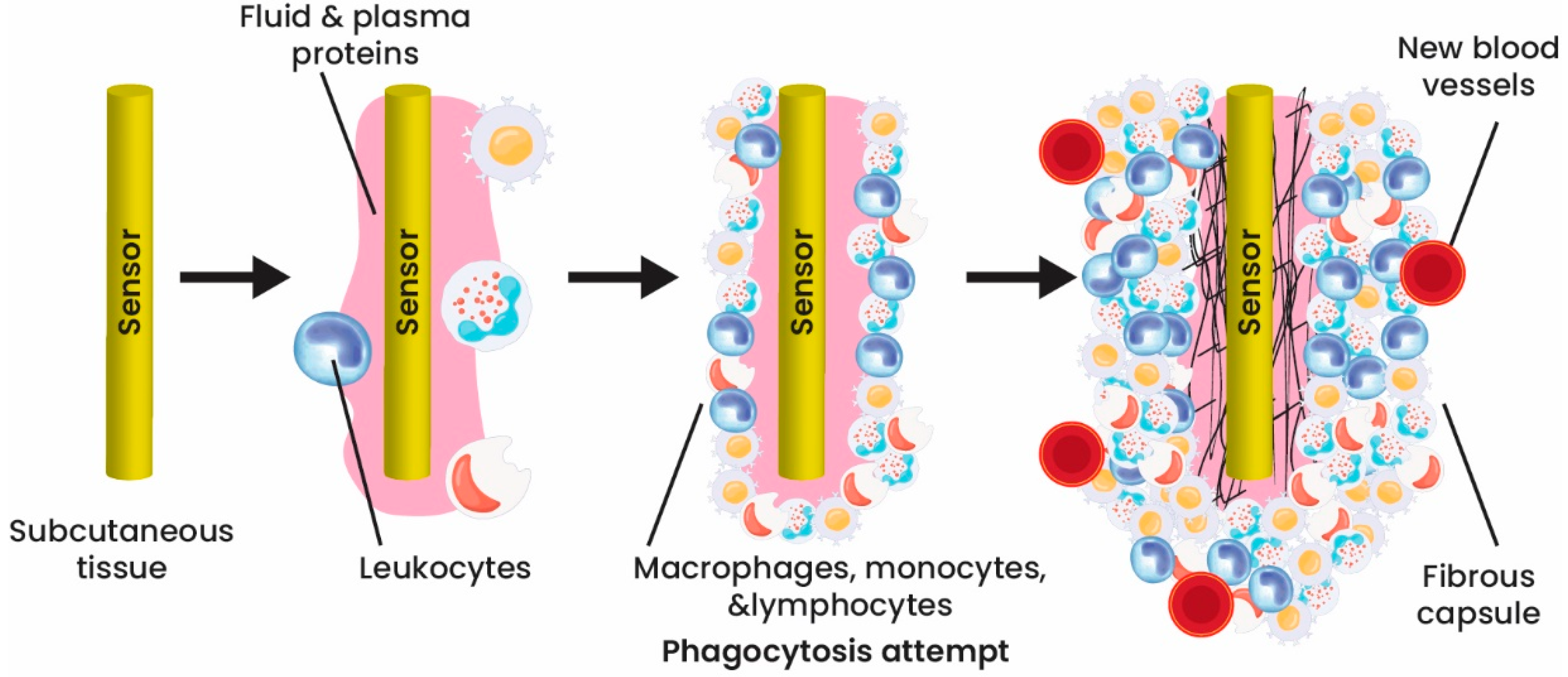
Creating implantable biosensors comes with significant obstacles, including the foreign-body response, stability, and biosensor response, as well as the need for continuous monitoring, power supply, and data transmission. Overcoming these obstacles requires meeting specific criteria, such as utilizing more adaptable and biocompatible biomaterials, achieving miniaturization, and ensuring reliability. The implementation of these design parameters is essential in the development of implantable biosensors [46][47]

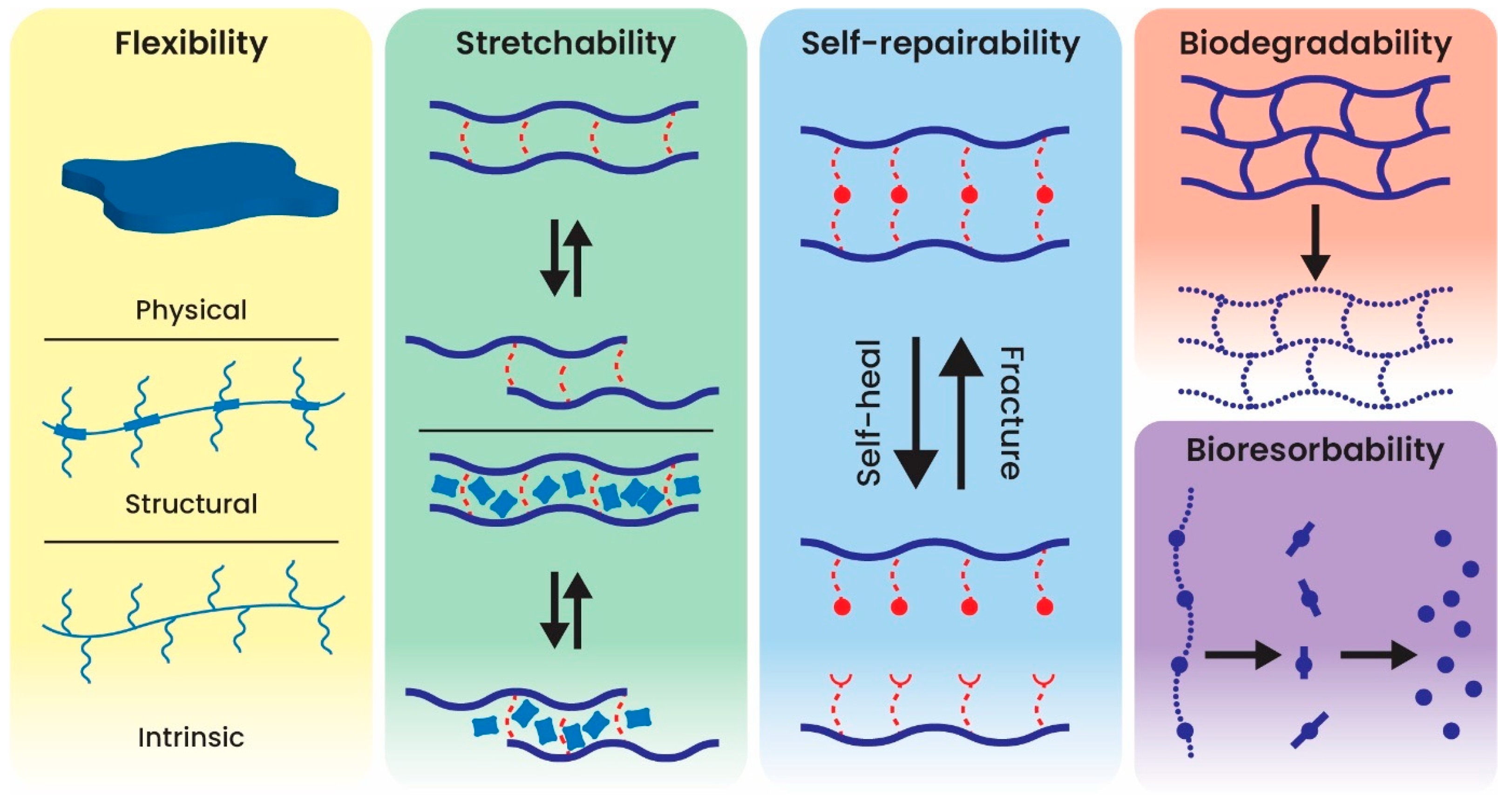
Various concerns should be considered and thought of in advance to design and then apply an ideal implantable biosensor. As aforementioned, biocompatibility of the design materials to avoid any unfavorable reactions in the body is the first and most crucial factor [48]. With developing science, many implantable sensors have shown minor cell injury. However, the materials used in long-term working biosensors (years or even a lifetime) must be biodegradable and biocompatible. For short-term strategies (e.g., digestible biosensors), high biocompatibility is demanded for both the instrument and the degraded products.

Besides biocompatibility, the device should show lasting stability, accuracy, selectivity, miniaturization, downscaled power, and portability. The label-free electrochemical implantable biosensors with the promising features cited earlier have gotten tremendous attention in this field. Integrating nanomaterials and nanotechnology in the biosensor field could improve biosensors’ performance and functionality and solve the main problematic issues in designing biosensors [49]

This entry is adapted from the peer-reviewed paper 10.3390/app13074630

References

  1. Clark, L.C., Jr.; Lyons, C. Electrode systems for continuous monitoring in cardiovascular surgery. Ann. N. Y. Acad. Sci. 1962, 102, 29–45.
  2. Boutry, C.M.; Beker, L.; Kaizawa, Y.; Vassos, C.; Tran, H.; Hinckley, A.C.; Pfattner, R.; Niu, S.; Li, J.; Claverie, J.; et al. Biodegradable and flexible arterial-pulse sensor for the wireless monitoring of blood flow. Nat. Biomed. Eng. 2019, 3, 47–57.
  3. Lee, J.; Cho, H.R.; Cha, G.D.; Seo, H.; Lee, S.; Park, C.K.; Kim, J.W.; Qiao, S.; Wang, L.; Kang, D.; et al. Flexible, sticky, and biodegradable wireless device for drug delivery to brain tumors. Nat. Commun. 2019, 10, 5205.
  4. Xiao, X.; Wang, J.; Liu, C.; Carlisle, J.A.; Mech, B.; Greenberg, R.; Guven, D.; Freda, R.; Humayun, M.S.; Weiland, J.; et al. In vitro and in vivo evaluation of ultrananocrystalline diamond for coating of implantable retinal microchips. J. Biomed. Mater. Res. B Appl. Biomater. 2006, 77, 273–281.
  5. Ben-Menachem, E.; Manon-Espaillat, R.; Ristanovic, R.; Wilder, B.J.; Stefan, H.; Mirza, W.; Tarver, W.B.; Wernicke, J.F. Vagus nerve stimulation for treatment of partial seizures: 1. A controlled study of effect on seizures. First International Vagus Nerve Stimulation Study Group. Epilepsia 1994, 35, 616–626.
  6. Cribier, A.; Eltchaninoff, H.; Bash, A.; Borenstein, N.; Tron, C.; Bauer, F.; Derumeaux, G.; Anselme, F.; Laborde, F.; Leon, M.B. Percutaneous transcatheter implantation of an aortic valve prosthesis for calcific aortic stenosis: First human case description. Circulation 2002, 106, 3006–3008.
  7. Kujawa, S.G.; Liberman, M.C. Adding insult to injury: Cochlear nerve degeneration after “temporary” noise-induced hearing loss. J. Neurosci. 2009, 29, 14077–14085.
  8. Rose, E.A.; Gelijns, A.C.; Moskowitz, A.J.; Heitjan, D.F.; Stevenson, L.W.; Dembitsky, W.; Long, J.W.; Ascheim, D.D.; Tierney, A.R.; Levitan, R.G.; et al. Long-term use of a left ventricular assist device for end-stage heart failure. N. Engl. J. Med. 2001, 345, 1435–1443.
  9. Sanders, G.D.; Hlatky, M.A.; Owens, D.K. Cost-effectiveness of implantable cardioverter-defibrillators. N. Engl. J. Med. 2005, 353, 1471–1480.
  10. Song, E.; Li, J.; Won, S.M.; Bai, W.; Rogers, J.A. Materials for flexible bioelectronic systems as chronic neural interfaces. Nat. Mater. 2020, 19, 590–603.
  11. Byun, S.H.; Sim, J.Y.; Zhou, Z.; Lee, J.; Qazi, R.; Walicki, M.C.; Parker, K.E.; Haney, M.P.; Choi, S.H.; Shon, A.; et al. Mechanically transformative electronics, sensors, and implantable devices. Sci. Adv. 2019, 5, eaay0418.
  12. Lim, H.R.; Kim, H.S.; Qazi, R.; Kwon, Y.T.; Jeong, J.W.; Yeo, W.H. Advanced Soft Materials, Sensor Integrations, and Applications of Wearable Flexible Hybrid Electronics in Healthcare, Energy, and Environment. Adv. Mater. 2020, 32, e1901924.
  13. Kang, J.; Son, D.; Wang, G.N.; Liu, Y.; Lopez, J.; Kim, Y.; Oh, J.Y.; Katsumata, T.; Mun, J.; Lee, Y.; et al. Tough and Water-Insensitive Self-Healing Elastomer for Robust Electronic Skin. Adv. Mater. 2018, 30, e1706846.
  14. Kang, J.; Tok, J.B.H.; Bao, Z.A. Self-healing soft electronics. Nat. Electron. 2019, 2, 144–150.
  15. Liu, Y.; Li, J.; Song, S.; Kang, J.; Tsao, Y.; Chen, S.; Mottini, V.; McConnell, K.; Xu, W.; Zheng, Y.Q.; et al. Morphing electronics enable neuromodulation in growing tissue. Nat. Biotechnol. 2020, 38, 1031–1036.
  16. Jeong, J.W.; McCall, J.G.; Shin, G.; Zhang, Y.; Al-Hasani, R.; Kim, M.; Li, S.; Sim, J.Y.; Jang, K.I.; Shi, Y.; et al. Wireless Optofluidic Systems for Programmable In Vivo Pharmacology and Optogenetics. Cell 2015, 162, 662–674.
  17. Kang, S.K.; Murphy, R.K.; Hwang, S.W.; Lee, S.M.; Harburg, D.V.; Krueger, N.A.; Shin, J.; Gamble, P.; Cheng, H.; Yu, S.; et al. Bioresorbable silicon electronic sensors for the brain. Nature 2016, 530, 71–76.
  18. Nelson, B.D.; Karipott, S.S.; Wang, Y.; Ong, K.G. Wireless Technologies for Implantable Devices. Sensors 2020, 20, 4604.
  19. Guillou, P.J.; Quirke, P.; Thorpe, H.; Walker, J.; Jayne, D.G.; Smith, A.M.; Heath, R.M.; Brown, J.M.; for the MRC CLASICC Trial Group. Short-term endpoints of conventional versus laparoscopic-assisted surgery in patients with colorectal cancer (MRC CLASICC trial): Multicentre, randomised controlled trial. Lancet 2005, 365, 1718–1726.
  20. Liu, J.; Fu, T.M.; Cheng, Z.; Hong, G.; Zhou, T.; Jin, L.; Duvvuri, M.; Jiang, Z.; Kruskal, P.; Xie, C.; et al. Syringe-injectable electronics. Nat. Nanotechnol. 2015, 10, 629–636.
  21. Montgomery, M.; Ahadian, S.; Davenport Huyer, L.; Lo Rito, M.; Civitarese, R.A.; Vanderlaan, R.D.; Wu, J.; Reis, L.A.; Momen, A.; Akbari, S.; et al. Flexible shape-memory scaffold for minimally invasive delivery of functional tissues. Nat. Mater. 2017, 16, 1038–1046.
  22. Qazi, R.; Gomez, A.M.; Castro, D.C.; Zou, Z.; Sim, J.Y.; Xiong, Y.; Abdo, J.; Kim, C.Y.; Anderson, A.; Lohner, F.; et al. Wireless optofluidic brain probes for chronic neuropharmacology and photostimulation. Nat. Biomed. Eng. 2019, 3, 655–669.
  23. Whyte, W.; Roche, E.T.; Varela, C.E.; Mendez, K.; Islam, S.; O’Neill, H.; Weafer, F.; Shirazi, R.N.; Weaver, J.C.; Vasilyev, N.V.; et al. Sustained release of targeted cardiac therapy with a replenishable implanted epicardial reservoir. Nat. Biomed. Eng. 2018, 2, 416–428.
  24. Lu, T.; Ji, S.; Jin, W.; Yang, Q.; Luo, Q.; Ren, T.L. Biocompatible and Long-Term Monitoring Strategies of Wearable, Ingestible and Implantable Biosensors: Reform the Next Generation Healthcare. Sensors 2023, 23, 2991.
  25. Mendelson, Y. Biomedical Sensors. In Introduction to Biomedical Engineering; Enderle, J.D., Bronzino, J.D., Eds.; Elsevier: Amsterdam, The Netherlands, 2012; pp. 609–666.
  26. Long, N.; Yu, B.; Moussy, Y.; Moussy, F. Strategies for testing long-term transcutaneous amperometric glucose sensors. Diabetes Technol. Ther. 2005, 7, 927–936.
  27. Frost, M.C.; Meyerhoff, M.E. Implantable chemical sensors for real-time clinical monitoring: Progress and challenges. Curr. Opin. Chem. Biol. 2002, 6, 633–641.
  28. Zhang, X. Real time and in vivo monitoring of nitric oxide by electrochemical sensors—From dream to reality. Front. Biosci. 2004, 9, 3434–3446.
  29. Zhou, D.D.; Greenberg, R.J. Microsensors and microbiosensors for retinal implants. Front. Biosci.-Landmark 2005, 10, 166–179.
  30. Gerritsen, M.; Kros, A.; Sprakel, V.; Lutterman, J.A.; Nolte, R.J.; Jansen, J.A. Biocompatibility evaluation of sol-gel coatings for subcutaneously implantable glucose sensors. Biomaterials 2000, 21, 71–78.
  31. Lee, S.; Ozlu, B.; Eom, T.; Martin, D.C.; Shim, B.S. Electrically conducting polymers for bio-interfacing electronics: From neural and cardiac interfaces to bone and artificial tissue biomaterials. Biosens. Bioelectron. 2020, 170, 112620.
  32. Ramanavicius, S.; Ramanavicius, A. Conducting Polymers in the Design of Biosensors and Biofuel Cells. Polymers 2021, 13, 49.
  33. Sharkawy, A.A.; Klitzman, B.; Truskey, G.A.; Reichert, W.M. Engineering the tissue which encapsulates subcutaneous implants. III. Effective tissue response times. J. Biomed. Mater. Res. 1998, 40, 598–605.
  34. Updike, S.J.; Shults, M.; Rhodes, R. Principles of long-term fully implanted sensors with emphasis on radiotelemetric monitoring of blood glucose from inside a subcutaneous foreign body capsule (FBC). In Biosensors in the Body: Continuous In Vivo Monitoring; Wiley: Hoboken, NJ, USA, 1997; pp. 117–137.
  35. Srinivasan, S.; Sawyer, P.N. Role of surface charge of the blood vessel wall, blood cells, and prosthetic materials in intravascular thrombosis. J. Colloid Interface Sci. 1970, 32, 456–463.
  36. Ainslie, K.M.; Desai, T.A. Microfabricated implants for applications in therapeutic delivery, tissue engineering, and biosensing. Lab Chip 2008, 8, 1864–1878.
  37. Popat, K.C.; Eltgroth, M.; Latempa, T.J.; Grimes, C.A.; Desai, T.A. Decreased Staphylococcus epidermis adhesion and increased osteoblast functionality on antibiotic-loaded titania nanotubes. Biomaterials 2007, 28, 4880–4888.
  38. Martin, F.; Walczak, R.; Boiarski, A.; Cohen, M.; West, T.; Cosentino, C.; Shapiro, J.; Ferrari, M. Tailoring width of microfabricated nanochannels to solute size can be used to control diffusion kinetics. J. Control. Release 2005, 102, 123–133.
  39. Yang, X.; Zhou, T.; Zwang, T.J.; Hong, G.; Zhao, Y.; Viveros, R.D.; Fu, T.M.; Gao, T.; Lieber, C.M. Bioinspired neuron-like electronics. Nat. Mater. 2019, 18, 510–517.
  40. Wang, L.; Xie, S.; Wang, Z.; Liu, F.; Yang, Y.; Tang, C.; Wu, X.; Liu, P.; Li, Y.; Saiyin, H.; et al. Functionalized helical fibre bundles of carbon nanotubes as electrochemical sensors for long-term in vivo monitoring of multiple disease biomarkers. Nat. Biomed. Eng. 2020, 4, 159–171.
  41. Curry, E.J.; Le, T.T.; Das, R.; Ke, K.; Santorella, E.M.; Paul, D.; Chorsi, M.T.; Tran, K.T.M.; Baroody, J.; Borges, E.R.; et al. Biodegradable nanofiber-based piezoelectric transducer. Proc. Natl. Acad. Sci. USA 2020, 117, 214–220.
  42. Morais, J.M.; Papadimitrakopoulos, F.; Burgess, D.J. Biomaterials/tissue interactions: Possible solutions to overcome foreign body response. AAPS J. 2010, 12, 188–196.
  43. Quinn, C.P.; Pathak, C.P.; Heller, A.; Hubbell, J.A. Photo-crosslinked copolymers of 2-hydroxyethyl methacrylate, poly(ethylene glycol) tetra-acrylate and ethylene dimethacrylate for improving biocompatibility of biosensors. Biomaterials 1995, 16, 389–396.
  44. Wisniewski, N.; Reichert, M. Methods for reducing biosensor membrane biofouling. Colloids Surf. B Biointerfaces 2000, 18, 197–219.
  45. Norton, L.W.; Koschwanez, H.E.; Wisniewski, N.A.; Klitzman, B.; Reichert, W.M. Vascular endothelial growth factor and dexamethasone release from nonfouling sensor coatings affect the foreign body response. J. Biomed. Mater. Res. A 2007, 81, 858–869.
  46. Webb, R.C.; Bonifas, A.P.; Behnaz, A.; Zhang, Y.; Yu, K.J.; Cheng, H.; Shi, M.; Bian, Z.; Liu, Z.; Kim, Y.S.; et al. Ultrathin conformal devices for precise and continuous thermal characterization of human skin. Nat. Mater. 2013, 12, 938–944.
  47. Rebelo, R.; Barbosa, A.I.; Correlo, V.M.; Reis, R.L. An Outlook on Implantable Biosensors for Personalized Medicine. Engineering 2021, 7, 1696–1699.
  48. Sadik, O.A.; Aluoch, A.O.; Zhou, A. Status of biomolecular recognition using electrochemical techniques. Biosens. Bioelectron. 2009, 24, 2749–2765.
  49. Erdem, A.; Karadeniz, H.; Caliskan, A. Single-Walled Carbon Nanotubes Modified Graphite Electrodes for Electrochemical Monitoring of Nucleic Acids and Biomolecular Interactions. Electroanalysis 2009, 21, 464–471.
More
This entry is offline, you can click here to edit this entry!
ScholarVision Creations