Cell migration is an essential process from embryogenesis to cell death. This is tightly regulated by numerous proteins that help in proper functioning of the cell. In diseases like cancer, this process is deregulated and helps in the dissemination of tumor cells from the primary site to secondary sites initiating the process of metastasis. For metastasis to be efficient, cytoskeletal components like actin, myosin, and intermediate filaments and their associated proteins should co-ordinate in an orderly fashion leading to the formation of many cellular protrusions-like lamellipodia and filopodia and invadopodia. Knowledge of this process is the key to control metastasis of cancer cells that leads to death in 90% of the patients. The focus of this review is giving an overall understanding of these process, concentrating on the changes in protein association and regulation and how the tumor cells use it to their advantage. Since the expression of cytoskeletal proteins can be directly related to the degree of malignancy, knowledge about these proteins will provide powerful tools to improve both cancer prognosis and treatment.
- cytoskeleton
- cancer
- actin
- actin binding proteins
- myosin
- tubulin
- intermediate filaments
- RhoGTPase
- PP2a
- PKA
1. Introduction
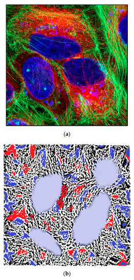
The cytoskeleton of eukaryotes is a dynamic complex three-dimensional network of filamentous proteins present within the cytoplasm. It consists of microfilaments mainly composed of G actin, intermediate filaments made up of keratins and vimentins and microtubules composed of α and β-tubulin [8] (Figure 1a and b). Cytoskeleton plays an essential role in controlling the cell shape by providing mechanical support, enabling cell motility and facilitating intra cellular transport. It also serves as a scaffold for signaling cascades by providing sites for localization and anchoring of signaling molecules. Dysregulation of cytoskeleton leads to numerous diseases including cancer [9].

Figure 1. (a) Immunofluorescent image of HeLa cells showing actin (green) tubulin (red tubulin antibody labelled with Cy3 secondary) and nucleus (blue). PFA fixed HeLa cells were stained for actin (Phalloidin 488) and alpha tubulin (Alexa 647) and image was taken using airy scan mode in Zeiss 800 confocal microscope using 63× objective. Scale bar = 5 µm. (b) Graphical representation of the cytoskeletal components of a cell like actin, tubulin, and intermediate filaments.
2. Data, Model, Management, Applications or Influences or …
Successful metastasis depends on cell invasion, migration, host immune escape, extravasation, and angiogenesis. The process of cell invasion and migration relies on the dynamic changes taking place in the cytoskeletal components; actin, tubulin and intermediate filaments. This is possible due to the plasticity of the cytoskeleton and coordinated action of all the three, is crucial for the process of metastasis from the primary site. Changes in cellular architecture by internal clues will affect the cell functions leading to the formation of different protrusions like lamellipodia, filopodia, and invadopodia that help in cell migration eventually leading to metastasis, which is life threatening than the formation of neoplasms.
This entry is adapted from the peer-reviewed paper 10.3390/biology9110385

