Neuroblastoma (NB) is an extracranial tumor of the peripheral nervous system arising from neural crest cells. It is the most common malignancy in infants and the most common extracranial solid tumor in children. The treatment for high-risk NB involves chemotherapy and surgical resection followed by high-dose chemotherapy with autologous stem-cell rescue and radiation treatment. However, those with high-risk NB are susceptible to relapse and the long-term side effects of standard chemotherapy. Polyphenols, including the sub-class of flavonoids, contain more than one aromatic ring with hydroxyl groups.
- neuroblastoma
- cancer
- flavonoids
- polyphenols
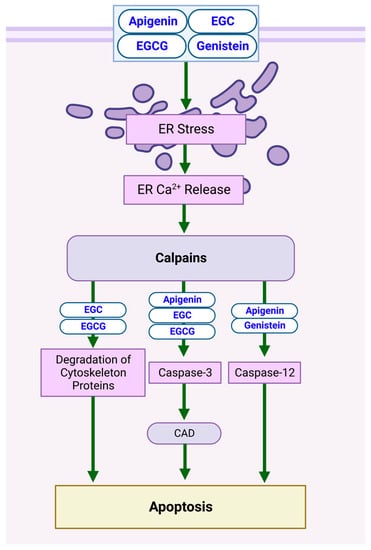
1. Calpain-Dependent Apoptotic Pathway

| Compound | Cell Line | Incubation Period | Concentration(s) | Biomarker Changes | Reference |
|---|---|---|---|---|---|
| Flavonoids | |||||
| Apigenin | SH-SY5Y | 24 h | 50 µM | ↑ Intracellular free [Ca2+] ↑ Calpain activation ↑ Caspase-12, -3 ↑ CAD |
[3] |
| EGC | SH-SY5Y | 24 h | 50 µM | ↑ Intracellular free [Ca2+] ↑ Calpain activation ↑ Cytoskeletal protein degradation ↑ Caspase-3 ↑ CAD |
[3] |
| EGCG | SH-SY5Y | 24 h | 50 µM | ↑ Intracellular free [Ca2+] ↑ Calpain activation ↑ Cytoskeletal protein degradation ↑ Caspase-3 ↑ CAD |
[3] |
| Genistein | SH-SY5Y | 24 h | 100 µM | ↑ Intracellular free [Ca2+] ↑ Calpain activation ↑ Caspase-12 |
[3] |
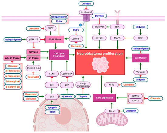
2. Anti-Proliferative Pathways

| Compound | Cell Line | Incubation Period | Concentration(s) | Biomarker Changes and Effects | Reference | |
|---|---|---|---|---|---|---|
| Flavonoids | ||||||
| Apigenin | NUB-7 and LAN-5 | 24 h | 10, 50, 100, 150, 200 µM IC50: 35 μM in NUB-7 IC50: 22 μM in LAN-5 |
↑ p53 ↑ p21WAF−1/CIP−1 |
↓ Proliferation | [10] |
| DEDC | SH-SY5Y | 24 h | 7.5 µg/mL | ↑ p53 mRNA ↑ p21 mRNA ↓ Cyclin-B1 |
[9] | |
| Didymin | CHLA-90 and SK-N-BE2 (p53-mutant) l + SMS-KCNR and LAN-5 (p53 wild-type) | 24 h | 50 μmol/L | ↓ P13K ↓ Akt |
↓ Proliferation | [11] |
| ↓ Vimentin | ↓ Motility of tumor cells | |||||
| ↓ N-Myc transcription | ||||||
| ↑ RKIP | ↓ MAPK pathway ↓ Proliferation |
|||||
| Isoliquiritigenin | SH-SY5Y | 24 h | 10–100 µM IC50: 25.4 µM |
↑ pERK1/2 | ↓ Cell migration ↓ Proliferation ↑ S + G2/M-phase arrest |
[12] |
| Rutin | LAN-5 | 24 h | 0, 25, 50, 100 μM | ↑ G2/M-phase arrest | [13] | |
| Quercetin | Neuro2a (mouse cell line) | 24 h | 10, 20, 40, 80, 120 μM IC50: 40 µM |
↑ p27 | ↓ Cyclin–CDK complex binding | [7] |
| ↓ Bcl-w | ↓ Tumor-cell-gene expression | |||||
| Quercetin | Neuroblastoma X glioma NG 108-15 cells (mouse cell line) | 48 h | 10 µM, 20 µM IC50: 10 µM |
↓ K+-channel activity | ↓ Cell growth | [8] |
| Non-Flavonoid Polyphenols | ||||||
| Curcumin | SK-N-SH | 24 h | 8, 16, 32 µM | ↓ CDC2 ↓ Cyclin B1 |
↑ G2/M-phase arrest | [14] |
| Curcumin | GI-L-IN, HTLA-230, SH-SY5Y, LAN5, SK-NBE2c, and IMR-32 | 18–72 h | 0.1–25 µM | ↓ NFκβ activator protein (AP-1) ↓ STAT3, STAT5 activation |
↓ Cell growth | [15] |
| Curcumin | NUB-7, LAN-5, IMR-32 and SK-N-BE(2) | 2–8 days | 0–100 µM * * Significantly inhibited proliferation in the range of 5–10 µM |
↑ p53 translocation from cytoplasm to nucleus ↑ p21WAF−1/CIP−1 |
↑ G1-, G2/M-, and S-phase arrest | [19] |
| Honokiol | Neuro-2a (mouse cell line) and NB41A3 | 72 h | 2.5, 5, 10, 20, 30, 40, 50, 60, 80, 100 µM LC50: 63.3 µM |
↑ Sub-G1-phase arrest | [16] | |
| Prenyl hydroxy-coumarins | Neuro-2a (mouse cell line) | 24, 48, 72 h | 6.25–200 µg/mL | ↑ Sub-G1-phase arrest | [20] | |
| Resveratrol | B103 (rat cell line) | 48 h | 5–20 µM IC50: 17.86 µM |
↓ Cyclin D1 | ↑ G1-phase arrest | [17] |
| Resveratrol | B65 (rat dopaminergic cell line) | 24 h | 25, 50, 100 µM | ↓ pAkt ↓ Cyclin D, E, A ↓ CDK2 ↑ p53 ↑ NFκβ |
↑ S-phase arrest | [18] |
| Resveratrol | NUB-7, LAN-5, IMR-32 and SK-N-BE(2) | 2–8 days | 25–160 µM | ↑ p53 translocation from cytoplasm to nucleus ↑ p21WAF−1/CIP−1 |
↑ G1-, G2/M-, and S-phase arrest | [30] |
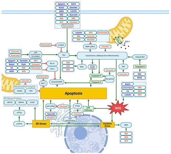
3. Mitochondrial and ER-Stress-Related Apoptotic Pathways

| Compound | Cell Line | Incubation Period | Concentration(s) | Biomarker Changes | Reference |
|---|---|---|---|---|---|
| Flavonoids | |||||
| DEDC | SH-SY5Y | 24 h | 7.5 µg/mL | ↓ Phosphor-STAT3 expression (ROS mediated) | [3] |
| Genistein | SK-N-DZ | 24 h | 10 µM | ↑ TNF-α ↑ FasL ↑ TRADD ↑ FADD |
[3] |
| EGC | SH-SY5Y | 24 h | 50 µM | ↑ Caspase-8 activation ↑ Proteolytic cleavage of Bid to tBid ↑ Bax oligomerization |
[3] |
| EGCG | SH-SY5Y | 24 h | 100 µM | ↑ Caspase-8 activation ↑ Proteolytic cleavage of Bid to tBid ↑ Bax oligomerization |
[3] |
| Rutin | LAN-5 | 24 h | 25, 50, 100 μM | ↑ TNF-α secretion | [13] |
| Non-Flavonoid Polyphenols | |||||
| Curcumin | LAN-5 | 3, 5, 24 h | 5, 10, 15, 20 µM | ↑ Bad ↑ PTEN ↑ ROS |
[31] |
| Honokiol | Neuro-2a (mouse cell line) | 30, 60, 120 µM | 24, 48, 72 h | ↑ RIP3 ↑ ROS |
[32] |
This entry is adapted from the peer-reviewed paper 10.3390/biom13030563
References
- Momeni, H.R. Role of Calpain in Apoptosis. Cell J. (Yakhteh) 2011, 13, 65.
- Martinez, J.A.; Zhang, Z.; Svetlov, S.I.; Hayes, R.L.; Wang, K.K.; Larner, S.F. Calpain and Caspase Processing of Caspase-12 Contribute to the ER Stress-Induced Cell Death Pathway in Differentiated PC12 Cells. Apoptosis 2010, 15, 1480–1493.
- Das, A.; Banik, N.L.; Ray, S.K. Mechanism of Apoptosis with the Involvement of Calpain and Caspase Cascades in Human Malignant Neuroblastoma SH-SY5Y Cells Exposed to Flavonoids. Int. J. Cancer 2006, 119, 2575–2585.
- Abotaleb, M.; Samuel, S.M.; Varghese, E.; Varghese, S.; Kubatka, P.; Liskova, A.; Büsselberg, D. Flavonoids in Cancer and Apoptosis. Cancers 2018, 11, 28.
- Ray, S.K.; Fidan, M.; Nowak, M.W.; Wilford, G.G.; Hogan, E.L.; Banik, N.L. Oxidative Stress and Ca2+ Influx Upregulate Calpain and Induce Apoptosis in PC12 Cells. Brain Res. 2000, 852, 326–334.
- Sergeev, I.N. Genistein Induces Ca2+-Mediated, Calpain/Caspase-12-Dependent Apoptosis in Breast Cancer Cells. Biochem. Biophys. Res. Commun. 2004, 321, 462–467.
- Sugantha Priya, E.; Selvakumar, K.; Bavithra, S.; Elumalai, P.; Arunkumar, R.; Raja Singh, P.; Brindha Mercy, A.; Arunakaran, J. Anti-Cancer Activity of Quercetin in Neuroblastoma: An in Vitro Approach. Neurol. Sci. 2014, 35, 163–170.
- Rouzaire-Dubois, B.; Gérard, V.; Dubois, J.M. Involvement of K+ Channels in the Quercetin-Induced Inhibition of Neuroblastoma Cell Growth. Pflügers Archiv. 1993, 423, 202–205.
- Liu, H.; Jiang, C.; Xiong, C.; Ruan, J. DEDC, a New Flavonoid Induces Apoptosis via a ROS-Dependent Mechanism in Human Neuroblastoma SH-SY5Y Cells. Toxicol. Vitr. 2012, 26, 16–23.
- Torkin, R.; Lavoie, J.F.; Kaplan, D.R.; Yeger, H. Induction of Caspase-Dependent, P53-Mediated Apoptosis by Apigenin in Human Neuroblastoma. Mol. Cancer 2005, 4, 1–11.
- Singhal, J.; Nagaprashantha, L.D.; Vatsyayan, R.; Ashutosh; Awasthi, S.; Singhal, S.S. Didymin Induces Apoptosis by Inhibiting N-Myc and Upregulating RKIP in Neuroblastoma. Cancer Prev. Res. 2012, 5, 473–483.
- Escobar, S.J.d.M.; Fong, G.M.; Winnischofer, S.M.B.; Simone, M.; Munoz, L.; Dennis, J.M.; Rocha, M.E.M.; Witting, P.K. Anti-Proliferative and Cytotoxic Activities of the Flavonoid Isoliquiritigenin in the Human Neuroblastoma Cell Line SH-SY5Y. Chem. Biol. Interact. 2019, 299, 77–87.
- Chen, H.; Miao, Q.; Geng, M.; Liu, J.; Hu, Y.; Tian, L.; Pan, J.; Yang, Y. Anti-Tumor Effect of Rutin on Human Neuroblastoma Cell Lines through Inducing G2/M Cell Cycle Arrest and Promoting Apoptosis. Sci. World J. 2013, 2013, 269165.
- Ye, Z.; Chen, D.; Zheng, R.; Chen, H.; Xu, T.; Wang, C.; Zhu, S.; Gao, X.; Zhang, J.; Li, D.; et al. Curcumin Induced G2/M Cycle Arrest in SK-N-SH Neuroblastoma Cells through the ROS-Mediated P53 Signaling Pathway. J. Food Biochem. 2021, 45, e13888.
- Pisano, M.; Pagnan, G.; Dettori, M.A.; Cossu, S.; Caffa, I.; Sassu, I.; Emionite, L.; Fabbri, D.; Cilli, M.; Pastorino, F.; et al. Enhanced Anti-Tumor Activity of a New Curcumin-Related Compound against Melanoma and Neuroblastoma Cells. Mol. Cancer 2010, 9, 1–12.
- Lin, J.W.; Chen, J.T.; Hong, C.Y.; Lin, Y.L.; Wang, K.T.; Yao, C.J.; Lai, G.M.; Chen, R.M. Honokiol Traverses the Blood-Brain Barrier and Induces Apoptosis of Neuroblastoma Cells via an Intrinsic Bax-Mitochondrion-Cytochrome c-Caspase Protease Pathway. Neuro Oncol. 2012, 14, 302–314.
- Rahman, M.A.; Kim, N.H.; Kim, S.H.; Oh, S.M.; Huh, S.O. Anti-proliferative and Cytotoxic Effects of Resveratrol in Mitochondria-Mediated Apoptosis in Rat B103 Neuroblastoma Cells. Korean J. Physiol. Pharm. 2012, 16, 321–326.
- Pizarro, J.G.; Verdaguer, E.; Ancrenaz, V.; Junyent, F.; Sureda, F.; Pallàs, M.; Folch, J.; Camins, A. Resveratrol Inhibits Proliferation and Promotes Apoptosis of Neuroblastoma Cells: Role of Sirtuin 1. Neurochem. Res. 2011, 36, 187–194.
- Liontas, A.; Yeger, H. Curcumin and Resveratrol Induce Apoptosis and Nuclear Translocation and Activation of P53 in Human Neuroblastoma. Anticancer Res. 2004, 24, 987.
- Sargolzaei, J.; Sadeghian, H.; Golahmadi, S.; Soukhtanloo, M. Cytotoxic Effects of Hydroxy Coumarin Derivations to Neuroblastoma N2a Cells. Iran. J. Pharm. Sci. 2020, 16, 95–106.
- Kubatka, P.; Kapinová, A.; Kello, M.; Kruzliak, P.; Kajo, K.; Výbohová, D.; Mahmood, S.; Murin, R.; Viera, T.; Mojžiš, J.; et al. Fruit Peel Polyphenols Demonstrate Substantial Anti-Tumour Effects in the Model of Breast Cancer. Eur. J. Nutr. 2016, 55, 955–965.
- Kapinova, A.; Stefanicka, P.; Kubatka, P.; Zubor, P.; Uramova, S.; Kello, M.; Mojzis, J.; Blahutova, D.; Qaradakhi, T.; Zulli, A.; et al. Are Plant-Based Functional Foods Better Choice against Cancer than Single Phytochemicals? A Critical Review of Current Breast Cancer Research. Biomed. Pharmacother. 2017, 96, 1465–1477.
- Hosseini, A.; Ghorbani, A. Cancer Therapy with Phytochemicals: Evidence from Clinical Studies. Avicenna J. Phytomed. 2015, 5, 84.
- Kubatka, P.; Kello, M.; Kajo, K.; Samec, M.; Liskova, A.; Jasek, K.; Koklesova, L.; Kuruc, T.; Adamkov, M.; Smejkal, K.; et al. Rhus Coriaria L. (Sumac) Demonstrates Oncostatic Activity in the Therapeutic and Preventive Model of Breast Carcinoma. Int. J. Mol. Sci. 2021, 22, 183.
- Morandi, F.; Bensa, V.; Calarco, E.; Pastorino, F.; Perri, P.; Corrias, M.V.; Ponzoni, M.; Brignole, C. The Olive Leaves Extract Has Anti-Tumor Effects against Neuroblastoma through Inhibition of Cell Proliferation and Induction of Apoptosis. Nutrients 2021, 13, 2178.
- Ahrens, S.; Appl, B.; Trochimiuk, M.; Dücker, C.; Feixas Serra, G.; Oliver Grau, A.; Reinshagen, K.; Pagerols Raluy, L. Kigelia Africana Inhibits Proliferation and Induces Cell Death in Stage 4 Neuroblastoma Cell Lines. Biomed. Pharmacother. 2022, 154, 113584.
- Roomi, M.W.; Kalinovsky, T.; Roomi, N.W.; Niedzwiecki, A.; Rath, M. Inhibition of the SK-N-MC Human Neuroblastoma Cell Line in Vivo and in Vitro by a Novel Nutrient Mixture. Oncol. Rep. 2013, 29, 1714–1720.
- Mazurakova, A.; Samec, M.; Koklesova, L.; Biringer, K.; Kudela, E.; Al-Ishaq, R.K.; Pec, M.; Giordano, F.A.; Büsselberg, D.; Kubatka, P.; et al. Anti-Prostate Cancer Protection and Therapy in the Framework of Predictive, Preventive and Personalised Medicine—Comprehensive Effects of Phytochemicals in Primary, Secondary and Tertiary Care. EPMA J. 2022, 13, 461–486.
- Chakrabarti, M.; Ai, W.; Banik, N.L.; Ray, S.K. Overexpression of MiR-7-1 Increases Efficacy of Green Tea Polyphenols for Induction of Apoptosis in Human Malignant Neuroblastoma SH-SY5Y and SK-N-DZ Cells. Neurochem. Res. 2013, 38, 420–432.
- ISSN 1791-7530 (Online)|Anti-cancer Research|The ISSN Portal. Available online: https://portal.issn.org/resource/ISSN/1791-7530 (accessed on 9 November 2022).
- Picone, P.; Nuzzo, D.; Caruana, L.; Messina, E.; Scafidi, V.; Di Carlo, M. Curcumin Induces Apoptosis in Human Neuroblastoma Cells via Inhibition of AKT and Foxo3a Nuclear Translocation. Free. Radic. Res. 2014, 48, 1397–1408.
- Zhang, J.; Liu, Q.; Shi, L.; Qin, P.; Wang, Q. Honokiol Triggers Receptor-Interacting Protein Kinase 3-Mediated Cell Death of Neuroblastoma Cells by Upregulating Reactive Oxygen Species. Mol. Med. Rep. 2017, 16, 8525–8529.
- George, J.; Banik, N.L.; Ray, S.K. Genistein Induces Receptor and Mitochondrial Pathways and Increases Apoptosis during BCL-2 Knockdown in Human Malignant Neuroblastoma SK-N-DZ Cells. J. Neurosci. Res. 2010, 88, 877–886.
- Seervi, M.; Xue, D. Mitochondrial Cell Death Pathways in Caenorhabiditis Elegans. Curr. Top. Dev. Biol. 2015, 114, 43–65.
