Plagiochilin A functions as an inhibitor of the termination phase of cytokinesis: the membrane abscission stage. This unique, innovative mechanism of action, coupled with its marked anticancer action, notably against prostate cancer cells, make plagiochilin A an interesting lead molecule for the development of novel anticancer agents. There are known options to increase its potency, as deduced from structure–activity relationships. The analysis shed light on this family of bryophyte species and the little-known group of bioactive terpenoid plagiochilins. Plagiochilin A and derivatives shall be further exploited for the design of novel anticancer targeting the cytokinesis pathway.
1. Introduction
Bryophytes are non-vascular plants which include thalloid and leafy liverworts, mosses and hornworts. These three lineages form a unique part of the vegetation. They are small-sized, structurally simple diversified plants able to adapt to most ecosystems on Earth [
1,
2]. Bryophytes and tracheophytes (non-vascular and vascular plants, respectively) derive from an ancestral land plant and diverged during the Cambrian, some 500 million years ago [
3]. Bryophytes are collectively divided into three main groups: Bryophyta (mosses), Marchantiophyta (liverworts) and Anthocerotophyta (hornworts). They represent the second-largest group of land plants after angiosperms. Liverworts are particularly abundant, with some 7300 extant species [
4]. The first representations of liverworts date from late antiquity [
5].
Leafy (or scaly) liverworts are particularly abundant and diversified (order: Jungermanniales). They grow commonly on moist soil or damp rocks (such as thallose liverworts). In 2016, a worldwide checklist for liverworts and hornworts included 7486 species in 398 genera representing 92 families from the two phyla [
6]. The genus
Plagiochila (Plagiochilaceae) represents one of the largest groups of leafy liverworts, with more than 500 species distributed worldwide and a broad geographical amplitude, mostly in the humid tropics [
7]. World Flora Online refers to 556 accepted names of
Plagiochila species and more than 950 species including synonyms and unchecked species [
8]. Another recent study refers to 1600 validly published
Plagiochila names [
9].
Despite their number and high adaptative capacities, the medicinal use of bryophytes remains relatively limited, probably because of their small size, lack of conspicuous organs such as colored fruits and flowers, and the difficulty of identification. There are, however, species with medicinal properties, such as
Conocephalum conicum (L.) Dumort.,
Polytrichum commune and
Marchantia chenopoda L. [
10]. In the genus
Plagiochila, a few species have also been used ethnomedicinally, such as
P. beddomei Steph. used in the form of paste by tribe Melghat Region (India) for treating skin diseases [
11,
12] and
P. disticha (Lehm. & Lindenb.) Lindenb used traditionally in Peru to treat rheumatism or to regulate menstruation [
13].
Diverse bioactive products have been isolated from
Plagiochila species, including antitumor agents [
14], antifungal molecules [
15,
16], insecticidal compounds [
17] and antimicrobial products [
18]. Most of the isolated bioactive compounds are terpenoids such as the antifungal products plagicosins A-N, or alkaloids such as plagiochianins A-B from the Chinese liverworts
P. fruticosa Mitt. and
P. duthiana Steph., respectively [
18,
19]. However, the leading product isolated from
Plagiochila species is without doubt the sesquiterpenoid plagiochilin A, first isolated from several
Plagiochila species in the 1970s, together with its congeners plagiochilins B and C, and precursors plagiochilide and plagiochilal [
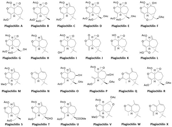
20]. Over the past 43 years, different analogues have been isolated leading to a series of 24 derivatives, designated plagiochilins A-to-X, and related compounds (
Figure 1). The present review deals the identification of these compounds and their pharmacological properties. Information about their mechanism of action is often very limited, but important observations have been made, leading to the identification of potential targets for some of these compounds, in particular for the leader product plagiochilin A (
Figure 2).
Figure 1. History of plagiochilins discovery. The 24 plagiochilins (A–W) have been identified and structurally characterized aver a period of 30 years. They are produced by several Plagiochila species, such as those indicated (a non-exhaustive list).
Figure 2. Structures of the 24 plagiochilins (A-to-W).
2. Pharmacological Properties of Plagiochilins A and C
The pharmacological effects of the plagiochilins have been rarely investigated. Nevertheless, a few types of bioactivities have been reported with plagiochilins A and C. The data in
Table 1 illustrate the potential of the two compounds, but there is no systematic study comparing activities of all compounds, or activities of a given compound across multiple indications or pathologies. Initially, it was shown that plagiochilin A displays a modest antifeedant action against the armyworm
Spodoptera exempta Walker (Lepidoptera: Noctuidae) which is an episodic migratory pest of cereal crops in sub-Saharan Africa. The level of activity is quite modest [
35]. Another study has investigated the insecticidal activity of natural products isolated from
P. diversifolia, but in that case the only plagiochilin tested was plagiochilin B and no activity was reported. In this case, a marked insecticidal action was observed with another epoxide-containing compound called fusicogigantone B, a fusicoccane-type diterpenoid [
31]. Both fusicogigantones A and B, isolated from
P. bursata and
P. diversifolia, respectively, have been shown to inhibit the growth of another species of Lepidoptera (
Spodoptera frugiperda) [
17,
31]. Plagiochilin A was shown to display a noticeable antiprotozoal activity, reducing the growth of the amastigote form of
Leishmania amazonensis, with an IC
50 value of 7.1 µM. However, the level of activity is quite modest compared to that of the control drug amphotericin B (IC
50 = 0.13 µM). In the same study, no activity was observed against the fungus
Mycobacterium tuberculosis [
13].
In a more interesting way, plagiochilin A was characterized as an antiproliferative agent, reducing the growth of different cultured cancer cell lines. Both plagiochilins A and C display marked antiproliferative activities and there are known options to further increase their anticancer potency. An option is to introduce a methoxy group at position C-3, as observed with the derivative methoxyplagiochilin A2 which has been shown to be more potent than plagiochilin C against H460 lung cancer cells (IC
50 = 6.7 and 13.1 µM, respectively) [
58]. Another option is to introduce a side chain at the C-14/C-15 position, either an octanoyl side chain or a dodecadienoate side chain, for example. In both cases, the resulting compounds were found to be 60 times more potent against P-388 leukemia cells than the parent compound plagiochilin A [
38]. The extraordinary potency of plagiochilin A-15-yl n-octanoate raises questions (solubility, stability) and opens perspectives. The octanoate moiety may serve only as a “lipophilic carrier” (bioavailability enhancement), and may not be directly implicated in the target interaction. Novel C-12/C-13-substituted derivatives of plagiochilin A should be designed.
Plagiochilin A exhibits antiproliferative activities against different types of cancer cells. The growth inhibition GI
50 values ranged from 1.4 to 6.8 µM with a range of tumor cell lines, including prostate (DU145), breast (MCF-7), lung (HT-29) and leukemia (K562) cells for example [
13]. The level of activity against DU145 prostate cancer cell is interesting (GI
50 = 1.4 µM) because it is superior to that observed with the reference anticancer drug fludarabine phosphate (GI
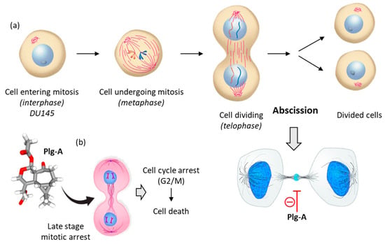
50 = 3.0 µM). The sensitivity of prostate cancer cells toward plagiochilin A warranted further investigation. In 2018, Bates and coworkers analyzed the effect of plagiochilin A on the cell cycle progression of DU145 cells and their capacity to complete cytokinesis, the part of the cell division process during which the cytoplasm of a single eukaryotic cell divides into two daughter cells. Interestingly, it was observed that the compound (at 5 µM for 24–48 h) could block cell division by preventing completion of cytokinesis, and thereby inducing cell death [
59]. The treated DU145 cells accumulated at the G2/M phase, notably cells still connected with intercellular bridges, corresponding to a late cytokinesis stage, the so-called membrane abscission stage (stained with an α-anti-tubulin antibody). The compound induced specific mitotic figures and reduced significantly the number and size of DU145 cell colonies. The failure of the cells to complete cytokinesis triggered apoptosis [
59]. Altogether, these data indicated that plagiochilin A exerts an effect on the cytoskeleton, with a rearrangement of α-tubulin characteristic of cytokinetic membrane abscission, which is a spatially and temporally regulated process [
59] (
Figure 3).
Figure 3. Mechanism of action of plagiochilin A (Plg-A). (
a) The cell division process, to illustrate DU145 prostate cancer cells undergoing mitosis. Prior to cell division, at the telophase stage of mitosis, the two cells are connected by an intercellular bridge (with a central midbody). Plg-A inhibits cell division by preventing completion of cytokinesis, particularly at the final abscission stage. (
b) Inhibition of the late stage of cytokinesis leads to cell cycle arrest (G2/M) and subsequently to inhibition of cell colony formation and induction of cell death [
59].
3. Hypothesized Mechanism of Action of Plagiochilin A
The mechanics implicated in the regulation of abscission is relatively well-known. This process leads to the physical cut of the intercellular bridge which connects two daughter cells and concludes cell division. The process is tightly regulated in cells, with intervention of multiple protein effectors including different kinases (e.g., PLK4, Aurora B) and proteins containing microtubule-interacting and trafficking (MIT) domains [
60,
61,
62]. The process opens perspectives to comprehend the mechanism of action of plagiochilin A. the compound may target MIT-containing proteins, or more directly it may associate with alpha-tubulin during mitosis, for example. There exist small molecules which induced cytokinesis failure at the point of abscission, such as a series of dynamin GTPase inhibitors called dynoles [
63,
64]. These products induce apoptosis following cytokinesis failure, as observed with plagiochilin A. Therefore, we can imagine that the natural product acts as an inhibitor of termination of cytokinesis (abscission) by blocking one or several proteins implicated in the process, or by directly altering the microtubule-organizing center which recruits α- and β-tubulins for microtubule nucleation. In this context, one of the potential mechanisms could be a direct binding of the compound to α-tubulin, in particular to the pironetin-binding site which is known to accommodate compounds bearing a dihydro-pyrone moiety [
65,
66]. This moiety can be found in plagiochilin Q, for example, and recently, we have shown that natural products with a 5,6-dihydro-α-pyrone unit (cryptoconcatones) can function as α-tubulin-binding agents [
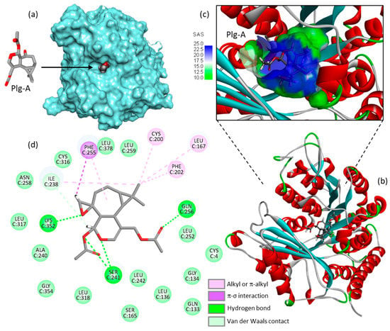
67]. Based on these considerations, we have initiated binding studies of plagiochilins to α-tubulin and the first information obtained by molecular docking look interesting. For the docking analysis, the high-resolution crystal structure of the reference product pironetin (a dihydropyrone derivative with an α,β-unsaturated lactone acting as a plant growth regulator) bound to α/β-tubulin dimer was used as a template (PDB: 5FNV) and the binding of plagiochilin A to the pironetin site was modeled. Apparently, plagiochilin A could form stable complexes with α-tubulin, via binding to the pironetin site, as represented in
Figure 4. These are preliminary, but promising information. We are now comparing various plagiochilins for their capacity to bind to α-tubulin, using molecular modeling. The mechanism whereby plagiochilin A specifically blocks abscission warrant further investigation. The compound is an atypical inhibitor of cytokinesis. The panoply of plagiochilins shall be further exploited to identify the best inhibitors and to delineate the structure–activity relationships in the series. The modeling analysis shall help also to delineate the mechanism of action of other aromadendrane derivatives, such as the related natural products hanegokedial, ovalifolienal, ovalifolienalone (from
P. semidecurrens) and others [
30,
68].
Figure 4. Molecular model of plagiochilin A (Plg-A) bound to the pironetin site of α-tubulin (PDB: 5FNV). (
a) Plg-A fits into a central, deep cavity of the protein. (
b) Ribbon model of α-tubulin with bound Plg-A, with α-helices (in red) and β-sheets (in cyan). (
c) A close-up view of Plg-A inserted into the binding cavity, with the solvent-accessible surface (SAS) surrounding the drug binding zone (color code indicated). (
d) Binding map contacts for Plg-A bound to α-tubulin (color code indicated). The docking model was kindly provided by Prof. Gérard Vergoten (University of Lille, France). The docking analysis was performed as recently described [
65].
This entry is adapted from the peer-reviewed paper 10.3390/life13030758


