Novel nano-engineering protocols have been actively synergized with fluorescence spectroscopic techniques to yield higher intensity from radiating dipoles, through the process termed plasmon-enhanced fluorescence (PEF). Consequently, the limit of detection of analytes of interest has been dramatically improvised on account of higher sensitivity rendered by augmented fluorescence signals. Metallic thin films sustaining surface plasmon polaritons (SPPs) have been creatively hybridized with such PEF platforms to realize a substantial upsurge in the global collection efficiency in a judicious technology termed surface plasmon-coupled emission (SPCE). This Editorial Review by Dr. Seemesh Bhaskar, University of Illinois Urbana-Champaign, provides a spotlight on the latest developments in SPCE substrate engineering to the broad audience of photo-plasmonics, spectroscopy, micro- & nanotechnology, life sciences, thin films and point-of-care diagnostics.
- surface plasmon coupled emission
- luminescence
- nano-engineering
- smartphone diagnostics
- photonics
- plasmonics
- biosensing
- point-of-care diagnostics
- materials
1. Introduction
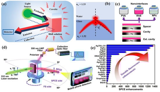
2. Surface Plasmon Coupled Emission (SPCE) Technology
This entry is adapted from the peer-reviewed paper 10.3390/mi14030574
References
- Yao, J.; Yang, M.; Duan, Y. Chemistry, Biology, and Medicine of Fluorescent Nanomaterials and Related Systems: New Insights into Biosensing, Bioimaging, Genomics, Diagnostics, and Therapy. Chem. Rev. 2014, 114, 6130–6178.
- Yoshida, M.; Chida, H.; Kimura, F.; Yamamura, S.; Tawa, K. Multi-Color Enhanced Fluorescence Imaging of a Breast Cancer Cell with A Hole-Arrayed Plasmonic Chip. Micromachines 2020, 11, 604.
- Badshah, M.A.; Koh, N.Y.; Zia, A.W.; Abbas, N.; Zahra, Z.; Saleem, M.W. Recent Developments in Plasmonic Nanostructures for Metal Enhanced Fluorescence-Based Biosensing. Nanomaterials 2020, 10, 1749.
- Xiong, Y.; Huang, Q.; Canady, T.D.; Barya, P.; Liu, S.; Arogundade, O.H.; Race, C.M.; Che, C.; Wang, X.; Zhou, L.; et al. Photonic Crystal Enhanced Fluorescence Emission and Blinking Suppression for Single Quantum Dot Digital Resolution Biosensing. Nat. Commun. 2022, 13, 4647.
- Cao, S.-H.; Cai, W.-P.; Liu, Q.; Li, Y.-Q. Surface Plasmon–Coupled Emission: What Can Directional Fluorescence Bring to the Analytical Sciences? Annu. Rev. Anal. Chem. 2012, 5, 317–336.
- Bhaskar, S.; Kowshik, N.C.S.S.; Chandran, S.P.; Ramamurthy, S.S. Femtomolar Detection of Spermidine Using Au Decorated SiO2 Nanohybrid on Plasmon-Coupled Extended Cavity Nanointerface: A Smartphone-Based Fluorescence Dequenching Approach. Langmuir ACS J. Surf. Colloids 2020, 36, 2865–2876.
- Li, J.-F.; Li, C.-Y.; Aroca, R.F. Plasmon-Enhanced Fluorescence Spectroscopy. Chem. Soc. Rev. 2017, 46, 3962–3979.
- Dutta Choudhury, S.; Badugu, R.; Lakowicz, J.R. Directing Fluorescence with Plasmonic and Photonic Structures. Acc. Chem. Res. 2015, 48, 2171–2180.
- Bhaskar, S.; Visweswar Kambhampati, N.S.; Ganesh, K.M.; Sharma P, M.; Srinivasan, V.; Ramamurthy, S.S. Metal-Free, Graphene Oxide-Based Tunable Soliton and Plasmon Engineering for Biosensing Applications. ACS Appl. Mater. Interfaces 2021, 13, 17046–17061.
- Lakowicz, J.R.; Ray, K.; Chowdhury, M.; Szmacinski, H.; Fu, Y.; Zhang, J.; Nowaczyk, K. Plasmon-Controlled Fluorescence: A New Paradigm in Fluorescence Spectroscopy. Analyst 2008, 133, 1308–1346.
- Bhaskar, S.; Das, P.; Moronshing, M.; Rai, A.; Subramaniam, C.; Bhaktha, S.B.N.; Ramamurthy, S.S. Photoplasmonic Assembly of Dielectric-Metal, Nd2O3-Gold Soret Nanointerfaces for Dequenching the Luminophore Emission. Nanophotonics 2021, 10, 3417–3431.
- Che, C.; Xue, R.; Li, N.; Gupta, P.; Wang, X.; Zhao, B.; Singamaneni, S.; Nie, S.; Cunningham, B.T. Accelerated Digital Biodetection Using Magneto-Plasmonic Nanoparticle-Coupled Photonic Resonator Absorption Microscopy. ACS Nano 2022, 16, 2345–2354.
- Arathi, P.J.; Seemesh, B.; Ramanathan, V. Disulphide Linkage: To Get Cleaved or Not? Bulk and Nano Copper Based SERS of Cystine. Spectrochim. Acta. A. Mol. Biomol. Spectrosc. 2018, 196, 229–232.
- Xiong, Y.; Li, N.; Che, C.; Wang, W.; Barya, P.; Liu, W.; Liu, L.; Wang, X.; Wu, S.; Hu, H.; et al. Microscopies Enabled by Photonic Metamaterials. Sensors 2022, 22, 1086.
- Chauhan, N.; Xiong, Y.; Ren, S.; Dwivedy, A.; Magazine, N.; Zhou, L.; Jin, X.; Zhang, T.; Cunningham, B.T.; Yao, S.; et al. Net-Shaped DNA Nanostructures Designed for Rapid/Sensitive Detection and Potential Inhibition of the SARS-CoV-2 Virus. J. Am. Chem. Soc. 2022.
- Rahman, M.A.; Kim, D.; Arora, D.; Huh, J.-Y.; Byun, J.Y. Structural Colors on Al Surface via Capped Cu-Si3N4 Bilayer Structure. Micromachines 2023, 14, 471.
- Lakowicz, J.R. Radiative Decay Engineering 5: Metal-Enhanced Fluorescence and Plasmon Emission. Anal. Biochem. 2005, 337, 171–194.
- Tran, N.H.T.; Trinh, K.T.L.; Lee, J.-H.; Yoon, W.J.; Ju, H. Fluorescence Enhancement Using Bimetal Surface Plasmon-Coupled Emission from 5-Carboxyfluorescein (FAM). Micromachines 2018, 9, 460.
- Bauch, M.; Toma, K.; Toma, M.; Zhang, Q.; Dostalek, J. Plasmon-Enhanced Fluorescence Biosensors: A Review. Plasmonics 2014, 9, 781–799.
- Zhang, L.; Miao, G.; Zhang, J.; Liu, L.; Gong, S.; Li, Y.; Cui, D.; Wei, Y.; Yu, D.; Qiu, X.; et al. Development of a Surface Plasmon Resonance and Fluorescence Imaging System for Biochemical Sensing. Micromachines 2019, 10, 442.
- Singh, S.; Singh, P.K.; Umar, A.; Lohia, P.; Albargi, H.; Castañeda, L.; Dwivedi, D.K. 2D Nanomaterial-Based Surface Plasmon Resonance Sensors for Biosensing Applications. Micromachines 2020, 11, 779.
- Jahani, S.; Jacob, Z. All-Dielectric Metamaterials. Nat. Nanotechnol. 2016, 11, 23–36.
- Zhang, D.; Xiang, Y.; Chen, J.; Cheng, J.; Zhu, L.; Wang, R.; Zou, G.; Wang, P.; Ming, H.; Rosenfeld, M.; et al. Extending the Propagation Distance of a Silver Nanowire Plasmonic Waveguide with a Dielectric Multilayer Substrate. Nano Lett. 2018, 18, 1152–1158.
- Badugu, R.; Mao, J.; Zhang, D.; Descrovi, E.; Lakowicz, J.R. Fluorophore Coupling to Internal Modes of Bragg Gratings. J. Phys. Chem. C 2020, 124, 22743–22752.
- Lakowicz, J.R. Radiative Decay Engineering: Biophysical and Biomedical Applications. Anal. Biochem. 2001, 298, 1–24.
- Lakowicz, J.R.; Shen, Y.; D’Auria, S.; Malicka, J.; Fang, J.; Gryczynski, Z.; Gryczynski, I. Radiative Decay Engineering: 2. Effects of Silver Island Films on Fluorescence Intensity, Lifetimes, and Resonance Energy Transfer. Anal. Biochem. 2002, 301, 261–277.
- Lakowicz, J.R. Radiative Decay Engineering 3. Surface Plasmon-Coupled Directional Emission. Anal. Biochem. 2004, 324, 153–169.
- Gryczynski, I.; Malicka, J.; Gryczynski, Z.; Lakowicz, J.R. Radiative Decay Engineering 4. Experimental Studies of Surface Plasmon-Coupled Directional Emission. Anal. Biochem. 2004, 324, 170–182.
- Badugu, R.; Nowaczyk, K.; Descrovi, E.; Lakowicz, J.R. Radiative Decay Engineering 6: Fluorescence on One-Dimensional Photonic Crystals. Anal. Biochem. 2013, 442, 83–96.
- Badugu, R.; Descrovi, E.; Lakowicz, J.R. Radiative Decay Engineering 7: Tamm State-Coupled Emission Using a Hybrid Plasmonic–Photonic Structure. Anal. Biochem. 2014, 445, 1–13.
- Zhu, L.; Badugu, R.; Zhang, D.; Wang, R.; Descrovi, E.; Lakowicz, J.R. Radiative Decay Engineering 8: Coupled Emission Microscopy for Lens-Free High-Throughput Fluorescence Detection. Anal. Biochem. 2017, 531, 20–36.
- Chowdhury, M.H.; Ray, K.; Geddes, C.D.; Lakowicz, J.R. Use of Silver Nanoparticles to Enhance Surface Plasmon-Coupled Emission (SPCE). Chem. Phys. Lett. 2008, 452, 162–167.
- Tran, H.N.Q.; Le, N.D.A.; Le, Q.N.; Law, C.S.; Lim, S.Y.; Abell, A.D.; Santos, A. Spectral Engineering of Tamm Plasmon Resonances in Dielectric Nanoporous Photonic Crystal Sensors. ACS Appl. Mater. Interfaces 2022, 14, 22747–22761.
- Bhaskar, S.; Ramamurthy, S.S. Mobile Phone-Based Picomolar Detection of Tannic Acid on Nd2O3 Nanorod–Metal Thin-Film Interfaces. ACS Appl. Nano Mater. 2019, 2, 4613–4625.
- Rai, A.; Bhaskar, S.; Ganesh, K.M.; Ramamurthy, S.S. Engineering of Coherent Plasmon Resonances from Silver Soret Colloids, Graphene Oxide and Nd2O3 Nanohybrid Architectures Studied in Mobile Phone-Based Surface Plasmon-Coupled Emission Platform. Mater. Lett. 2021, 304, 130632.
- Cao, S.-H.; Cai, W.-P.; Liu, Q.; Xie, K.-X.; Weng, Y.-H.; Huo, S.-X.; Tian, Z.-Q.; Li, Y.-Q. Label-Free Aptasensor Based on Ultrathin-Linker-Mediated Hot-Spot Assembly To Induce Strong Directional Fluorescence. J. Am. Chem. Soc. 2014, 136, 6802–6805.
- Rai, A.; Bhaskar, S.; Reddy, N.; Ramamurthy, S.S. Cellphone-Aided Attomolar Zinc Ion Detection Using Silkworm Protein-Based Nanointerface Engineering in a Plasmon-Coupled Dequenched Emission Platform. ACS Sustain. Chem. Eng. 2021, 9, 14959–14974.
- Tran, N.H.T.; Trinh, K.T.L.; Lee, J.-H.; Yoon, W.J.; Ju, H. Reproducible Enhancement of Fluorescence by Bimetal Mediated Surface Plasmon Coupled Emission for Highly Sensitive Quantitative Diagnosis of Double-Stranded DNA. Small 2018, 14, 1801385.
- Rathnakumar, S.; Bhaskar, S.; Rai, A.; Saikumar, D.V.V.; Kambhampati, N.S.V.; Sivaramakrishnan, V.; Ramamurthy, S.S. Plasmon-Coupled Silver Nanoparticles for Mobile Phone-Based Attomolar Sensing of Mercury Ions. ACS Appl. Nano Mater. 2021, 4, 8066–8080.
- Bhaskar, S.; Das, P.; Srinivasan, V.; Bhaktha B.N., S.; Ramamurthy, S.S. Bloch Surface Waves and Internal Optical Modes-Driven Photonic Crystal-Coupled Emission Platform for Femtomolar Detection of Aluminum Ions. J. Phys. Chem. C 2020, 124, 7341–7352.
- Rai, A.; Bhaskar, S.; Ramamurthy, S.S. Plasmon-Coupled Directional Emission from Soluplus-Mediated AgAu Nanoparticles for Attomolar Sensing Using a Smartphone. ACS Appl. Nano Mater. 2021, 4, 5940–5953.
- Bhaskar, S.; Singh, A.K.; Das, P.; Jana, P.; Kanvah, S.; Bhaktha B.N., S.; Ramamurthy, S.S. Superior Resonant Nanocavities Engineering on the Photonic Crystal-Coupled Emission Platform for the Detection of Femtomolar Iodide and Zeptomolar Cortisol. ACS Appl. Mater. Interfaces 2020, 12, 34323–34336.
- Rai, A.; Bhaskar, S.; Ganesh, K.M.; Ramamurthy, S.S. Gelucire®-Mediated Heterometallic AgAu Nanohybrid Engineering for Femtomolar Cysteine Detection Using Smartphone-Based Plasmonics Technology. Mater. Chem. Phys. 2022, 279, 125747.
- Xie, K.-X.; Jia, S.-S.; Zhang, J.-H.; Wang, H.; Wang, Q. Amplified Fluorescence by Carbon Nanotube (CNT)-Assisted Surface Plasmon Coupled Emission (SPCE) and Its Biosensing Application. New J. Chem. 2019, 43, 14220–14223.
- Thao, N.T.; Hoang, T.X.; Phan, T.B.; Kim, J.Y.; Ta, H.K.T.; Trinh, K.T.L.; Tran, N.H.T. Metal-Enhanced Sensing Platform for the Highly Sensitive Detection of C-Reactive Protein Antibody and Rhodamine B with Applications in Cardiovascular Diseases and Food Safety. Dalton Trans. 2021, 50, 6962–6974.
- Weng, Y.-H.; Xu, L.-T.; Chen, M.; Zhai, Y.-Y.; Zhao, Y.; Ghorai, S.K.; Pan, X.-H.; Cao, S.-H.; Li, Y.-Q. In Situ Monitoring of Fluorescent Polymer Brushes by Angle-Scanning Based Surface Plasmon Coupled Emission. ACS Macro Lett. 2019, 8, 223–227.
- Cao, S.-H.; Weng, Y.-H.; Xie, K.-X.; Wang, Z.-C.; Pan, X.-H.; Chen, M.; Zhai, Y.-Y.; Xu, L.-T.; Li, Y.-Q. Surface Plasmon Coupled Fluorescence-Enhanced Interfacial “Molecular Beacon” To Probe Biorecognition Switching: An Efficient, Versatile, and Facile Signaling Biochip. ACS Appl. Bio Mater. 2019, 2, 625–629.
- Bhaskar, S.; Moronshing, M.; Srinivasan, V.; Badiya, P.K.; Subramaniam, C.; Ramamurthy, S.S. Silver Soret Nanoparticles for Femtomolar Sensing of Glutathione in a Surface Plasmon-Coupled Emission Platform. ACS Appl. Nano Mater. 2020, 3, 4329–4341.
- Pan, X.-H.; Cao, S.-H.; Chen, M.; Zhai, Y.-Y.; Xu, Z.-Q.; Ren, B.; Li, Y.-Q. In Situ and Sensitive Monitoring of Configuration-Switching Involved Dynamic Adsorption by Surface Plasmon-Coupled Directional Enhanced Raman Scattering. Phys. Chem. Chem. Phys. 2020, 22, 12624–12629.
- Rai, A.; Bhaskar, S.; Ganesh, K.M.; Ramamurthy, S.S. Cellphone-Based Attomolar Tyrosine Sensing Based on Kollidon-Mediated Bimetallic Nanorod in Plasmon-Coupled Directional and Polarized Emission Architecture. Mater. Chem. Phys. 2022, 285, 126129.
- Xie, K.-X.; Xu, L.-T.; Zhai, Y.-Y.; Wang, Z.-C.; Chen, M.; Pan, X.-H.; Cao, S.-H.; Li, Y.-Q. The Synergistic Enhancement of Silver Nanocubes and Graphene Oxide on Surface Plasmon-Coupled Emission. Talanta 2019, 195, 752–756.
- Rai, A.; Bhaskar, S.; Mohan, G.K.; Ramamurthy, S.S. Biocompatible Gellucire® Inspired Bimetallic Nanohybrids for Augmented Fluorescence Emission Based on Graphene Oxide Interfacial Plasmonic Architectures. ECS Trans. 2022, 107, 4527.
- Mulpur, P.; Podila, R.; Lingam, K.; Vemula, S.K.; Ramamurthy, S.S.; Kamisetti, V.; Rao, A.M. Amplification of Surface Plasmon Coupled Emission from Graphene–Ag Hybrid Films. J. Phys. Chem. C 2013, 117, 17205–17210.
- Rai, A.; Bhaskar, S.; Ganesh, K.M.; Ramamurthy, S.S. Hottest Hotspots from the Coldest Cold: Welcome to Nano 4.0. ACS Appl. Nano Mater. 2022, 5, 12245–12264.
- Bhaskar, S.; Thacharakkal, D.; Ramamurthy, S.S.; Subramaniam, C. Metal–Dielectric Interfacial Engineering with Mesoporous Nano-Carbon Florets for 1000-Fold Fluorescence Enhancements: Smartphone-Enabled Visual Detection of Perindopril Erbumine at a Single-Molecular Level. ACS Sustain. Chem. Eng. 2023, 11, 78–91.
- Rangełowa-Jankowska, S.; Jankowski, D.; Bogdanowicz, R.; Grobelna, B.; Bojarski, P. Surface Plasmon-Coupled Emission of Rhodamine 110 Aggregates in a Silica Nanolayer. J. Phys. Chem. Lett. 2012, 3, 3626–3631.
- Bhaskar, S.; Rai, A.; Mohan, G.K.; Ramamurthy, S.S. Mobile Phone Camera-Based Detection of Surface Plasmon-Coupled Fluorescence from Streptavidin Magnetic Nanoparticles and Graphene Oxide Hybrid Nanointerface. ECS Trans. 2022, 107, 3223.
- Wang, H.; Zhang, B.; Zhao, Y.; Chen, X.; Zhang, Z.; Song, H. Integrated Effects of Near-Field Enhancement-Induced Excitation and Surface Plasmon-Coupled Emission of Elongated Gold Nanocrystals on Fluorescence Enhancement and the Applications in PLEDs. ACS Appl. Electron. Mater. 2019, 1, 2116–2123.
- Xie, K.-X.; Liu, Q.; Song, X.-L.; Huo, R.-P.; Shi, X.-H.; Liu, Q.-L. Amplified Fluorescence by Hollow-Porous Plasmonic Assembly: A New Observation and Its Application in Multiwavelength Simultaneous Detection. Anal. Chem. 2021, 93, 3671–3676.
- Xu, L.-T.; Chen, M.; Weng, Y.-H.; Xie, K.-X.; Wang, J.; Cao, S.-H.; Li, Y.-Q. Label-Free Fluorescent Nanofilm Sensor Based on Surface Plasmon Coupled Emission: In Situ Monitoring the Growth of Metal–Organic Frameworks. Anal. Chem. 2022, 94, 6430–6435.
- Bhaskar, S.; Ramamurthy, S.S. High Refractive Index Dielectric TiO2 and Graphene Oxide as Salient Spacers for > 300-Fold Enhancements. In Proceedings of the 2021 IEEE International Conference on Nanoelectronics, Nanophotonics, Nanomaterials, Nanobioscience & Nanotechnology (5NANO), Kottayam, Indian, 29–30 April 2021; pp. 1–6.
- Xie, K.-X.; Li, Z.; Fang, J.-H.; Cao, S.-H.; Li, Y.-Q. Au-Ag Alloy Nanoshuttle Mediated Surface Plasmon Coupling for Enhanced Fluorescence Imaging. Biosensors 2022, 12, 1014.
- Chen, M.; Cao, S.-H.; Li, Y.-Q. Surface Plasmon–Coupled Emission Imaging for Biological Applications. Anal. Bioanal. Chem. 2020, 412, 6085–6100.
- Bhaskar, S.; Rai, A.; Ganesh, K.M.; Reddy, R.; Reddy, N.; Ramamurthy, S.S. Sericin-Based Bio-Inspired Nano-Engineering of Heterometallic AgAu Nanocubes for Attomolar Mefenamic Acid Sensing in the Mobile Phone-Based Surface Plasmon-Coupled Interface. Langmuir 2022, 38, 12035–12049.
- Bek, A.; Jansen, R.; Ringler, M.; Mayilo, S.; Klar, T.A.; Feldmann, J. Fluorescence Enhancement in Hot Spots of AFM-Designed Gold Nanoparticle Sandwiches. Nano Lett. 2008, 8, 485–490.
- Xie, K.-X.; Liu, Q.; Jia, S.-S.; Xiao, X.-X. Fluorescence Enhancement by Hollow Plasmonic Assembly and Its Biosensing Application. Anal. Chim. Acta 2021, 1144, 96–101.
- Rai, A.; Bhaskar, S.; Battampara, P.; Reddy, N.; Sathish Ramamurthy, S. Integrated Photo-Plasmonic Coupling of Bioinspired Sharp-Edged Silver Nano-Particles with Nano-Films in Extended Cavity Functional Interface for Cellphone-Aided Femtomolar Sensing. Mater. Lett. 2022, 316, 132025.
- Choudhury, S.D.; Badugu, R.; Ray, K.; Lakowicz, J.R. Surface-Plasmon Induced Polarized Emission from Eu(III)—A Class of Luminescent Lanthanide Ions. Chem. Commun. 2014, 50, 9010–9013.
- Bhaskar, S.; Ramamurthy, S.S. Synergistic Coupling of Titanium Carbonitride Nanocubes and Graphene Oxide for 800-Fold Fluorescence Enhancements on Smartphone Based Surface Plasmon-Coupled Emission Platform. Mater. Lett. 2021, 298, 130008.
- Bhaskar, S.; Jha, P.; Subramaniam, C.; Ramamurthy, S.S. Multifunctional Hybrid Soret Nanoarchitectures for Mobile Phone-Based Picomolar Cu2+ Ion Sensing and Dye Degradation Applications. Phys. E Low-Dimens. Syst. Nanostruct. 2021, 132, 114764.
- Bhaskar, S.; Srinivasan, V.; Ramamurthy, S.S. Nd2O3-Ag Nanostructures for Plasmonic Biosensing, Antimicrobial, and Anticancer Applications. ACS Appl. Nano Mater. 2023, 6, 1129–1145.
- Srinivasan, V.; Manne, A.K.; Patnaik, S.G.; Ramamurthy, S.S. Cellphone Monitoring of Multi-Qubit Emission Enhancements from Pd-Carbon Plasmonic Nanocavities in Tunable Coupling Regimes with Attomolar Sensitivity. ACS Appl. Mater. Interfaces 2016, 8, 23281–23288.
- Rai, B.; Sarma, P.V.; Srinivasan, V.; Shaijumon, M.M.; Ramamurthy, S.S. Engineering of Exciton–Plasmon Coupling Using 2D-WS2 Nanosheets for 1000-Fold Fluorescence Enhancement in Surface Plasmon-Coupled Emission Platforms. Langmuir 2021, 37, 1954–1960.
- Rai, B.; Malmberg, R.; Srinivasan, V.; Ganesh, K.M.; Kambhampati, N.S.V.; Andar, A.; Rao, G.; Sanjeevi, C.B.; Venkatesan, K.; Ramamurthy, S.S. Surface Plasmon-Coupled Dual Emission Platform for Ultrafast Oxygen Monitoring after SARS-CoV-2 Infection. ACS Sens. 2021, 6, 4360–4368.
- Venkatesh, S.; Badiya, P.K.; Ramamurthy, S.S. Low-Dimensional Carbon Spacers in Surface Plasmon-Coupled Emission with Femtomolar Sensitivity and 1000-Fold Fluorescence Enhancements. Chem. Commun. 2015, 51, 7809–7811.
- Srinivasan, V.; Vernekar, D.; Jaiswal, G.; Jagadeesan, D.; Ramamurthy, S.S. Earth Abundant Iron-Rich N-Doped Graphene Based Spacer and Cavity Materials for Surface Plasmon-Coupled Emission Enhancements. ACS Appl. Mater. Interfaces 2016, 8, 12324–12329.
- Rai, B.; Bukka, S.; Srinivasan, V.; Matsumi, N.; Ramamurthy, S.S. 30 Seconds Procedure for Decoration of Titania Nanotube with Noble Metals as Metal-Dielectric Spacer Materials towards Tunable Purcell Factor and Plasmon-Coupled Emission Enhancement. Phys. E Low-Dimens. Syst. Nanostruct. 2021, 134, 114868.
- Andar, A.; Hasan, M.-S.; Srinivasan, V.; Al-Adhami, M.; Gutierrez, E.; Burgenson, D.; Ge, X.; Tolosa, L.; Kostov, Y.; Rao, G. Wood Microfluidics. Anal. Chem. 2019, 91, 11004–11012.
- Arora, D.; Tan, H.R.; Wu, W.-Y.; Chan, Y. 2D-Oriented Attachment of 1D Colloidal Semiconductor Nanocrystals via an Etchant. Nano Lett. 2022, 22, 942–947.
- Xiong, Y.; Huang, Q.; Canady, T.D.; Barya, P.; Liu, S.; Arogundade, O.H.; Race, C.M.; Che, C.; Wang, X.; Zhou, L.; et al. Photonic Crystal Enhanced Quantum Dot Biosensor for Cancer-Associated MiRNA Detection. In Proceedings of the 2022 IEEE Sensors, Dallas, TX, USA, 30 October–2 November 2022; pp. 1–4.
- Biswas, S.; Prasanna Kar, G.; Arora, D.; Bose, S. A Unique Strategy towards High Dielectric Constant and Low Loss with Multiwall Carbon Nanotubes Anchored onto Graphene Oxide Sheets. RSC Adv. 2015, 5, 24132–24138.
- Nagar, A.; Pradeep, T. Clean Water through Nanotechnology: Needs, Gaps, and Fulfillment. ACS Nano 2020, 14, 6420–6435.
- Krämer, J.; Kang, R.; Grimm, L.M.; De Cola, L.; Picchetti, P.; Biedermann, F. Molecular Probes, Chemosensors, and Nanosensors for Optical Detection of Biorelevant Molecules and Ions in Aqueous Media and Biofluids. Chem. Rev. 2022, 122, 3459–3636.
- Mulpur, P.; Vemu, S.K.; Lingam, K.; Srinivasan, V.; Sathish Ramamurthy, S.; Kamisetti, V.; Rao, A.M. Ultra-Amplification of Surface Plasmon Coupled Emission Using an Engineered Graphene-Silver Thin Film Hybrid. In Proceedings of the 2012 International Conference on Fiber Optics and Photonics (PHOTONICS), Tamilnadu, India, 9–12 December 2012; pp. 1–3.
- Bhaskar, S.; Das, P.; Srinivasan, V.; Bhaktha, S.B.N.; Ramamurthy, S.S. Plasmonic-Silver Sorets and Dielectric-Nd2O3 Nanorods for Ultrasensitive Photonic Crystal-Coupled Emission. Mater. Res. Bull. 2022, 145, 111558.
- Hutter, T.; Huang, F.M.; Elliott, S.R.; Mahajan, S. Near-Field Plasmonics of an Individual Dielectric Nanoparticle above a Metallic Substrate. J. Phys. Chem. C 2013, 117, 7784–7790.