The p21 Activated Kinases (PAKs) are serine threonine kinases and play important roles in many biological processes, including cell growth, survival, cytoskeletal organization, migration, and morphology. Recently, PAKs have emerged in the process of liver disorders, including liver cancer, hepatic ischemia-reperfusion injury, hepatitis, and liver fibrosis, owing to their effects in multiple signaling pathways in various cell types. Activation of PAKs promotes liver cancer growth and metastasis and contributes to the resistance of liver cancer to radiotherapy and chemotherapy, leading to poor survival of patients. PAKs also play important roles in the development and progression of hepatitis and other pathological processes of the liver such as fibrosis and ischemia-reperfusion injury.
1. Introduction
The p21 Activated Kinases (PAKs) are serine-threonine kinases containing six members, and play important roles in a variety of biological processes such as cell growth, survival, cytoskeletal organization, migration, and morphology [
1]. These kinases act downstream of the Ras-related Rho GTPases cell division control protein 42 (CDC42) and RAC, and are involved in multiple signaling pathways such as phosphoinositide 3-kinase (PI3K)/protein kinase B (AKT) and Wnt/β-catenin pathways [
2,
3]. PAKs can be divided into two groups based on structural and biological properties. Group I PAKs include PAK1-3, and group II PAKs contain PAK4-6. PAKs have been involved in many physiological and pathological processes. Their roles in cancer have been extensively investigated. Increasing evidence implicates PAK effects in liver disorders including liver cancer, hepatitis, liver fibrosis, and hepatic ischemia-reperfusion injury (IRI) [
4,
5,
6,
7].
2. Structure and Activation of PAKs Family
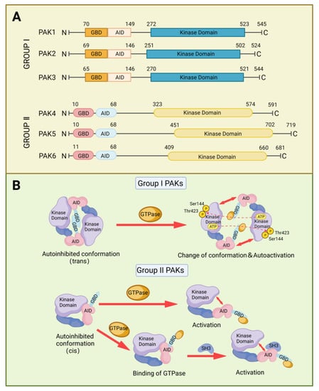
PAK proteins contain a kinase domain at the C-terminal, and an autoinhibitory domain (AID) and a GTPase-binding domain (GBD) at the N-terminus [
8]. The AID of Group I PAKs overlaps with the GBD and binds in trans to the kinase domain of another PAK molecule to form a dimer, preventing the formation of an activation loop. The binding of Rho GTPases or other proteins to the GBD dissociates the binding of AID with the kinase domain and causes a conformational change, which in turn triggers autophosphorylation of Ser 144 and Thr 423 and activation of the kinase. Group II PAKs function as monomers and they have AID-like domains different from the AID in group I PAKs [
9]. There are two proposed models to explain the activation of group II PAKs. One model is the AID-like domain, which binds to the kinase domain in cis, forming an inactive conformation. When CDC42 or RAC binds to GBD, this conformation will change and release the kinase domain from auto-inhibition [
10]. The other model contains two phases for activation. In addition to the binding of Rho GTPases, it also requires the involvement of proteins that contain SH3 domains, which interact with the AID-like domains [
11] (
Figure 2).
Figure 2. The structure and activation of PAKs. (A) Two-dimensional diagram of PAKs structure. (B) Different activation mechanisms of Group I PAKs and Group II PAKs.
PAKs act in multiple signaling pathways that are crucial for physiological and pathological processes. Particularly, overexpression of PAKs in many types of cancers is correlated with cancer progression and therapeutical resistance. Both PAK1 and PAK4 can activate the PI3K/AKT pathway, one of the key pathways for cell growth and survival. PAKs promote chemoresistance of cancer cells through activating the PI3K/AKT pathway [
12]. PAK1 stimulates cell migration by activation of AKT [
13]. PAK4 bind to p85, the regulatory subunit of PI3K, which in turn increases the activity of PI3K and phosphorylation of AKT, thereby promoting the migration of cancer cells [
14]. On the other hand, PI3K/AKT can also regulate the activation of PAKs. It has been suggested that a positive feedback loop may exist between PAKs and PI3K/AKT in the progression of cancers [
15,
16,
17]. RAS/RAF/mitogen-activated protein kinase (MEK)/extracellular-regulated kinase (ERK) axis is a well-known pathway contributing to the growth, survival, and metabolism of cancer cells. PAKs can phosphorylate MEK and RAF at Ser298 and at Ser338, respectively, promoting the activation of RAS-mediated RAF/MEK/ERK signaling [
18]. In IκB/nuclear factor-κB (NF-κB) signaling, PAK1 promoted the phosphorylation and degradation of IκB, leading to the activation and nuclear translocation of NF-κB [
19]. Blockading PAK1-activated NF-κB inhibited the growth of HCC and alleviated the inflammatory response in acute pancreatitis [
20,
21]. PAKs can activate β-catenin signaling and stabilize the activation of β-catenin, contributing to the chemotherapy resistance of cancer [
22,
23,
24]. PAKs can form a complex with β-catenin in the nucleus, which can further stabilize β-catenin. PAK4 can phosphorylate β-catenin at Ser 675, preventing the ubiquitination and degradation of β-catenin [
25,
26]. PAKs regulate the activation of Lin-11/Isl-1/Mec-3 kinase and matrix metalloproteinases, which are associated with cancer cell metastasis [
27,
28].
3. PAKs in Liver Cancers
Liver cancer is the sixth most common cancer worldwide and the second most common cause of cancer-related death. Hepatocellular carcinoma (HCC) accounts for 85% of liver cancer [
33,
34]. The major risk factors for liver cancer are chronic infection with hepatitis B virus (HBV) or hepatitis C virus (HCV) [
34,
35,
36]. PAKs have been reported to be upregulated in many cancers including breast cancer, glioblastoma, pancreatic cancer, and HCC, promoting tumor growth and metastasis [
7,
37,
38]. Increased PAKs expression endowed tumors with chemoresistance, and thus PAKs become attractive therapeutical targets for cancer treatment and biomarkers to predict prognosis [
39,
40,
41]. The roles of PAKs in liver cancer, especially HCC, are discussed below (
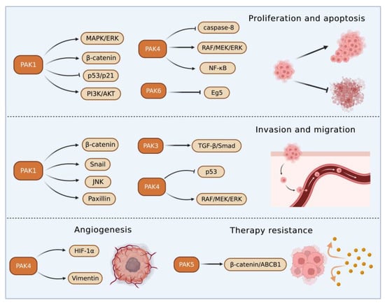
Figure 3).
Figure 3. The pathways affected by PAKs in liver cancer. PAKs modulate multiple signaling pathways (e.g., RAF/MEK/ERK, β-catenin) to promote the proliferation, invasion/migration, angiogenesis, and therapy resistance of cancer cells while inhibiting apoptosis.
3.1. Roles of Group I PAKs in Liver Cancer
PAK1 has been the most investigated among the I PAKs group. Overexpression of PAK1 was most frequently observed in HCC and associated with the growth and metastasis of HCC. PAK1 affects HCC progression via multiple pathways, PAK1 stimulated proliferation and inhibited apoptosis of HCC cells by activation of β-catenin signaling, and facilitated migration and invasion by upregulation of Snail [
42]. Genetic inhibition of PAK1 suppressed xenograft tumor growth through Snail-, β-catenin-, and p53/p21-mediated pathways [
42,
43]. Treatment of phorbol ester increased PAK1, which in turn promoted HCC metastasis by activation of paxillin and c-Jun N-terminal kinase (JNK) [
44]. PAK1 also promoted HCC metastasis by regulating the formation and degradation of actin stress fibers [
45]. PAK1 enhanced vasculogenic mimicry in HCC through the stabilization of hypoxia-inducible factor-1α and phosphorylation of Vimentin [
46]. Activation of PAK1-mediated epidermal growth factor receptor (EGFR) and vascular endothelial growth factor receptor (VEGFR) pathways inhibited anoikis, a form of programmed cell death, contributing to the anoikis resistance of HCC cells [
47,
48].
A natural product, resveratrol, inhibited HCC cell proliferation and survival by downregulation of PAK1, cyclin D1, mitogen-activated protein kinase (MAPK), and AKT [
49]. In a liver carcinogenesis animal model, diethylnitrosamine-induced HCC progression was correlated with increased levels of PAK1, cyclin D1, MAPK, and AKT [
35]. Myricetin induces apoptosis of HCC cells by abrogating RAS-activated PAK1 and inhibiting MAPK/ERK and PI3K/AKT signaling [
50].
KRAS or
BRAF mutations have been shown to promote hepatic vascular cavernomas [
51]. Although PAK1 has been reported to affect the activation level of RAS/MEK/ERK signaling [
52,
53], the role of PAK1 in hepatic vascular cavernomas is unknown.
MicroRNAs (miRNAs) are small, non-coding RNAs that bind to specific mRNA targets to regulate gene expression post-transcriptionally. Altered expression of miRNAs is associated with liver metabolic dysfunction and tumor development. LINC00460, a long intergenic non-protein coding RNA, promoted HCC progression by stimulation of PAK1 through suppressing miR-485-5p [
54].
PAK2 played a critical role in HCC metastasis. MiR-26a inhibited metastasis of HCC through downregulation of PAK2 [
57]. Sato et al. reported that PAK2 phosphorylation was associated with AKT activation in transforming growth factor-β (TGF-β)-induced migration of HCC cells [
58]. PAK3 is mainly expressed in the central nervous system for regulation of neuronal plasticity and spinogenesis [
59]. However, Gao and colleagues recently found that PAK3 was upregulated in HCC and stimulated the epithelial mesenchymal transition (EMT) of tumor cells through activating the TGF-β/Smad pathway [
60].
3.2. Roles of Group II PAKs in Liver Cancer
PAK4 enhanced HCC cell survival by modulating caspase-8 and NF-κB pathways [61]. In an HCC transgenic mouse model, miR-199-3p inhibited HCC growth by targeting the PAK4/RAF/MEK/ERK pathway [62,63]. On the contrary, an increased cyclin-dependent kinase 5 (CDK5) regulatory subunit-associated protein 3 in HCC accelerated tumor metastasis bound to and activated PAK4 [64]. PAK4 promoted migration and invasion of HCC cells through direct phosphorylation of p53 at S215 [65]. MiR-1271 suppressed HCC growth and metastasis through downregulation of RAF/MEK/ERK signaling, achieved by inhibiting Zic2/PAK4. PAK4 was overexpressed in metastatic tumor tissues compared with primary tumor and normal tissues, and PAK4 expression was correlated with worse survival of HCC patients. The results from multivariate analyses of 615 HCC patients showed that PAK4 was an independent indicator of poor prognosis [66]. MiR-433 was reduced in HCC tissues. MiR-433 repressed the viability of HCC cells through directly binding to the sequence at 3’-UTR of PAK4 mRNA, which in turn inhibited PAK4 expression [67]. MiRNAs are key upstream regulators that directly modulate the translation of multiple oncogenic proteins including PAK4. A miRNA cocktail therapy targeting PAK4, mechanistic target of rapamycin (mTOR), and RAS homolog gene family member C (RHOC), showed remarkable anti-HCC effects in patient-derived xenografts [68].
4. PAKs in Hepatic Ischemia/Reperfusion Injury
Hepatic IRI commonly occurs in the process of hemorrhagic shock, liver surgery, and liver transplantation [
78,
79]. The underlying mechanisms of hepatic IRI relate to the elevated oxidative stress and activation of immune-metabolic responses, which are harmful to normal cellular structures and functions, and result in liver damage [
80]. PAKs participate in the modulation of immune responses and inflammation [
81,
82], and PAKs have been reported to play a critical role in hepatic IRI [
6,
83].
Neuregulin-1/PAK1 axis was found to decrease IRI of liver grafts with or without steatosis through increasing vascular endothelial growth factor-α and insulin growth factor-1 levels, respectively [
83]. PAK4 was upregulated in hepatic IRI in mice and humans to promote hepatic hypoxia-reoxygenation-induced damage through phosphorylating nuclear factor erythroid 2-related factor 2 and suppressing its transcriptional activity [
6]. Genetic knockout or pharmacological inhibition of PAK4 reduced the inflammation and necrosis of hepatocytes [
6]. These results suggest that PAK4 inhibition may protect the liver from IRI-induced damage.
5. PAKs in Hepatitis
Hepatitis is defined as inflammation of the liver, and its etiologies include virus infection, parasitic invasion, alcohol abuse, autoimmunity imbalance, and metabolic disorder. The main characteristic of hepatitis is represented by the infiltration of inflammatory cells, which induces apoptosis and necrosis of hepatocytes leading to liver damage and dysfunctions [84,85,86,87].
In hepatitis B, phosphorylation of PAK1 induced the translocation of RAF-1 to mitochondrial, which in turn facilitated the anti-apoptotic effect of HBV X protein in hepatocyte [88]. In fact, HBV X protein can activate PAK1, which protected HCC from anoikis and promoted the metastasis of HCC [56]. Results from the PCR showed that the PAK3 gene was the preferential integration site of HBV DNA and the integration potentially changed the expression of PAK3 [5].
Different from the promoting role of PAK1 in HBV, PAK1 had potentially inhibited HCV replication, and its antiviral effects were independent of interferon regulatory factor 3 but dependent on the mTOR-activated PI3K/AKT and ERK [91]. HCV protected the infected hepatocytes from cell death by suppressing apoptosis and inflammatory reaction partially through the upregulation of PAK2 [92].
In a mouse model infected with
Schistosoma japonicum, PAK1 in Kupffer cells was elevated and facilitated the differentiation of CD4
+ T cells to T helper 17 cells via the NF-κB/interferon regulatory factor 1/interleukin-6 pathway, leading to aggravated hepatic inflammation [
95]. Increased PAK1 expression in Kupffer cells was also noticed in patients with autoimmune hepatitis, and the PAK1 expression was found to be associated with disease progression [
96]. The increased expression of PAK6 found in alcoholic hepatitis may contribute to tumorigenesis [
97].
6. PAKs in Liver Fibrosis
Liver fibrosis is implicated in the interaction between injured hepatocytes, inflammatory cells, and hepatic myofibroblasts. In response to injury, hepatocytes will express more profibrogenic miRNAs and proteins such as TGFβ and Notch, thus initiating fibrosis [
98]. The transformation from hepatic stellate cells to myofibroblasts that produce extracellular matrix (ECM) provides a protection to liver tissue from injury, and there is a balance between ECM production and degradation. However, the excessive accumulation of ECM produced by consistently activated myofibroblasts will impair the normal physiological structure and function of the liver, resulting in the occurrence of liver fibrosis [
99].
PAK1 and PAK3 were upregulated in activated hepatic myofibroblasts and promoted fibrotic effects by enhancing the expression of integrin β-1, which is essential for myofibroblasts activation and ECM production [
100].
7. Therapeutic Effects of PAK Inhibitors in Liver Disorders
The main strategy for treating HBV or HCV-related liver diseases is long-term administration of antiviral drugs such as entecavir, disoproxil, and IFN-α. Different from HCV, HBV has the characteristic of escaping innate immunity, thus immune therapy is also a vital treatment for HBV [
102,
103]. For metabolism-related liver diseases, limiting alcohol consumption, changing eating patterns and diet composition, and reducing insulin resistance and improving lipid metabolism are effective regimens [
104,
105].
Liver resection and transplantation are considered as curative treatments for HCC. However, only a small proportion of patients can have these surgical operations at the diagnosis stage and the recurrence after surgery is high. Most HCC patients are diagnosed at advanced stages of the disease and can only have non-surgical treatments, including chemo-, radio-, and immune-therapies. The first-line therapy for advanced HCC includes broad-spectrum tyrosine kinase inhibitors, such as sorafenib and lenvatinib, and combinational therapies of immunotherapy and anti-angiogenesis therapy [
106].
PAKs play key roles in liver disorders and thus are considered useful targets for the treatment of liver diseases. PAK inhibitors have been developed and can be divided into two categories depending on the binding sites within PAK molecules, ATP-competitive inhibitors, and allosteric PAK inhibitors [
109]. ATP-competitive PAK inhibitors block PAK phosphorylation by targeting the ATP-binding pocket within the kinase domain. This type of PAK inhibitors is further classified into aminopyrazole-based inhibitors, aminopyrimidine-based inhibitors, indolocarbazole-based inhibitors, 2-amino pyrido[2,3-d] pyrimidine-7(8H)-one-based inhibitors, and other ATP-competitive inhibitors. Compared with ATP-competitive PAK inhibitors, allosteric PAK inhibitors have the potential to be more selective and display discriminative inhibitory activity among PAK family proteins [
110,
111].
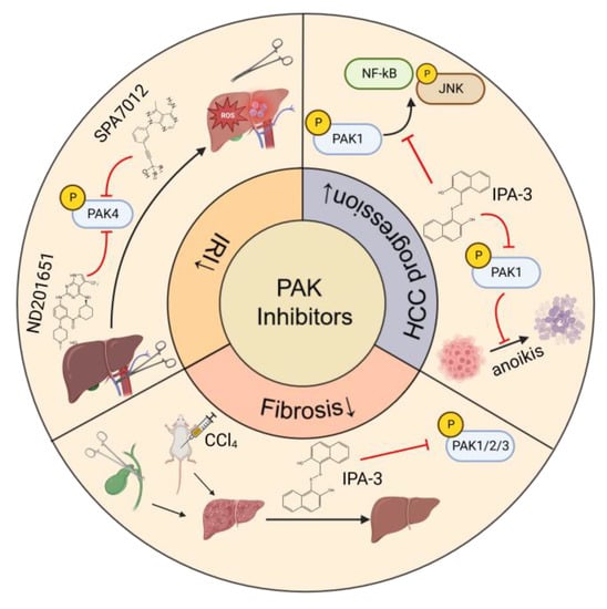
The studies of the effects of PAK inhibitors in liver disorders are limited (
Figure 4). PAK4 was upregulated and played a critical role in the process of hepatic IRI. Application of an ATP-competitive PAK4 inhibitor ND201651 reduced hepatic IRI [
6]. Another novel specific PAK4 inhibitor, SPA7012, a pyrazolo [3,4-d] pyrimidine derivative, also showed an anti-inflammatory effect on the liver during hepatic IRI [
112]. More experimental evidence is necessary for its clinical application.
Figure 4. PAK inhibitors applied in liver disorders. Abbreviations: HCC, hepatocellular carcinoma; IRI, ischemia-reperfusion injury; PAK, p21 Activated Kinase; ROS, reactive oxygen species.
IPA-3 is a non-ATP-competitive allosteric inhibitor targeting PAK1 phosphorylation. Since group I PAKs share the same inhibitory domain in the N-terminal sequence, IPA-3 also suppresses the activation of PAK2 and PAK3 [
113]. Administration of IPA-3 remarkably improved the bile duct ligation and CCl
4-induced fibrosis in mice without hepatotoxicity [
100]. IPA-3 suppressed HCC growth by inhibition of PAK1-mediated JNK phosphorylation and NF-κB intranuclear localization [
21].
8. Conclusions
PAKs play pivotal roles in the progression of liver disorders including liver cancer, hepatic IRI, hepatitis, and liver fibrosis. Elevated PAKs expression in liver cancer stimulates tumor growth and metastasis, contributes to therapeutical resistance, and is associated with the poor prognosis of patients. The studies of the effects of PAK1 and PAK4 in hepatic IRI have provided a useful guide for the potential application of PAK inhibitors in clinical settings. PAK1, the most extensively studied PAKs, plays important roles in the occurrence of hepatitis and liver fibrosis through affecting hepatocytes, immune cells, and myofibroblasts. It is worth noting that PAK1 promotes the development of HBV, hepatitis E virus, parasite, and autoimmune-related hepatitis while inhibiting HCV-induced hepatitis. With the increased occurrence of metabolism-related liver disease such as NAFLD, it is becoming important to explore the association between PAKs and liver metabolism and mechanisms involved.
This entry is adapted from the peer-reviewed paper 10.3390/cancers15020551