1. Introduction
(Snippets) Fluorescence spectroscopy has revealed great promise with myriad probes and devices demonstrating a rich spectrum of applications related to biological and chemical sensing, topographical analysis, immunoassays, optofluidics, forensics, microscopy, single molecule detection, environmental health monitoring as well as myriad point-of-care (POC) diagnostic technologies [
1,
2,
3,
4]. In an attempt to obtain enhanced signal intensities in traditional fluorescence-based analytical detection methodologies, it has been synergized with the metallic/plasmonic nanomaterials garnering active optoelectronic functionalities [
5,
6,
7,
8,
9,
10]. Such explorations have significantly advanced the frontier areas of biosensing research with several economical and industrial applications. This is on account of the ability of researchers to tailor the excitation and emission intensities of fluorescent moieties by placing them in the proximal vicinity of the plasmonic nanoparticles (NPs) sustaining localized surface plasmon resonances (LSPR) [
10,
11,
12,
13]. The high-gradient electromagnetic (EM) field intensity provided by such LSPRs assist augmented sensitivity in analyte detection on account of substantial modification in the local density of states (LDoS) [
10,
14,
15,
16]. Moreover, it has been observed that the resonant charge density perturbations in plasmonic NPs interact with the fluorophores in the near-field, and consequently, the emitters assist in the generation of plasmons that radiate into the far-field, carrying the emission characteristics of the fluorophores [
10,
17]. From this perspective, the resulting hybrid system of metal-fluorophore generates an efficient plasmophore (plasmon + fluorophore), transmitting the optical features of the individual counterparts. Furthermore, such an increase in the fluorescence intensity is attributed to the high radiative decay rate, robust photostability as well as the decrease in the lifetimes, ensuing an associated upsurge in the global quantum yield. Such investigations where the light (emission)–matter (nanomaterial) interactions assist in optical trapping, tuning, control, evaluation and manipulation of the resultant fluorescence intensity have developed into a mature field termed ‘plasmon-enhanced fluorescence (PEF)’ [
8,
10,
18,
19,
20]. These explorations have supported the comprehension of diverse novel phenomena in the sub-fields of nanophotonics, such as metal-dependent plasmonics [
3], graphene-based plasmonics [
21], dielectric-dependent metamaterials [
22] and photonic crystals (PCs) [
23,
24], to name a few.
Nevertheless, in spite of the abovementioned application potential of PEF technologies, the far-reaching capabilities of the fluorescence-based analytical detection systems are compromised on account of the omnidirectional (isotropic) emission and allied low-signal collection efficiency (<1%), photobleaching and high background noise [
8,
10,
17]. In order to overcome these limitations, Lakowicz and co-workers developed an innovative technology termed surface plasmon coupled emission (SPCE) in a series of research credentials termed radiative decay engineering, ‘one to eight’ [
17,
25,
26,
27,
28,
29,
30,
31]. SPCE platform is a prism coupling technique where the fluorescence is coupled to the surface plasmon polaritons (SPPs) of the metal thin film assisting in the realization of >50% signal collection efficiency, on account of exceptional directionality of emission. Further to the high p-polarized attribute of the emission signal (reinforced by the SPPs of the metal thin film), the SPCE fosters a 10–15-fold enhancement in the signal vis-à-vis conventional fluorescence, with high background suppression and spectral resolution [
28]. In an attempt to further increase the fluorescence enhancements observed in the SPCE framework, Chowdhury et al. demonstrated the utility of plasmonic AgNPs as active spacer material [
32]. This has assisted in the realization of 60-fold SPCE enhancements; following which, several other nano-architectures with numerous sizes, shapes and assemblies have been examined in the SPCE platform for achieving amplified SPCE enhancements [
33,
34,
35,
36,
37,
38,
39,
40]. Such synergy of fluorescence spectroscopy and applied nano-research with effective nano-engineering strategies has advanced the spectro-plasmonic modalities in the SPCE platform with newer applications and processes including, but not limited to: ultra-high sensitivity [
41,
42,
43], CNT-assisted augmented coupling [
44], cardiovascular disease and food biomarker monitoring [
45], fluorescent polymer brushes for large angle studies [
46], interfacial molecular beacon-related explorations [
47], cavity-void plasmon coupling in nano-assemblies sustaining Bragg and Mie plasmons [
48], adsorption-desorption analysis [
49], lightning-rod effect [
50], graphene π-plasmon hybrid coupling [
51,
52,
53,
54], mesoporous carbon florets for photon cascading in nanocavity [
55], lower-to-higher aggregates coupling [
56], magneto-plasmonics [
57], PLEDs [
58], simultaneous multianalyte sensing [
59] and other cost-effective biosensing applications [
60,
61,
62,
63,
64,
65,
66].
2. Surface Plasmon Coupled Emission (SPCE) Technology
(Snippets) Following the pioneering work by Lakowicz and co-workers, SPCE technology has been implemented in the advancement of several biosensing platforms [
82,
83,
84]. This section provides a brief overview of the SPCE platform and the associated nanointerfaces.
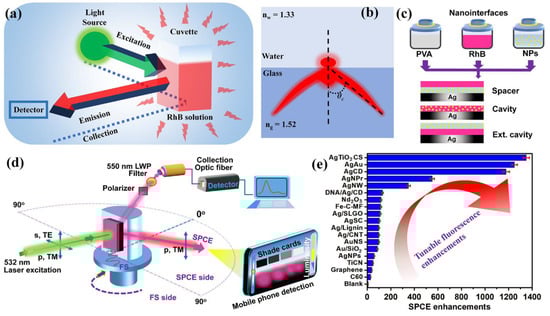
Figure 1 showcases a typical configuration in which the fluorescence is captured using a cuvette in a conventional fluorescence spectrophotometer. Traditionally, the detectors are placed at 90° in order to avoid the direct light from the irradiation source, as well as any other interference [
8,
10,
11]. In this regard, as the detector is placed in a fixed location at one particular angle, the collection efficiency is drastically lowered as the light emitted is isotropic from the cuvette. Furthermore, conventional fluorescence spectroscopy has several drawbacks: (i) low signal collection efficiency; (ii) poor resolution of emission peaks; (iii) lower sensitivity; (iv) requirement of cumbersome equipment; (v) omnidirectional emission property with negligible recognition of low quantum yield emitters [
8,
10,
11,
17]. For the radiating dipoles placed at the glass-water interface, the emission in the relatively HRI region (glass, n
g = 1.52) develops into a partially directional emission (
Figure 1b). This is due to the effect of critical angle (
θC), at which the evanescent field is generated at the interface, presenting an off-normal (non-isotropic) and partially directional and not polarized emission [
10]. While these are the preliminary observations with regard to the emission, as discussed in detail elsewhere [
10], the emission pattern can be channelized into sharply directional and polarized emissions using SPCE and PCCE platforms (
Figure 1d).
Figure 1. Conceptual schematic of (
a) fluorescence emission (of RhB) recorded by conventional fluorescence spectrophotometer, (
b) the angular dispersion of fluorescence emission as observed in the water-glass interface. The angle shown is critical angle (
θC) of emission. Adapted from [
11]. (
c) Schematic of the spacer, cavity and ext. cavity nanointerfaces. (
d) Optical setup used for SPCE experimental work with reverse Kretschmann (RK) configuration. The detection system is carried out using the conventional Ocean Optics detector, as well as the mobile phone-based detection system. Adapted from [
64]. (
e) Tunable enhancements in the fluorescence enhancements observed using different nanomaterials and nanohybrids in SPCE platform. (Acronyms: AgTiO
2 CS: silver titanium dioxide cryosoret; AgAu: silver-gold nanohybrid; AgCD: silver NP decorated-carbon dots; AgNPrs: silver nanoprisms; AgNW: silver nanowire; DNA/Ag/CD: DNA based AgCD composite; Nd
2O
3: neodymium (III) oxide; Ag/SLGO: silver NPs decorated on single layer graphene oxide; Ag/lignin: lignin-based AgNPs; Ag/CNT: carbon nanotubes decorated with AgNPs; AuNS: gold nanostars; Au/SiO
2: gold NPs decorated on silica NPs; TiCN: titanium carbonitride; C60: carbon allotrope or buckminsterfullerene, (C60-Ih) [5, 6] fullerene).
The generally explored nanointerfaces in the SPCE platform are shown in
Figure 1c, presenting the spacer, cavity and extended (ext.) cavity nanointerfaces, and the SPCE platform is schematically shown in
Figure 1d [
11,
33,
34,
35,
36]. The nanointerfaces are often fabricated using the spin-coating methodology, wherein the nanomaterial and the fluorophores of interest are doped in a polymer matrix and spin coated over the SPCE platform. The SPCE enhancements depend on several characteristics of the nanomaterials used, and also significantly depend on the nanointerfaces utilized. In the spacer nanointerface, in principle, the nanomaterial functions as an active spacer material between the radiating dipoles (fluorescent moieties) and the SPPs of the metallic thin film [
61,
64,
67]. In the cavity nanointerface, the infinitesimal nanogaps generated between the nanomaterial and the metal thin film sustain plasmonic hotspots where the radiating dipoles are sandwiched [
34,
37,
39]. Furthermore, as the name suggests, the cavity hotspots in the cavity nanointerface are extended to a defined distance in the ext. cavity interface [
6].
While the spacer and ext. cavity nanointerfaces, as observed in
Figure 1c, are constituted by two separate nanolayers, the cavity nanointerface is a single nanolayer. Consequently, the surface-induced quenching effects are significantly observed in the cavity nanointerface compared to the other two. By and large, the performance of these architectural designs has been examined with different nanomaterials and a comprehensive analysis of such explorations would demand the usage of meta-analysis and associated artificial intelligence and machine learning tools to comprehend the opto-electronic response of nanomaterials generated from a combination of elements from different parts of the periodic table [
54,
64,
71]. In a typical SPCE experiment, the SPPs are generated by illumination at an appropriate angle, which can satisfy the phase matching conditions at the metallo-dielectric nanointerface [
9,
10,
11]. The evanescent field is generated via both the Kretschmann-Raether (KR) and Reverse Kretschmann (RK) configurations, although the latter is more conducive for large-scale production and incorporation of the SPCE platform in biosensing approaches [
9]. This is on account of the fundamental difference between the two technologies in terms of the laser excitation and emission collection attributes. While the excitation and emission are performed from the curved surface of the prism in the KR configuration, the excitation is carried out from the flat surface of the prism (or from the sample side) in the case of RK optical configuration [
9,
10,
11]. In a typical experiment, the nano-engineered SPCE substrate is affixed over the prism using an index matching fluid, as shown in
Figure 1d. The prism is then mounted on a rotating stage and the emission is collected using appropriate optical filters and polarizers using an optic fiber. The final detection and the analysis of the SPCE emission signal is carried out using two detection systems: (i) the exorbitant Ocean Optics detector system; (ii) the cost-effective smartphone-based detection platform. This departure from conventional detection systems towards hand held devices has been recently pursued on account of the advantages of the latter in terms of easy transportability, unparalleled data acquisition ability, superior computing and ever-refining premium quality camera technologies [
34,
35,
37,
39].
In order to enhance the sensitivity of the detection devices, different nano-engineering techniques have been investigated and explored over the SPCE platform using myriad nanomaterials, including metallic nanomaterials (Ag, Au, Pt, Cu), dielectric nanomaterials (Nd
2O
3, SiO
2, TiO
2, TiC, TiN, TiCN), ferromagnetic nanomaterials (Fe
2O
3, Nd
2O
3-Ag or NdAg nanohybrids), homometallic and heterometallic, bi-, tri-, tetra-metallic nanohybrids, as well as graphene Dirac fermions and other 2-dimensional material-sustaining partially propagating plasmons, etc. [
9,
10,
55,
85,
86,
87,
88,
89,
90,
91]. The EM field intensity in the spatial regions of nanogaps between the NPs and the metallic thin film is dependent on several factors, such as the shape (rods, triangles, urchins, spheres, cubes stars, and wires), size (<10 nm, 10 nm–100 nm, >100 nm), architecture (core-shell, decorated), surface roughness, nature of adjacently situated plasmonic and/or dielectric NPs as well as the immediate environment and its refractive index [
92,
93,
94,
95,
96,
97]. Extensive theoretical analysis of the utility of such nanomaterials for efficient photo-plasmonic hotspot generation have been carried out using discrete dipole approximation (DDA) [
10], finite-difference time-domain (FDTD) [
9,
48,
71,
74] and COMSOL Multiphysics simulations [
34,
98] to obtain a comprehensive understanding of the hotspot behavior. These explorations have assisted in the realization of new opto-electronic phenomena at nano-dimensions, such as Casimir force, Rabi splitting, Fabry-Perot photonic mode-coupling, Fano resonance, quantum confinement and the Purcell effect in the SPCE platform, rendering scientific insights into physicochemical interactions at advanced interfaces [
9,
11,
55]. These research studies have resulted in the development of intriguing biosensing platforms, thereby supporting translational photonics research in addition to providing newer insights from the basic (simulations) and applied research perspectives.
Acknowledgments
S.B. specially thanks Prof. Sai Sathish Ramamurthy, founder, Surface plasmon-based Translational and Advanced Research (STAR) laboratory, Central Research Instruments Facility (CRIF), SSSIHL for scientific insights, inspiration and support. S.B. acknowledges support and valuable study inputs from Prof. Shivakiran Bhaktha, B.N, Photonic Systems Laboratory, Indian Institute of Technology Kharagpur, and Prof. Chandramouli Subramaniam, NanoStructured Materials (NSM) Lab, IIT-Bombay. S.B. also thanks Prof. Roopa Reddy, Prof. Nagananda G.S., Prof. Swetha Seshagiri, and Prof. Narendra Reddy, the Center for Incubation Innovation Research and Consultancy (CIIRC), Bangalore, for their scientific insights and support. SB thanks the enormous support, motivation and guidance from Prof. Brian T. Cunningham, Department of Electrical and Computer Engineering, Illinois Cancer Center, Woese Institute for Genomic Biology, Holonyak Micro and Nanotechnology Laboratory (HMNTL), UIUC, as well the support from all the member of Nanosensors group, HMNTL, UIUC, United States. S.B. also thanks the support rendered by all the members of STAR lab, CRIF, SSSIHL and NanoStructured Materials (NSM) Lab, IIT-Bombay for their timely support in associated research activities.
Dedication
Dedicated to Bhagawan Sri Sathya Sai Baba on the occasion of his 97th Birthday Celebrations and to all the first responders for their service and social support offered right through the COVID-19 pandemic.
Photograph of the contributor in the Special Issue "Photon-Driven Technologies: Exploring the Latest Advancements" (left) and performing SPCE experiment (right).
Brief biography: Seemesh Bhaskar originally hails from a remote place in Karnataka state (India) named 'Bellippady Nilaya', Kavu. He obtained his Ph.D. from STAR Lab, Central Research Instruments Facility (CRIF), SSSIHL, India and further completed Post-Doctoral research at Indian Institute of Technology (IIT) Bombay. He is currently serving as Research Associate at Holonyak Micro and Nanotechnology Laboratory, Carl R. Woese Institute for Genomic Biology, University of Illinois at Urbana-Champaign (UIUC). He has pursued research internships and projects at SASTRA University and Indian Institute of Science, Bangalore as part of the DST-Inspire program. His collaboration with research groups at various universities such as IIT-Bombay, IIT-Kharagpur, IIT-Gandhinagar, CIIRC have resulted in 30+ publications in reputed national and international peer-reviewed journals, and 10+ conference proceedings. He is the recipient of the prestigious DST-Inspire scholarship & fellowship, AWSAR, K. V. Rao Scientific Society (KVRSS), Young Achiever, M.Sc. Chemistry Gold medal and All-rounder Gold medal awards. His research work focuses on building effective nano-engineering protocols for photo-plasmonic biosensing at advanced interfaces and is the pioneer in the development of Photonic Crystal-Coupled Emission (PCCE) platform (2020), Graphene oxide plasmon-coupled Soliton Emission (GraSE) (2021), CryoSoret Nano-Engineering (CSNE) (2022) and FerroPlasmon-on-Mirror (FPoM) (2023) technologies.
This entry is adapted from the peer-reviewed paper 10.3390/mi14030574