The Virtuous Galleria mellonella as Scientific Model: History
Please note this is an old version of this entry, which may differ significantly from the current revision.
Subjects: Microbiology
Contributor: , , ,

The first research on the insect Galleria mellonella was published 85 years ago, and the larva is now widely used as a model to study infections caused by bacterial and fungal pathogens, for screening new antimicrobials, to study the adjacent immune response in co-infections or in host-pathogen interaction, as well as in a toxicity model. The immune system of the G. mellonella model shows remarkable similarities with mammals. Furthermore, results from G. mellonella correlate positively with mammalian models and with other invertebrate models. Unlike other invertebrate models, G. mellonella can withstand temperatures of 37 °C, and its handling and experimental procedures are simpler. Despite having some disadvantages, G. mellonella is a virtuous in vivo model to be used in preclinical studies, as an intermediate model between in vitro and mammalian in vivo studies, and is a great example on how to apply the bioethics principle of the 3Rs (Replacement, Reduction, and Refinement) in animal experimentation. 

  • Galleria mellonella
  • infection model
  • toxicity model
  • 3Rs
  • antimicrobial therapy
  • drug screening

1. Introduction

The insect larva of Galleria mellonella has been widely used in the last few decades as an infection model to study bacterial and fungal infections and for assessing the efficacy of novel antimicrobial drugs, before proceeding to preclinical studies in mammals, reinforcing the application of the 3Rs principles (replacement, reduction, and refinement) in animal experimentation. The G. mellonella model offers several advantages over mammalian models: it does not require ethical approval, it is easily manipulated, it is inexpensive, it allows large-scale breeding, and it allows a rapid experimental execution, associated with the ability to incubate caterpillars at temperatures between 25 °C and 37 °C and the simple inoculation of the pathogens to be tested [1][2][3].
Historically, G. mellonella was described in the 4th century BC by Aristoteles as a honeybee pest in the book Historia Animallium (Book VIII, Chapter 27) [4]. The first publication in PubMed was in 1938, describing the genetic characteristics of G. mellonella following X-ray treatment [5]. Then, in 1951, this insect was used for the first time as a toxicity model for an antituberculosis drug [6]. Further studies in 1957 and 1961 used it to test antifungal agents [7][8], and since the 60s G. mellonella was validated as an infection model for many bacterial species, Salmonella enterica serovar Typhimurium being the first one [9], in addition to several fungi [10]. Although viruses can only infect specific hosts, leading researchers to use mammalian models for viruses related studies, some insect and mammalian viruses, such as the Tipula iridescent virus and the Nodamura virus [11][12], have been validated in the G. mellonella model. Since 2000, the use of the G. mellonella model has been exponentially increasing, being widely used for infection studies in the scope of human and veterinary pathogens [3][4]. The G. mellonella model was also successfully used to study the effects of co-infection in 2004 [13]. The G. mellonella transcriptome was sequenced in 2011 and its genome was sequenced in 2018, revealing over 14,000 genes [14]. Genome sequencing and studies on the immune response at the proteomic, epigenetic, and transcriptomic levels have launched new areas of research [15][16][17]. Its wide application is due to G. mellonella susceptibility to a high number of bacteria, and, according to Champion et al. [18] to twenty-nine species of fungi, seven viruses, one species of parasite, and sixteen biological toxins [18].

2. Characterization of Galleria mellonella

The great wax moth or the honeycomb moth, Galleria mellonella (Linnaeus, 1758), is an insect moth from the Phylum Arthropoda, Class Insects, order Lepidoptera and family Pyralidae (snout moths) [19]. It is a ubiquitous parasite of the honeybees Apis mellifera and Apis cerana and of their hives, and it can be found in beehives, bumblebee nests, wasp nests, or in stored waxes. In nature, caterpillars feed on honey, and also pollen, beeswax, pupa skins, cocoons, and feces from the hive [20].
The G. mellonella larvae can cause galleriasis within the hives, a phenomenon in which the hatching larvae create tunnels through the honeycombs containing larva and honey stores. The larvae cover these tunnels with silk entangling and starving the emerging bees, leading to colony loss and a reduction in the size of the migratory bee swarms [21]. The tunnels make holes through which honey leaks out [20][21], resulting in massive destruction of the combs. G. mellonella adults and larvae are carriers of two viruses, the Israeli acute paralysis virus and the black queen cell virus, that can potentially infect and kill honeybees [20][21].
Galleria mellonella is distributed worldwide, being present on all continents, especially in mountain range areas, except Antarctica [20], coinciding with the occurrence of its host bees. It has been detected in seventy-seven countries and in several islands, and it is anticipated that the pest may spread further, especially due to climate change [20].
Galleria mellonella is a typical holometabolous insect undergoing four developmental stages in its life cycle: the egg, larva, pupa, and adult [20][22][23]. The duration of a complete cycle is approximately six to eight weeks at 29–39 °C and high humidity [23], with four to six generations per year. Complete metamorphosis is affected by abiotic factors such as temperature, diet, and relative humidity, and by biotic factors such as competition for food and cannibalism [20][24]. The 50–150 eggs layered by the female between the cracks of the honeycomb are spheroidal-shaped and white to light pink in color. The development into larvae is temperature dependent, and lasts between three to eight days at 24–27 °C, and thirty days at 10–16 °C [25].
The larva body is divided into a head, a three-segmented thorax with six legs, and an abdomen of ten segments with eight prolegs and two anal prolegs [26]. The larvae dorsal region is called the “new immune tissue” because it is where the immune response is organized [27][28]. Within the inner cavity, there is the hemolymph, which is the larval circulatory system, and the fat body, a biosynthetic organ analogous to the mammalian liver. Within the fat body, the digestive system and the silk gland can be found. The ventral region corresponds to the nervous system and contains several ganglia [26][29].
Larvae at the last instar produce silk using their salivary glands to form cocoons that should be protected from the open air and excess humidity. At this moment, at the stage of pre-pupae, the larvae stop eating and become less mobile. Pupae are immobilized in cocoons and do not eat during this period for one to nine weeks until emerging as adults. The color of the cocoon depends upon the presence of colored pigments in the layer, varying from white to brown, and finally turning into a light golden-brown cocoon [22][23][26].
The G. mellonella larvae microbiota (the larvae microbial community) is dominated by a single Enterococcus taxon, presenting E. gallinarum or E. saccharolyticus as a main species, independently of the body site sampled, although other taxa, such as the Staphylococcus, Pseudomonas and Enterobacter species, were also already found [30]. Interestingly, commercially available larvae show a lower bacterial load than standardized research larvae, probably due to previous treatments with hormones and antibiotics [30][31]. However, there has been some disagreement regarding the microbial composition of G. mellonella, as some authors argue that it is not as simple as has been advocated [32]. According to Gohl et al. [32] the G. mellonella microbiome is diverse, dominated by the Proteobacteria and Firmicutes phylum, appears to have a resident microbiome represented by Ralstonia bacteria, and is affected by both diet and ontogeny [32].

3. Immune System of Galleria mellonella

As observed in mammalians, the innate immune system of insects such as G. mellonella comprises the cellular and the humoral immune system, and is more advanced than other invertebrates such as nematodes [33].
The innate immune system is non-specific and is distributed throughout the body. It is the first line of defense against microbes, maintaining homeostasis and preventing infections [34]. The lack of adaptive immunity in G. mellonella and other insects is in fact an advantage because it allows the study of host-pathogen interactions without the interference of adaptive responses [35]. Obviously, if the study aims to understand the response of the adaptive immune system to a certain pathogen, insect models are not adequate. The innate immune response of G. mellonella shares several properties with the mammalian innate immune system (for example, phagocytosis in insects and mammals is believed to be very similar [34][36], therefore G. mellonella is a valuable in vivo model to be used in preclinical studies as an intermediate model between in vitro and mammalian in vivo studies [2][3][4].
In G. mellonella, there is considerable overlap between humoral and cellular defenses, as many humoral molecules affect cellular immune response, and cellular immune response is an important source of many humoral molecules [37]. The cellular immune response is mediated by hemocytes, which are phagocytic cells that present an analogous function to mammalian blood. They are found free in the hemolymph or attached to internal organs, such as the digestive tract, fat body, and surface of the insect heart [27][38][39]. Hemocytes recognize pathogenic microorganisms through the direct interaction of their surface receptors with pathogen molecules, or indirectly by the recognition of humoral immune receptors that bind to and opsonize the pathogen. Therefore, both humoral and cell surface receptors are involved in these recognition events. These receptors are known as pathogen recognition receptors (PRRs), and are able to recognize pathogen-associated molecular patterns (PAMPs). PAMPs are conserved microbial components, including lipopolysaccharide (LPS), peptidoglycan, lipoteichoic acids (LTA), and β-1,3 glucan [4][40].
Hemocytes are involved in phagocytosis, encapsulation, and nodulation of the invading agent. Six types of hemocytes can be found in G. mellonella: plasmatocytes and granular cells, which are the most abundant hemocytes [41]; prohemocytes, which are progenitor cells that can differentiate into several cell types; coagulocytes, involved in hemolymph coagulation; spherulocytes, which transport and secrete several cuticular components; and oenocytoids, which are involved in the melanization pathway and, like mammal neutrophils, are able to secrete extracellular nucleic acid traps involved in pathogen sequestration and coagulation activation [2][31]. Encapsulation takes place when pathogens are too large to be phagocytosed and occurs without melanization. Nodulation begins with the attachment of granular cells to the microbes’ surface, triggering the release of multiple plasmatocyte spreading peptides, which will attach around the bacteria, fungal spores’ surface, or foreign targets, resulting in the formation of a smooth capsule [42][43]. This step is often followed by melanization [3].
The hemocytes’ concentration varies in response to pathogenic agents and during the G. mellonella life cycle [27][38][39]. Just after infection, there is an activation of the larval immune system regardless of the pathogenic agent, which may lead to a decrease in hemocyte count [44][45].

3.1. Opsonins

Galleria mellonella produces numerous opsonins, which are hemolymph proteins that identify and attach to conserved microbial elements, namely peptidoglycan recognition proteins (PGRPs), Apolipophorin-III (apoLp-III), hemolin, and G. mellonella cationic protein 8 (GmCP8) [46].
PGRPs are identical to mammalian opsonins and bind to peptidoglycan through a conserved domain homologous to a lysozyme of the T4 bacteriophage [47].
The apoLp-III is a major exchangeable lipid transport molecule that plays a crucial role in the innate immune response. It has a high affinity for hydrophobic ligands, such as LPS and LTA, and shows a multifunctional role, as it is able to stimulate the phagocytic activity of hemocytes and increase the production of the antimicrobial peptide cecropin [48]. More recently, it was shown that apoLp-III acts synergistically with G. mellonella lysozyme, increasing its antimicrobial activity against Gram-negative bacteria [49]. The apoLp-III is also involved in pathogen differentiation, including between Gram-positive and Gram-negative bacteria, and between yeasts and filamentous fungi, and in the establishment of an adequate immune response [50]. The apoLpIII shows high homology with mammalian apolipoprotein E, which is involved in LPS detoxification, and in phagocytosis stimulation [3][51].
Hemolin is a member of the immunoglobulin superfamily, being able to bind to LPS and LTA [52]. Hemolin is expressed in several organs, including the silk gland of the larvae and its fat body, being up-regulated during bacterial and fungal infections [53], or after exposure to low doses of β-1,3-glucan [54]. Both hemolin and GmCP8 stimulate phagocytosis by hemocytes [3][4].

3.2. Antimicrobial Peptides

Antimicrobial peptides, or host defense peptides, are usually 10–40 residue polypeptides (although some may have more than one hundred amino acids) produced by virtually all multicellular organisms. They belong to the immune system of all classes of life, providing a broad-spectrum first line of defense against the dissemination of bacteria, virus, protozoa, and fungi [55]. AMPs’ antimicrobial activity results from their cationic charge, which is associated with the presence of lysine, tryptophan, and arginine residues, and from their hydrophobic and amphipathic properties, which allow the peptides to attack the microbial membranes by interacting with their lipids [56][57].
Insect AMPs are mainly found in the fat body, hemocytes, digestive tract, salivary glands, and reproductive tract of G. mellonella, both in non-infected larvae and in infected larvae, in response to microbial invasion. Some AMPs are identical to molecules characterized in mammals [2][3]. About twenty known or putative AMPs have been identified in G. mellonella, namely two cecropins-like peptides, seven moricins, defensins-like peptides, gloverin, G. mellonella proline-rich peptides, G. mellonella anionic peptides, lysozymes, x-tox, an inducible serine protease inhibitor, heliocin-like peptide, apolipophoricin, and a metalloproteinase inhibitor (IMPI) [2][4].
Cecropins A- and D-like peptides are linear peptides active against Gram-positive and Gram-negative bacteria. Moricins are α-helical peptides that are particularly active against filamentous fungi [58], but also against yeasts and Gram-positive and Gram-negative bacteria [4]. Defensin-like peptides are cysteine-rich cationic peptides that act by forming voltage-dependent ion channels in the cytoplasmic membrane resulting in ion leakage and cell lysis [59]. Defensins include galiomycin (active against filamentous fungi and yeast but without antibacterial activity), Galleria defensin gallerimycin (active against entomopathogenic fungi but not active against yeast), and defensin-like peptides (which acts against Gram-positive bacteria and fungi) [59][60]. Gloverin is rich in glycine and it also presents cysteine residues. It inhibits the synthesis of the outer membrane proteins, increasing membrane permeability and showing specific antimicrobial activity [61]. Gm proline-rich peptides 1 and 2 also appear to increase the membrane permeability of bacteria [60]. Gm anionic peptides 1 and 2 bind to positively charged regions of the bacterial membrane and are active against Gram-positive bacteria and fungi [40][60]. Lysozymes are muramidases, able to digest bacterial peptidoglycan and act against Gram-positive bacteria, some Gram-negative bacteria, and some fungi [40][59]. These enzymes are also able to induce apoptotic changes in Candida albicans cells [62]. X-tox is an atypical inducible defensin-like peptide that lacks detectable antimicrobial activity [3][63]. The expression of inducible serine protease inhibitor 2, Heliocin-like peptide, and apolipophoricin is induced in response to infection [59][60].

3.3. Melanine

Melanization is the synthesis and deposition of melanin around microbes, allowing to encapsulate microbes within the hemolymph, followed by hemolymph coagulation and opsonization [2][27].
The formation of melanin is catalyzed by phenoloxidase (PO), which is produced as the inactive zymogen pro-phenoloxidase (ProPO), an innate immunity protein of cellular and humoral defense, in hemocytes [64]. Melanization begins upon the recognition and phagocytosis of the pathogen, prompting the phenoloxidase pathway. The soluble PRRs coupled to target molecules on bacterial or fungal surfaces trigger the serine protease cascade that results in the cleavage of pro-PO to PO. The activated PO will lead to the non-enzymatic polymerization of quinines to form melanin around microbes and wounds [2][3].
The velocity at which G. mellonella melanization occurs depends on the virulence of the infecting pathogen [27]. As it starts, the surface of the larval cuticle becomes covered with black spots and, as the infection progresses, it can be completely melanized, leading to the obtaining of completely black and dead larva [3]. A more noticeable melanization occurs in the dorsal region of the larva, the larva’s new immune tissue, where the heart is located [27].

3.4. Extracellular Nucleic Acid

Two extracellular nucleic acids are involved in the entrapment and killing of pathogens and in enhancing innate immune responses [65]. Neutrophil extracellular traps (NETs) trap and kill pathogens and are originated by neutrophils that release chromosomal DNA spiked with bactericidal proteins to form NETs upon stimulation with LPS or interleukin-8. Oenocytoids are hemocytes that represent a source of endogenously derived extracellular nucleic acids implicated in both sequestering the pathogen and activating hemolymph coagulation [3][31][65].

3.5. Influence of Diet on Larval Immune Health

Several authors already showed that the G. mellonella diet can influence its immune system by intervening in larvae development, causing its death, or increasing its susceptibility to infections [1][4][66]. According to Jorjão et al. [1], an energetic diet is associated with a shorter life cycle, increased caterpillar weight, and immune system activation, by enhancing hemolymph volume and a high concentration of hemocytes. This type of diet also lengthens larval survival to bacterial (S. aureus and E. coli) and fungal (C. albicans) infections.

4. Galleria mellonella In Vivo Model—Experimental Design Considerations
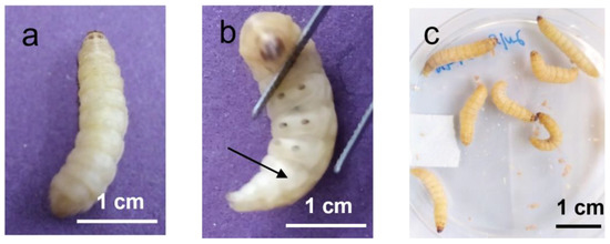
Differences in the supplier, breeding conditions, age, weight, nutrition, maintenance, and handling of G. mellonella larvae, and the presence of antibiotics and hormones that can alter their metabolism, might easily result in differences in mortality rates generating inconsistent experimental responses in an infection model [4][67]. This might explain conflicting results with some reference strains that induced different larvae mortality rates in studies performed in distinct research labs. The standardization of research protocols parameters would help to provide reliable results and improve inter-laboratory comparisons between results from G. mellonella experiments [1][2][31].
Before starting in vivo tests, it is useful to watch some video components of articles done by experienced researchers in order to better understand experimental procedures in G. mellonella larva [68][69].
It is recognized that the use of G. mellonella reared in the lab is preferable instead of commercially available larvae. Commercial larvae are available from a wide range of independent breeders and are sold as food for reptiles and birds in captivity or as fishing bait. At least ten larvae for each experimental condition should be used in order to reduce the effect of heterogeneity between individuals, and at least two or three independent experiments should be performed. G. mellonella should be placed at 25 °C in the dark, from egg to last instar larvae, and fed on a natural diet of beeswax and pollen grains. Individuals selected for experiments should include healthy worms of the final instar larval stage, which develop from the egg in about five weeks, weighing 250 ± 25 mg, having a length of 2–2.5 cm, and a creamy color [3][26][31].
Before infection, microbial inoculums should be washed to prevent virulence factors secreted during their in vitro growth from being introduced within the larvae. It is also recommended to have control larvae injected with a placebo inoculum, such as Phosphate Buffer Saline (PBS) or sodium chloride (0.9%), to detect potential physical trauma due to the injection [3][70]. Although most authors use microbial inoculums of 10 µL and 20 µL, smaller microbial inoculum up to 5 µL may also be used [71].
The most common infection route is through the hemocoel (the body cavity containing the hemolymph), by inoculation through the last left pro-leg of the abdominal region [72], the surface of which should be previously sanitized with 70% (v/v) alcohol using a cotton swab. The injection into the last proleg will allow for more space to insert the needle and a fair distribution of the inoculum throughout the body. Further injections e.g., for drug screening, should be given in the opposite upper prolegs (Figure 1).
Figure 1. (a) Dorsal and (b) ventral views of Galleria mellonella larva at the last instar stage. The larva is divided into a head, a thorax with three segments, and an abdomen with 10 segments. The segment with the last proleg (arrow) is the fifth, counting from the anal segment with two non-visible anal prolegs; (c) Several larvae within a petri dish (original).
 
The larva can be immobilized in different ways: by using different restrain devices, such as a microscope blade, a blue pipette tip, or by putting the larva inside a yellow tip [73] (Figure 2); between the fingers of the researcher; or between two sponges, protecting the researcher’s fingers [74].
Figure 2. Restraining devices for handling Galleria mellonella larva. (a) using a microscope blade to immobilize the larva; (b) using a blue pipette tip to turn the left proleg to make it more visible; (c) using a yellow tip to immobilize the larva (original).
 
The injection is performed manually, using a fine dosage syringe (up to 1 mL) and a fine needle (up to 0.50 µm), or using an automatic syringe pump which controls the dose injected into each larva. The needle must be inserted parallel to the body so as not to rupture the digestive tract [74].
Oral infection is also a possibility by using a force-feeding method [69], through which the microbial agent is inoculated into the digestive tract and ends up in the intestine with the food bolus. The intestinal microbiota is separated from the bolus by a protecting matrix which prevents the intestinal epithelium from contamination [74].
For drug screening, antimicrobial agents can be administered in different treatment regimens, including different times and number of doses. The larvae can be incubated at diverse temperatures that are necessary for the expression of virulence factors [75]. Most studies usually perform the administration of the antimicrobial agent between 30 min to 2 h after infecting the larvae with the pathogen, immediately after infection, or even before infection [3].

5. Gram-Positive and Gram-Negative Bacteria Causing Disease in Humans

Human diseases can be caused by polymicrobial infections or by human microbiome dysbiosis. Therefore, detailed studies on the host response during co-infection, including the complex interactions between microbes and also between them and the host, are very important [76].
Two studies on co-infection in cystic fibrosis were performed by using the G. mellonella model. The first one comprised a co-infection by the P. aeruginosa Liverpool epidemic strain (LES) and Anginosus group streptococci (AGS) [77]. The authors showed that the virulence of LES phenotypic variants can be synergistically enhanced by the presence of oral commensal streptococci, and that this synergy is dependent not only on AGS species but also on the genotype and phenotype of P. aeruginosa [77]. The second study focused on a P. aeruginosa and Aspergillus fumigatus co-infection [78]. It concluded that infecting the larvae with sublethal concentrations of A. fumigatus to mimic colonization resulted in significantly higher mortality rates in larvae injected with P. aeruginosa 24 h later [78]. Interestingly, Reece et al. [79] noted that G. mellonella inoculation with mixed biofilm co-cultures resulted in a reduced biofilm development, and quantification by species-specific qPCR revealed that both pathogens had mutually antagonistic effects on each other [79]. In opposition, Scott et al. [78] found that the presence of P. aeruginosa supports Aspergillus growth by producing volatile sulfur compounds that provide a sulfur source to the fungus, suggesting that this synergism may increase virulence in co-infection [78].

6. Comparison with Other Invertebrate and Mammalian Models

Traditionally, two other invertebrate models are widely described to study the virulence of bacteria or to test the effect of antimicrobial compounds: Caenorhabditis elegans (Phylum nematoda, class Chromadorea) and Drosophila melanogaster (Phylum Arthropoda, class of Insects) [80]. The G. mellonella model has several advantages over these two models and mammalian models, but it is not so well established because it had only started to be widely used more recently [31][80].
Galleria mellonella length (between 3–30 mm) is significantly higher than those from D. melanogaster and C. elegans, (3 mm and 1 mm, respectively), facilitating the operation and handling of the larvae [4][26]. G. mellonella can withstand temperatures of 37 °C, unlike the C. elegans and D. melanogaster models, making it a considerable gain for investigating human pathogens, considering that the expression of many virulence genes is under temperature control [75]. Furthermore, G. mellonella exhibits immune phagocytes, unlike C. elegans, which is unable to phagocytize microorganisms, conferring on G. mellonella a major advantage for the study of several bacteria and fungi [31][67]. G. mellonella also shows advantages in studies on the virulence of intracellular bacteria such as Legionella pneumophila [68][81], since in the G. mellonella model, but not in C. elegans, bacteria can penetrate from the intestinal lumen to the intestinal epithelial cells [81]. In addition, the use of Drosophila requires more specialized equipment and experience than does Galleria, and wild-type Drosophila is resistant to fungi [82].
Due to the more recent implementation of the G. mellonella model regarding other invertebrate models, there is a lack of stock centers for its commercialization, and of databases such as Flybase (for D. melanogaster) or WormBase (C. elegans), and only a few microarrays, libraries of RNA interference and mutant strains are available for G. mellonella [3][4][31]. Unlike C. elegans and D. melanogaster, the G. mellonella genome (GenBank NTHM00000000) is still a shotgun project that has not been fully analyzed [14]. The lack of mutant and transgenic strains and access to microarrays or RNA interference libraries limit studies on many biological processes and specific diseases. Also, the lack of full genome sequencing and analysis does not allow a detailed molecular study understanding of the host response, and the comparison between genomic, transcriptomic, and proteomic data [31][83]. There is also a lack of standardized experimental procedures or subjective interpretation of scoring parameters, which prevents a direct comparison of different published studies using the G. mellonella model [3][4][31]. It is expected that scientific advances will solve these drawbacks soon.
Mammals, such as mouse (Phylum Chordata, Class Mammalia), rabbits and non-human primates, have been used as models to study human pathogens longer than G. mellonella [31][80], and for that reason they have gathered a large set of reliable information and to clearly indicate what happens in humans. They also have adaptative immunity, unlike invertebrates [3][84]. Invertebrates are insensitive to pain due to the absence of nociceptors, and consequently there are no restrictive ethical rules [85]. On the other hand, there are ethical concerns and higher costs associated with the in vivo experiments in mammalian models.

7. Conclusions

Research on the insect Galleria mellonella was first published in the 1930s, and the larva is now widely used, mainly as a model of infection and for drug screening against bacteria and fungi that cause disease in humans. The model is also used as a toxicity model, and as to study the adjacent immune response in co-infections or in host-pathogen interactions. The limitations of the Galleria mellonella model are mainly related to the unavailability of stock centers, commune databases, standardized procedures, mutant and transgenic strains, microarrays or RNA interference libraries, and full genome sequencing and analysis. Regardless, the Galleria mellonella model is a virtuous model, because despite having some disadvantages that mainly come from the fact that it has only recently been commonly used. Furthermore, there is a positive correlation between the results from G. mellonella with mammalian models and with other invertebrate models. Unlike D. melanogaster and C. elegans, G. mellonella can withstand temperatures of 37 °C, and its handling, as well as microbial inoculation and drug administration, are simpler. In conclusion, Galleria mellonella is a valuable in vivo model to be used in preclinical studies as an intermediate model between in vitro and mammalian in vivo studies, and is an excellent example of the application of the bioethics principle of the 3Rs in animal experimentation.

This entry is adapted from the peer-reviewed paper 10.3390/antibiotics12030505

References

  1. Jorjão, A.L.; Oliveira, L.D.; Scorzoni, L.; Figueiredo-Godoi, L.M.A.; Cristina A Prata, M.; Jorge, A.O.C.; Junqueira, J.C. From moths to caterpillars: Ideal conditions for Galleria mellonella rearing for in vivo microbiological studies. Virulence 2018, 9, 383–389.
  2. Pereira, M.F.; Rossi, C.C.; da Silva, G.C.; Rosa, J.N.; Bazzolli, D.M.S. Galleria mellonella as an infection model: An in-depth look at why it works and practical considerations for successful application. Pathog. Dis. 2020, 78, ftaa056.
  3. Tsai, C.J.; Loh, J.M.; Proft, T. Galleria mellonella infection models for the study of bacterial diseases and for antimicrobial drug testing. Virulence 2016, 7, 214–229.
  4. Cutuli, M.A.; Petronio Petronio, G.; Vergalito, F.; Magnifico, I.; Pietrangelo, L.; Venditti, N.; Di Marco, R. Galleria mellonella as a consolidated in vivo model hosts: New developments in antibacterial strategies and novel drug testing. Virulence 2019, 10, 527–541.
  5. Smith, T.L. Genetical Studies on the Wax Moth Galleria mellonella Linn. Genetics 1938, 23, 115–137.
  6. Ingrao, F.; Belli, N. Galleria mellonella test in the study of the antituberculosis activity of thiosemicarbazones. Lotta Contro La Tuberc. 1951, 21, 282–287.
  7. Morellini, M.; Avegno, R.P. Antimycotic drugs evaluated with the Galleria mellonella test. Ann. Ist. Carlo Forlanini 1957, 17, 149–154.
  8. Smissman, E.E.; Beck, S.D.; Boots, M.R. Growth Inhibition of Insects and a Fungus by Indole-3-Acetonitrile. Science 1961, 133, 462.
  9. Kurstak, E.; Vega, C.E. Bacterial infection due to Salmonella typhimurium in an invertebrate, Galleria mellonella L. Can. J. Microbiol. 1968, 14, 233–237.
  10. Fuchs, B.B.; Mylonakis, E. Using non-mammalian hosts to study fungal virulence and host defense. Curr. Opin. Microbiol. 2006, 9, 346–351.
  11. Younghusband, H.B.; Lee, P.E. Virus-cell studies of tipula iridescent virus in Galleria mellonella L. I. Electron microscopy of infection and synthesis of tipula iridescent virus in hemocytes. Virology 1969, 38, 247–254.
  12. Garzon, S.; Charpentier, G.; Kurstak, E. Morphogenesis of the nodamura virus in the larbae of the lepidopteran Galleria mellonella (L.). Arch. Virol. 1978, 56, 61–76.
  13. Fargues, J.; Bon, M.C. Influence of temperature preferences of two Paecilomyces fumosoroseus lineages on their co-infection pattern. J. Invertebr. Pathol. 2004, 87, 94–104.
  14. Lange, A.; Beier, S.; Huson, D.H.; Parusel, R.; Iglauer, F.; Frick, J.S. Genome Sequence of Galleria mellonella (Greater Wax Moth). Genome Announc. 2018, 6, e01220-17.
  15. Heitmueller, M.; Billion, A.; Dobrindt, U.; Vilcinskas, A.; Mukherjee, K. Epigenetic Mechanisms Regulate Innate Immunity against Uropathogenic and Commensal-Like Escherichia coli in the Surrogate Insect Model Galleria mellonella. Infect. Immun. 2017, 85, e00336-17.
  16. Sheehan, G.; Kavanagh, K. Proteomic analysis of the responses of candida albicans during infection of Galleria mellonella larvae. J. Fungi 2019, 5, 7.
  17. Mukherjee, K.; Vilcinskas, A. Development and immunity-related microRNAs of the lepidopteran model host Galleria mellonella. BMC Genom. 2014, 15, 705.
  18. Champion, O.L.; Wagley, S.; Titball, R.W. Galleria mellonella as a model host for microbiological and toxin research. Virulence 2016, 7, 840–845.
  19. Scoble, M.J. The Lepidoptera: Form, Function, and Diversity; Oxford University Press: Oxford, UK, 1992; p. 404.
  20. Kwadha, C.A.; Ong’amo, G.O.; Ndegwa, P.N.; Raina, S.K.; Fombong, A.T. The Biology and Control of the Greater Wax Moth, Galleria mellonella. Insects 2017, 8, 61.
  21. Gulati, R.; Kaushik, H.D. Enemies of honeybees and their management—A review. Agric. Rev. 2004, 25, 189–200.
  22. Smith, T.L. External Morphology of the Larva, Pupa, and Adult of the Wax Moth, Galleria Mellonella L. J. Kans. Entomol. Soc. 1965, 38, 287–310.
  23. Ellis, J.; Graham, J.; Mortensen, A. Standard methods for wax moth research. J. Apic. Res. 2013, 52, 1–17.
  24. Nielsen, R.A.; Brister, C.D. Greater Wax Moth: Behavior of Larvae. Ann. Entomol. Soc. Am. 1979, 726, 811–815.
  25. Charrière, J.-D.; Imdorf, A. Protection of honey combs from wax moth damage. Am. Bee J. 1999, 139, 627–630.
  26. Singkum, P.; Suwanmanee, S.; Pumeesat, P.; Luplertlop, N. A powerful in vivo alternative model in scientific research: Galleria mellonella. Acta Microbiol. Immunol. Hung. 2019, 66, 31–55.
  27. Pereira, M.F.; Rossi, C.C.; Vieira de Queiroz, M.; Martins, G.F.; Isaac, C.; Bossé, J.T.; Li, Y.; Wren, B.W.; Terra, V.S.; Cuccui, J.; et al. Galleria mellonella is an effective model to study Actinobacillus pleuropneumoniae infection. Microbiology 2015, 161, 387–400.
  28. Hillyer, J.F.; Pass, G. The Insect Circulatory System: Structure, Function, and Evolution. Annu. Rev. Entomol. 2020, 65, 121–143.
  29. Durieux, M.F.; Melloul, É.; Jemel, S.; Roisin, L.; Dardé, M.L.; Guillot, J.; Dannaoui, É.; Botterel, F. Galleria mellonella as a screening tool to study virulence factors of Aspergillus fumigatus. Virulence 2021, 12, 818–834.
  30. Allonsius, C.N.; Van Beeck, W.; De Boeck, I.; Wittouck, S.; Lebeer, S. The microbiome of the invertebrate model host Galleria mellonella is dominated by Enterococcus. Anim. Microbiome 2019, 1, 7.
  31. Ménard, G.; Rouillon, A.; Cattoir, V.; Donnio, P.Y. Galleria mellonella as a Suitable Model of Bacterial Infection: Past, Present and Future. Front. Cell Infect. Microbiol. 2021, 11, 782733.
  32. Gohl, P.; LeMoine, C.M.R.; Cassone, B.J. Diet and ontogeny drastically alter the larval microbiome of the invertebrate model. Can. J. Microbiol. 2022, 68, 594–604.
  33. Lemaitre, B.; Hoffmann, J. The host defense of Drosophila melanogaster. Annu. Rev. Immunol. 2007, 25, 697–743.
  34. Sheehan, G.; Garvey, A.; Croke, M.; Kavanagh, K. Innate humoral immune defences in mammals and insects: The same, with differences ? Virulence 2018, 9, 1625–1639.
  35. Kavanagh, K.; Sheehan, G. The use of Galleria mellonella larvae to identify novel antimicrobial agents against fungal species of medical interest. J. Fungi 2018, 4, 113.
  36. Tojo, S.; Naganuma, F.; Arakawa, K.; Yokoo, S. Involvement of both granular cells and plasmatocytes in phagocytic reactions in the greater wax moth, Galleria mellonella. J. Insect. Physiol. 2000, 46, 1129–1135.
  37. Lavine, M.D.; Strand, M.R. Insect hemocytes and their role in immunity. Insect Biochem. Mol. Biol. 2002, 32, 1295–1309.
  38. Browne, N.; Heelan, M.; Kavanagh, K. An analysis of the structural and functional similarities of insect hemocytes and mammalian phagocytes. Virulence 2013, 4, 597–603.
  39. Arteaga Blanco, L.A.; Crispim, J.S.; Fernandes, K.M.; de Oliveira, L.L.; Pereira, M.F.; Bazzolli, D.M.S.; Martins, G.F. Differential cellular immune response of Galleria mellonella to Actinobacillus pleuropneumoniae. Cell Tissue Res. 2017, 370, 153–168.
  40. Kordaczuk, J.; Sułek, M.; Mak, P.; Zdybicka-Barabas, A.; Śmiałek, J.; Wojda, I. Cationic protein 8 plays multiple roles in Galleria mellonella immunity. Sci. Rep. 2022, 12, 11737.
  41. Wu, G.; Liu, Y.; Ding, Y.; Yi, Y. Ultrastructural and functional characterization of circulating hemocytes from Galleria mellonella larva: Cell types and their role in the innate immunity. Tissue Cell 2016, 48, 297–304.
  42. Pech, L.L.; Strand, M.R. Granular cells are required for encapsulation of foreign targets by insect haemocytes. J. Cell Sci. 1996, 109, 2053–2060.
  43. Schmit, A.R.; Ratcliffe, N.A. The encapsulation of foreign tissue implants in Galleria mellonella larvae. J. Insect Physiol. 1977, 23, 175–184.
  44. Insua, J.L.; Llobet, E.; Moranta, D.; Pérez-Gutiérrez, C.; Tomás, A.; Garmendia, J.; Bengoechea, J.A. Modeling Klebsiella pneumoniae pathogenesis by infection of the wax moth Galleria mellonella. Infect. Immun. 2013, 81, 3552–3565.
  45. Barnoy, S.; Gancz, H.; Zhu, Y.; Honnold, C.L.; Zurawski, D.V.; Venkatesan, M.M. The Galleria mellonella larvae as an in vivo model for evaluation of Shigella virulence. Gut Microbes 2017, 8, 335–350.
  46. Kim, C.H.; Shin, Y.P.; Noh, M.Y.; Jo, Y.H.; Han, Y.S.; Seong, Y.S.; Lee, I.H. An insect multiligand recognition protein functions as an opsonin for the phagocytosis of microorganisms. J. Biol. Chem. 2010, 285, 25243–25250.
  47. Wang, Q.; Ren, M.; Liu, X.; Xia, H.; Chen, K. Peptidoglycan recognition proteins in insect immunity. Mol. Immunol. 2019, 106, 69–76.
  48. Park, S.Y.; Kim, C.H.; Jeong, W.H.; Lee, J.H.; Seo, S.J.; Han, Y.S.; Lee, I.H. Effects of two hemolymph proteins on humoral defense reactions in the wax moth, Galleria mellonella. Dev. Comp. Immunol. 2005, 29, 43–51.
  49. Zdybicka-Barabas, A.; Stączek, S.; Mak, P.; Skrzypiec, K.; Mendyk, E.; Cytryńska, M. Synergistic action of Galleria mellonella apolipophorin III and lysozyme against Gram-negative bacteria. Biochim. Biophys. Acta 2013, 1828, 1449–1456.
  50. Stączek, S.; Zdybicka-Barabas, A.; Mak, P.; Sowa-Jasiłek, A.; Kedracka-Krok, S.; Jankowska, U.; Suder, P.; Wydrych, J.; Grygorczuk, K.; Jakubowicz, T.; et al. Studies on localization and protein ligands of Galleria mellonella apolipophorin III during immune response against different pathogens. J. Insect Physiol. 2018, 105, 18–27.
  51. Riddell, D.R.; Graham, A.; Owen, J.S. Apolipoprotein E inhibits platelet aggregation through the L-arginine:nitric oxide pathway. Implications for vascular disease. J. Biol. Chem. 1997, 272, 89–95.
  52. Yu, X.Q.; Kanost, M.R. Binding of hemolin to bacterial lipopolysaccharide and lipoteichoic acid. An immunoglobulin superfamily member from insects as a pattern-recognition receptor. Eur. J. Biochem. 2002, 269, 1827–1834.
  53. Shaik, H.A.; Sehnal, F. Hemolin expression in the silk glands of Galleria mellonella in response to bacterial challenge and prior to cell disintegration. J. Insect Physiol. 2009, 55, 781–787.
  54. Mowlds, P.; Coates, C.; Renwick, J.; Kavanagh, K. Dose-dependent cellular and humoral responses in Galleria mellonella larvae following beta-glucan inoculation. Microbes Infect. 2010, 12, 146–153.
  55. Toke, O. Antimicrobial peptides: New candidates in the fight against bacterial infections. Biopolymers 2005, 80, 717–735.
  56. Diamond, G.; Beckloff, N.; Weinberg, A.; Kisich, K.O. The roles of antimicrobial peptides in innate host defense. Curr. Pharm. Des. 2009, 15, 2377–2392.
  57. Serrano, I. Antimicrobial Peptides. In Frontiers in Antimicrobial Agents—The Challenging of Antibiotic Resistance in the Development of New Therapeutics; Oliveira, M., Serrano, I., Eds.; Bentham Science Publishers: Sharjah, United Arab Emirates, 2015; Volume 1, pp. 33–68.
  58. Brown, S.E.; Howard, A.; Kasprzak, A.B.; Gordon, K.H.; East, P.D. The discovery and analysis of a diverged family of novel antifungal moricin-like peptides in the wax moth Galleria mellonella. Insect Biochem. Mol. Biol. 2008, 38, 201–212.
  59. Hoffmann, J.A.; Reichhart, J.M.; Hetru, C. Innate immunity in higher insects. Curr. Opin. Immunol. 1996, 8, 8–13.
  60. Cytryńska, M.; Mak, P.; Zdybicka-Barabas, A.; Suder, P.; Jakubowicz, T. Purification and characterization of eight peptides from Galleria mellonella immune hemolymph. Peptides 2007, 28, 533–546.
  61. Zitzmann, J.; Weidner, T.; Czermak, P. Optimized expression of the antimicrobial protein Gloverin from Galleria mellonella using stably transformed Drosophila melanogaster S2 cells. Cytotechnology 2017, 69, 371–389.
  62. Sowa-Jasiłek, A.; Zdybicka-Barabas, A.; Stączek, S.; Wydrych, J.; Skrzypiec, K.; Mak, P.; Deryło, K.; Tchórzewski, M.; Cytryńska, M. Galleria mellonella lysozyme induces apoptotic changes in Candida albicans cells. Microbiol. Res. 2016, 193, 121–131.
  63. Brown, S.E.; Howard, A.; Kasprzak, A.B.; Gordon, K.H.; East, P.D. A peptidomics study reveals the impressive antimicrobial peptide arsenal of the wax moth Galleria mellonella. Insect Biochem. Mol. Biol. 2009, 39, 792–800.
  64. Lu, A.; Zhang, Q.; Zhang, J.; Yang, B.; Wu, K.; Xie, W.; Luan, Y.X.; Ling, E. Insect prophenoloxidase: The view beyond immunity. Front. Physiol. 2014, 5, 252.
  65. Altincicek, B.; Stötzel, S.; Wygrecka, M.; Preissner, K.T.; Vilcinskas, A. Host-derived extracellular nucleic acids enhance innate immune responses, induce coagulation, and prolong survival upon infection in insects. J. Immunol. 2008, 181, 2705–2712.
  66. Banville, N.; Browne, N.; Kavanagh, K. Effect of nutrient deprivation on the susceptibility of Galleria mellonella larvae to infection. Virulence 2012, 3, 497–503.
  67. Champion, O.L.; Titball, R.W.; Bates, S. Standardization of G. mellonella Larvae to Provide Reliable and Reproducible Results in the Study of Fungal Pathogens. J. Fungi 2018, 4, 108.
  68. Harding, C.; Schroeder, G.; Collins, J.; Frankel, G. Use of Galleria mellonella as a Model Organism to Study Legionella pneumophila Infection. J. Vis. Exp. JoVE 2013, 22, e50964.
  69. Ramarao, N.; Nielsen-Leroux, C.; Lereclus, D. The insect Galleria mellonella as a powerful infection model to investigate bacterial pathogenesis. J. Vis. Exp. 2012, 11, e4392.
  70. Desbois, A.P.; Coote, P.J. Wax moth larva (Galleria mellonella): An in vivo model for assessing the efficacy of antistaphylococcal agents. J. Antimicrob. Chemother. 2011, 66, 1785–1790.
  71. Mil-Homens, D.; Barahona, S.; Moreira, R.N.; Silva, I.J.; Pinto, S.N.; Fialho, A.M.; Arraiano, C.M. Stress Response Protein BolA Influences Fitness and Promotes Salmonella enterica Serovar Typhimurium Virulence. Appl. Environ. Microbiol. 2018, 84, e02850-17.
  72. Cotter, G.; Doyle, S.; Kavanagh, K. Development of an insect model for the in vivo pathogenicity testing of yeasts. FEMS Immunol. Med. Microbiol. 2000, 27, 163–169.
  73. Fredericks, L.R.; Lee, M.D.; Roslund, C.R.; Crabtree, A.M.; Allen, P.B.; Rowley, P.A. The design and implementation of restraint devices for the injection of pathogenic microorganisms into Galleria mellonella. PLoS ONE 2020, 15, e0230767.
  74. Bismuth, H.; Aussel, L.; Ezraty, B. The greater wax moth, Galleria mellonella to study host-pathogen interactions. Med. Sci. 2019, 35, 346–351.
  75. Konkel, M.E.; Tilly, K. Temperature-regulated expression of bacterial virulence genes. Microbes Infect. 2000, 2, 157–166.
  76. Peters, B.M.; Jabra-Rizk, M.A.; O’May, G.A.; Costerton, J.W.; Shirtliff, M.E. Polymicrobial interactions: Impact on pathogenesis and human disease. Clin. Microbiol. Rev. 2012, 25, 193–213.
  77. Whiley, R.A.; Sheikh, N.P.; Mushtaq, N.; Hagi-Pavli, E.; Personne, Y.; Javaid, D.; Waite, R.D. Differential potentiation of the virulence of the Pseudomonas aeruginosa cystic fibrosis liverpool epidemic strain by oral commensal Streptococci. J. Infect. Dis. 2014, 209, 769–780.
  78. Scott, J.; Sueiro-Olivares, M.; Ahmed, W.; Heddergott, C.; Zhao, C.; Thomas, R.; Bromley, M.; Latgé, J.P.; Krappmann, S.; Fowler, S.; et al. Pseudomonas aeruginosa-Derived Volatile Sulfur Compounds Promote Distal Aspergillus fumigatus Growth and a Synergistic Pathogen-Pathogen Interaction That Increases Pathogenicity in Co-infection. Front. Microbiol. 2019, 10, 2311.
  79. Reece, E.; Doyle, S.; Greally, P.; Renwick, J.; McClean, S. Aspergillus fumigatus inhibits Pseudomonas aeruginosa in co-culture: Implications of a mutually antagonistic relationship on virulence and inflammation in the CF airway. Front. Microbiol. 2018, 9, 1205.
  80. Mukherjee, K.; Domann, E.; Hain, T. The Greater Wax Moth Galleria mellonella as an Alternative Model Host for Human Pathogens; Springer Science+Business Media: Berlin, Germany, 2010; Volume 2, pp. 3–14.
  81. Harding, C.R.; Schroeder, G.N.; Reynolds, S.; Kosta, A.; Collins, J.W.; Mousnier, A.; Frankel, G. Legionella pneumophila pathogenesis in the Galleria mellonella infection model. Infect. Immun. 2012, 80, 2780–2790.
  82. Lionakis, M.S. Drosophila and Galleria insect model hosts: New tools for the study of fungal virulence, pharmacology and immunology. Virulence 2011, 2, 521–527.
  83. Piatek, M.; Sheehan, G.; Kavanagh, K. Galleria mellonella: The Versatile Host for Drug Discovery, In Vivo Toxicity Testing and Characterising Host-Pathogen Interactions. Antibiotics 2021, 10, 1545.
  84. Jander, G.; Rahme, L.G.; Ausubel, F.M. Positive correlation between virulence of Pseudomonas aeruginosa mutants in mice and insects. J. Bacteriol. 2000, 182, 3843–3845.
  85. Eisemann, C.; Jorgensen, W.; Merritt, D.; Rice, M.; Cribb, B.; Webb, P.; Zalucki, M. Do insects feel pain?—A biological view. Experientia 1984, 40, 164–167.
More
This entry is offline, you can click here to edit this entry!
Video Production Service