1. Introduction
In the cell’s metabolic process, reactive oxygen and nitrogen species (ROS/RNS) are produced, which have high reactivity; they are free radicals. Free radicals come from redox reactions, radiolysis, photolysis, and hemolytic fission, where chemical bonds break and each newly created fragment preserves one of the bounded initial electrons [
[1]]. Even though they are reactive molecules, they also contribute to different cellular processes, such as protein phosphorylation, secondary messengers, the activation of transcription factors, immune responses, and apoptosis. The cellular organelles that contribute to the endogen generation of ROS/RNS are the mitochondria, through the electron transport chain that produces the primary ROS, the superoxide anion radical (O
2•−), which can further interact with other molecules to generate secondary ROS such as hydrogen peroxide (H
2O
2) and the hydroxyl radical (HO
•). Peroxisomes produce H
2O
2 under physiological conditions, and nicotine adenine dinucleotide phosphate (NAD(P)H) oxidase in phagocytes generates O
2•− through respiratory bursts to destroy bacteria [
[2]]. In parallel, RNS are also mainly produced under hypoxic conditions that activate the nitric oxide synthases in the mitochondria and phagocytic cells during respiratory bursts. Moreover, Toll-like receptors (TLR) such as TLR1, 2, and 4 can produce ROS by recruiting mitochondria to macrophage phagosomes [
[3]].
All those mechanisms are driven at the physiological level of ROS/RNS, known as oxidative eustress. Cellular antioxidant mechanisms maintain eustress, and when the formation of ROS/RNS overwhelms the cell’s antioxidant defense, molecular damage is produced, characterized as oxidative stress or distress [
[4]]. Exogenous factors contributing to the generation of ROS/RNS include exposure to environmental pollutants, such as heavy metals (Cd, Hg, Pb, Fe, and As), certain drugs (cyclosporine, tacrolimus, gentamycin, and bleomycin), chemical solvents, cooking (smoked meat, used oil, and fat), cigarette smoke, vaping, alcohol, and radiation [
[5]]. There is growing evidence that air pollution enhances oxidative stress and contributes to several diseases, from airway illnesses to DNA damage [
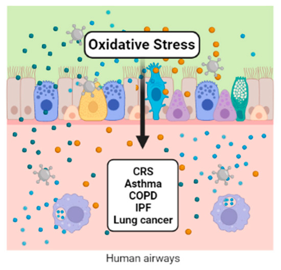
[6]]. The participation of oxidative stress in the pathophysiology of respiratory tract diseases is shown (
Figure 1).
Figure 1. Oxidative stress activates the lungs’ epithelial cells, generating inflammatory mediators that participate in the macrophage activation and the modulation of gene expression and transcription factors. All of them are implicated in numerous respiratory diseases.
2. Chronic Rhinosinusitis (CRS) and Nasal Polyps (NP)
Chronic rhinosinusitis (CRS) is a chronic inflammation of the nose and paranasal sinuses, with a wide range of clinical phenotypes. This heterogeneous disease has an incidence of approximately 5%, significantly impacting the patients’ quality of life and productivity. One-third of the world’s population with CRS has nasal polyps (CRSwNP) [
[7]]. A Type 2 inflammation mediated by the mast cells is present in CRS in response to increased oxidative stress. It has been suggested that air pollution causes an inflammatory change in the respiratory epithelium associated with CRS. However, there are few studies on the impact of air pollution and oxidative stress on the development of CRS. Recently, Patel and colleagues studied the relationship between levels of particulate air pollution (PM
2.5) and the pathogenesis of CRS. They found that exposure to ambient air pollutants may contribute to the pathogenesis of this disease. Ozone is another air component linked to higher tissue inflammation, eosinophilic aggregates, and Charcot–Leyden crystals in CRSwNP patients evaluated in one study [
[8]]. Another critical aspect investigated was whether socioeconomic status and exposure to airborne pollutants such as PM
2.5, black carbon (BC), and NO
2 increased the disease’s severity. The results showed that lower socioeconomic status predicted higher exposure to air pollution and increased disease severity in patients with CRS [
[9]].
A study evaluated occupational airborne exposure and the severity of CRS [
[10]]. The impact of exposure to vapors, gases, dust, fumes, fibers, and mist on 113 patients with CRSwNP, 96 with CRS without nasal polyps (CRSsNP), and 96 patients with aspirin-exacerbated respiratory disease (AERD) were evaluated. Patients exposed to these air contaminants required higher steroid doses than nonexposed patients. Contrary to other reports, this study found that PM
2.5 and BC did not have a high impact on disease severity. On the other hand, Zheng and colleagues (2020) studied the role of nicotinamide adenine dinucleotide phosphate (NADPH) oxidase in CRSwNP. This oxidase has been associated with the pathogenesis of CRSwNP. Zheng et al. found, by Western blotting and real-time PCR, that this oxidase is increased in the nasal polyps of patients. These findings suggest that oxidative stress plays a role in the pathogenesis of CRSwNP [
[11]]. The expression level of several oxidative stress and inflammation-related genes provided valuable information on the impact of air pollution on the nasal mucosa and nasal polyps of patients with CRS.
Recently, at the molecular level, the expression of oxidative stress- and inflammation-related genes in nasal polyps from patients with CRSwNP was evaluated. A significantly lower difference in the expression levels of transcription of antioxidant enzymes, including superoxide dismutase (SOD) and peroxiredoxin-2 (PRDX2), was reported, independent of age, sex, and smoking in patients with CRSwNP [
[12]]. These results correspond to reduced SOD capacity with the increase in oxidative stress. Additionally, an analysis of the advanced oxidation protein products (AOPP) and SOD showed the opposite effect in patients with NP. The level of AOPP from NP was higher than in the healthy control group. However, SOD activity was lower, indicating that oxidative stress plays a vital role in the development of nasal polyps [
[13]]. Another mechanism of regulating oxidative stress is mediated by the thioredoxin-interacting protein (TXNIP), which acts as a pro-oxidant protein by suppressing the activity of thioredoxin (TRX) and its antioxidant function [
[14]]. However, in nasal tissue samples from patients with CRSwNP, the protein and mRNA of TXNIP and TRX were significantly increased and decreased, respectively, compared with the control subjects [
[15]].
The transcription factors (TFs) are the primary regulators of gene expression. In this sense, essential TFs are related to oxidative stress, such as nuclear erythroid 2-related factor 2 (Nrf2), which regulates several antioxidant genes. For example, Nrf2 was necessary for an antioxidant pathway in a mouse model of rhinosinusitis. Knockout mice showed enhanced severity of eosinophilic sinonasal inflammation from disruption of the epithelial-specific Nrf2 pathway [
[16]] and enhanced susceptibility to eosinophilic sinonasal inflammation [
[17]]. This transcription factor has also been related to the stability of the sinonasal epithelial cell barrier function [
[18]]. Scavenger receptors (SRs) are a broad family of transmembrane receptors involved in a dysfunctional host–environment interaction through a reaction with ROS production. Lectin-like oxidized LDL receptor-1 (LOX-1) is one member of these transmembrane receptors. In 2020, Nishida and colleagues found a significant increase in the mRNA expression levels of LOX-1 in CRSwNP patients [
[19]].
Moreover, human sinuses are the primary source of NO in the airways. NO plays a role in regulating airway inflammation through the expression of NO synthase isoforms. Oxidative damage to the cellular components occurs when excessive amounts of NO are produced. Therefore, measuring NO levels can help diagnose CRS and sinonasal inflammation [
[20]]. Additionally, dupilumab, an anti-IL-4 receptor alpha monoclonal antibody, has been used recently in the treatment of CRSwNP. Patients with CRSwNP treated with dupilumab were evaluated through extended nitric oxide analyses (exhaled, FENO; bronchial, JawNO; alveolar, CalvNO components; nasal, nNO) where the results showed that nitric oxide significantly improved after 15 days of treatment [
[21]].
Some patients with CRSwNP suffer from bacterial airway infection and damage to the respiratory epithelia. TAS2R38 is an essential receptor in epithelial cells; its stimulation increases the production of NO, then the NO damages bacterial membranes, enzymes, and DNA, and increases the ciliary beat frequency. The expression of TAS2R38 in the cilia of human sinonasal epithelial cells is associated with susceptibility to CRS. Patients with advanced CRSwNP showed reduced TAS2R38 receptor expression in the sinonasal mucosa [
[22]]. Similar results were found in Italian patients with CRSwNP [
[23]]. Another critical receptor in the epithelial cells of the human airway is the bitter taste receptor (T2Rs). T2Rs can stimulate endothelial NO synthase (eNOS), whereby NO enhances mucociliary clearance with antibacterial effects on ciliated epithelial cells [
[24]]. This activation of T2Rs is associated with CRSwNP status and has been proposed as a biomarker [
[25]]. Oxidation can activate the calcium-activated kinase (CaMKII); the role of this kinase has been reported in several models of asthma, CRSwNP, cardiovascular disease, diabetes mellitus, and cancer [
[26]]. The expression of ox-CaMKII was measured in CRSwNP with other proteins such as indoleamine 2,3-dioxygenase 1, tryptophan 2,3-dioxygenase, and kynurenine. Oxidized CaMKII was increased in eosinophilic polyps [
[27]].
Nasal polyps (NP) are a common inflammatory mass affecting from 0.2% to 5.6% of the population [
[28]]. The etiology of NP is unclear because the factors involved in its occurrence include the genetic background, the immune system, anatomical differences, and environmental conditions. However, NP are associated with other chronic inflammatory respiratory diseases such as cystic fibrosis, AERD, respiratory allergies, and, as mentioned above, CRSwNP [
[29]]. Epidemiologically, there is an association between air pollution and the increased prevalence of these respiratory diseases [
[30]]. Exposure to air pollutants enhances the symptoms’ severity, resulting in an imbalanced concentration of free radicals and ROS such as NO
•, HO
•, O
2•−, and H
2O
2. In this regard, some studies have explored the association of the development and pathogenesis of NP with oxidative stress and air pollution, although information is limited.
Since the nasal epithelium is the first barrier of entry for inhaled particles such as pollutants, it plays a crucial role in the formation of NP. Oxidative stress damages the epithelium and causes mucosal edema due to impaired ion transport. The intracellular Na
+ increases, the Ca
2+ moves into the cell, and intracellular K
+ decreases [
[31]]. Moreover, chronic exposure to air contaminants affects the concentration of H
2O
2 and IL-8 in the nasal epithelium, a physiological defense mechanism [
[32]]. The increase in the cells’ permeability and the migration of inflammatory cells of the proliferative and secretory response is yet, another innate immune response mechanism. The release of cytokines by effector cells, and the activity of cyclooxygenase and lipoxygenase are also associated with the pathophysiology of NP [
[33]].
One of the most critical lines of defense against ROS are enzymes crucial for the activity of antioxidant, such as SOD, catalase, glutathione peroxidase, and thiol reductase [
[13]. The expression and activity of SOD, which catalyzes the dismutation of superoxide anions, is lower in NP than in healthy mucosa, which is correlated with lower antioxidant blood levels in NP patients [
[34],
[35],
[36]]. Different molecules are related to oxidative stress, for example, malondialdehyde (MDA) and free radicals are the products of lipid peroxidation of polyunsaturated fatty acids in cell membranes. These oxidant products have higher NP levels than control tissues [
[37],
[38]]. Another compound is nitric oxide, which is released in response to inflammation. Nitric oxide is involved in antiviral and bactericidal activity but inhibits cell proliferation, DNA synthesis, and collagen production. In NP, NO
• reacts with oxygen, producing peroxynitrite, which is associated with progressive epithelial injury. In patients with nasal polyposis, there is a lower concentration of NO
• compared with healthy patients, which is related to the downregulation of the nitric oxide metabolism, in which dismutase is crucial for the modulation of its activity [
[37]].
In the same way, SOD activity was decreased, and MDA increased in NP samples, as mentioned above. Another approach to studying the role of oxidative stress in NP is to examine how the apoptotic pathway is related. In 2021, Simsek and colleagues reported deficient apoptosis through the MAPK/JNK pathway in NP tissues, which may have a role in the pathogenesis and is consistent with previous reports [
[39]].
To date, oxidative stress is increased in patients with CRSwNP. However, this condition has a multifactorial etiology, and the role of air pollution is unclear. However, airborne pollutants may contribute to the pathogenesis of these diseases through the expression of several transcription factors and receptors in sinonasal epithelial cells. Some of them have been proposed as biomarkers.
3. Asthma
Asthma is a complex condition that is heterogeneous and is characterized by the critical role of chronic airway inflammation and oxidative stress. The eosinophils, lymphocytes, neutrophils, and mast cells generate inflammatory mediators and ROS/RNS that negatively affect the redox balance [
[40],
[41]]. Furthermore, these are the basis for identifying the actual Type 2 high and Type 2 low phenotypes [
[42]]. In Type 2 asthma patients, environmental factors favor the release of alarmins from the respiratory epithelium, which induces the differentiation of naïve T cells into Th2 cells. Damaged cells release interleukins such as IL−6, IL-1β, nitric oxide (NO
•), prostaglandin E2 (PGE2), and tumor necrosis factor α (TNFα); the principal marker in these patients is the sputum eosinophilia [
[43]]. T2-low asthma patients are characterized by sputum neutrophilia secondary to the activation of the NLRP3 inflammasome and elevated IL-1β; the activation of Th1 and/or Th17 cells associated with the imbalance of Th17/Treg cells seems to play an essential role in the pathology of asthma [
[44]]. The response to the combination of Th1, Th2, and Th17 and genetic predisposition induce permanent structural changes in T2-high and T2-low asthma patients [
[45],
[46]]. The process of airway remodeling is driven by subepithelial fibrosis, thickening of the sub-basement membrane, increased airway smooth muscle mass, angiogenesis, and mucous gland hyperplasia [
[45]].
An imbalance in the airway-reducing state is a determinant of the initiation and severity of asthma. The ability of an individual to ward off oxidative lung damage depends partly on their endogenous antioxidant systems and exogenous antioxidant intake [
[27]]. Several groups have shown that the levels of enzymatic antioxidants such as SOD, catalase, and glutathione peroxidases, as well as heme oxygenase-1 (HO-1), thioredoxins, peroxiredoxins, and glutaredoxins, are decreased in the bronchoalveolar lavage, sputum, and serum of asthmatic patients [
[40],
[47],
[48]].
Some factors increase the risk of the development of asthma. Among these, regular exposure through inhalation to oxidants derived from outdoor and indoor ambient air pollutants is on the list of factors that contributes to the progression of the disease [
[49],
[50],
[51]]. Since the relationship between oxidative stress and the inflammatory response depends on each other, and genetic predisposition could modify their balance, there is interest in the role of the inflammatory process as an activator of oxidative stress [
[52]]. Signaling pathways involving the inflammatory process and the oxidative response associated with the development of asthma are a current matter of evaluation. For example, the adenosine 5’ monophosphate-activated protein kinase (AMPK)/sirtuin 1 (Sirt1) and Nrf2/HO-1 pathway [
[53]] and the nitrogen-activated protein kinase (MAPK) pathway that includes extracellular signal-regulated kinases (ERKs), c-Jun N-terminal kinase (JNK), and p38 [
[54]] have been evaluated. Nrf2 potentiates the activity of the antioxidant response element (ARE) that synthesizes antioxidant proteins such as HO-1 [
[55]]. Multiple phytochemicals involved in the immune response activate the Nrf2/HO-1 signaling axis. In this sense, the Nrf2/HO-1, NF-κB, and MAPK pathways are relevant therapeutic molecular targets in asthma [
[56],
[57],
[58],
[59],
[60]].
The methodologies used to identify the molecular biomarkers associated with respiratory diseases are varied and range from the use of proteomics platforms to the use of real-time PCR. In 2021, Suzuki et al. evaluated the plasma proteome using an aptamer-base affinity proteomic platform (SOMAscan
®) in 34 subjects with stable COPD and 51 subjects with asthma, detecting 1238 proteins within which stress markers were found, such as myeloperoxidase (MPO), heme oxygenase 2 (HMOX2), superoxide dismutase (Cu-Zn) (SOD1), peroxiredoxin-1 (PRDX1), and glutathione-S-transferase P1 (GSTP1) [
[61]].
However, some markers are associated with oxidative stress-related cell damage, such as MDA, which can be measured by colorimetric techniques, high-performance liquid chromatography (HPLC), or LC/atmospheric pressure chemical ionization tandem mass spectrometry (LC/APCI–MS/MS). In asthmatic patients, the sputum measurement of MDA in the sputum discriminated between patients and controls with greater accuracy than the levels found in plasma, where it might be more difficult to evidence the redox imbalance due to comorbidities and lifestyle risk factors. In addition, 8-isoprostane and the oxidative DNA damage marker 8-oxo-7,8-dihydro-29-deoxyguanosine (8-OHdG) were also increased in the sputum from asthmatic patients compared with nonasthmatic controls in several studies [
[62]].
The mitochondria are the organelles that contributes the most to the generation of reactive oxygen species, and its contribution to oxidative stress in asthma has also been evaluated. The mitochondria are also susceptible to oxidative stress; under such conditions, they undergo an adaptive response through mitochondrial biogenesis. In 2021, Carpagnano et al. determined that the mitochondrial DNA/nuclear DNA (mtDNA/nDNA) ratio was a marker of mitochondrial oxidative stress in the exhaled breath condensate (EBC) of 53 patients with severe asthma, 11 patients with mild to moderate asthma, and 12 healthy subjects. They found higher levels of exhaled mtDNA/nDNA in severe asthmatic patients compared with the mild-moderate and healthy controls; this may be useful for differentiating the asthma phenotypes [
[63]].
It is crucial to take into account that the presence of oxidative stress is a factor that triggers asthma symptoms and contributes to the severity of the disease. Moreover, oxidative stress promotes corticosteroid insensitivity by disrupting glucocorticoid receptor (GR) signaling, leading to the sustained activation of proinflammatory pathways in immune cells and the airway’s structural cells [
[64],
[65]].
As already described in this section, many methodological strategies and various target molecules are related to oxidative stress. However, specific biomarkers with clinical applications in asthma have not yet been found.
This entry is adapted from the peer-reviewed paper 10.3390/ijms24010853