Environmental Implications Associated with the Development of Nanotechnology: History
Please note this is an old version of this entry, which may differ significantly from the current revision.

Nanotechnology involves materials at a scale ranging from 1 to 100 nm in one of its dimensions. It has brought advances in several areas such as electronics, pharmaceuticals, biotechnology, agriculture, cosmetics, and food. Nanostructures have a higher surface-to-volume ratio compared to bulk materials aside from exhibiting enhanced catalytic, mechanical, optical, electrical, tribological, thermal, and other properties. For this reason, nanomaterials have been widely studied and applied for the production of different products such as textiles, food coloring, pharmaceuticals, and cosmetics. Furthermore, the interaction of nanomaterials with biological systems and the environment still needs to be clarified. Moreover, some issues such as toxicity, bioaccumulation, and physicochemical transformations are found to be dependent on several factors such as size, capping agent, and shape, making the comparisons even more complex. 

  • nanomaterials
  • green synthesis
  • environment
  • risks
  • toxicity

1. Synthesis of Nanomaterials

Top–down and bottom–up methodologies have been adopted to synthesize NMs through different routes such as physical, chemical, and biological. Physical and chemical methods are widely used. However, they usually require toxic solvents and generate by-products that have high-risks to the environment and harm human and animal health, hence limited applications [1][2].
Recently, the biogenic approach has gained notoriety. In this synthesis method, biological systems or their products are used in the process. Microorganisms, plant extracts, or isolated biomolecules such as sugars, nucleic acids, and proteins are being investigated for this purpose [3][4][5][6][7][8][9][10][11][12][13][14][15]. It has no or low toxicity besides being an inexpensive and straightforward methodology [10][16][17].
Green chemistry aims to produce nanomaterials sustainably to reduce the maximum waste generated, energy consumption, and the concentration of precursors required. It replaces toxic compounds and solvents with benign or less toxic alternatives such as plant extracts. Another example consists of the use of water instead of organic solvents. Furthermore, reducing the steps during the synthesis process will spare wasting reagents and energy. Another essential factor to be considered is that more eco-friendly conditions of pH, temperature, and pressure are adopted during green synthesis and the establishment of protocols with reactions that generate few or no by-products. Thus, the synthesis needs to be sustainable throughout the process including the disposal [18][19][20][21].
One of the most studied routes is using plant extracts as reducing agents and surface stabilizers. Different plant parts have already been used such as fruits [22][23][24][25], roots [15][26][27], peels [13][28], seeds [29][30], leaves [11][31][32][33], and flowers [34]. In a bottom–up method, the nanostructure control can be achieved through multiple factors such as pressure, temperature, and the concentration of reagents, among others [35]. The synthesis process starts with a metal precursor, usually an inorganic salt. In a reaction medium, this salt suffers an ionic dissociation and biomolecules present such as proteins, sugars, flavonoids, and tannins could act to reduce these ions to a reduced state, assuming the state of its fundamental atom in a nucleation phase where a stable nucleus is formed by reducing the metallic precursor. Subsequently, the interaction with other nuclei begins to aggregate, followed by an increase in nuclei size during the growth phase. This phase can be interrupted by depleting the metallic precursor or coating molecules to prevent the aggregation of these nanostructures [16][36][37][38]. Agents capable of stabilizing nanostructures are essential for the formation of a colloidal solution without aggregates [39]. This occurs because the lack of bonds on the surface increases the Gibbs’ free energy, making this highly reactive and the nanostructure very unstable. As a result, it has a high contact surface area compared to its volume. In this way, due to dissatisfied bonds, the network parameter of the structure is reduced. Consequently, the atoms are closer together, changing the material’s properties as a whole [40]. Due to the Gibbs’ high energy, nanostructures tend to clump together to form larger particles. The synthesis process and reagents involved will directly influence the morphology and size of the resulting nanomaterial [39].

2. Applications

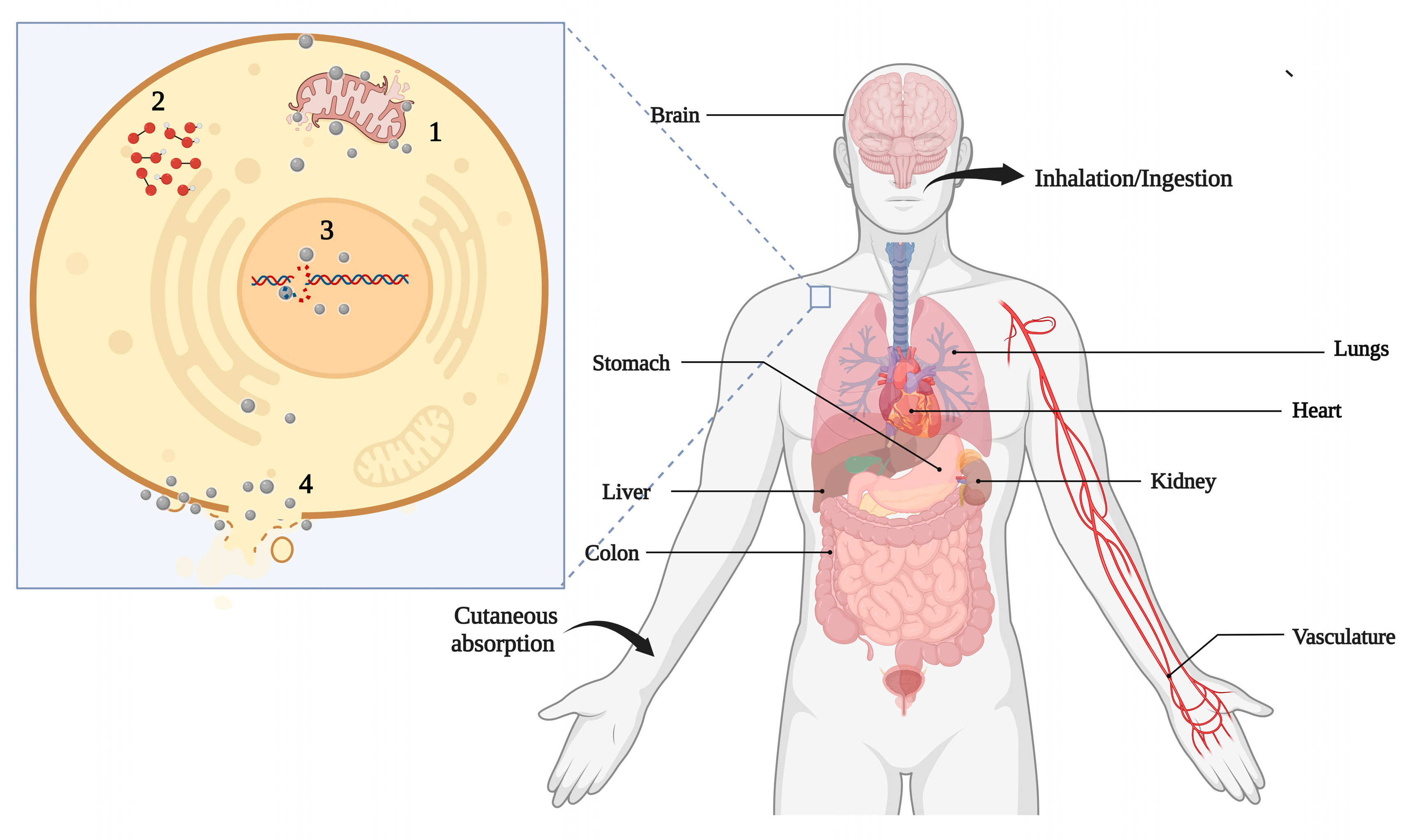
A variety of products containing NMs are already on the market and under development. However, through their production and utilization, these NMs can reach the environment and may cause contamination and pollution.

One of NM applications is cosmetics, mainly in sunscreens. TiO2 and ZnONPs are used for their absorbing properties of ultraviolet (UV) radiation and hypoallergenicity. To constitute a transparent emulsion, they must have nanometric dimensions [1][41]. Another application is nanoemulsions, which use NPs to achieve textures, optical, and tactile characteristics specific to each product [1]. The main effect of the possible risks of these products would be on the aquatic environment and living beings in that environment, and direct contact with the skin when using such products [1]. However, many of these products have been extensively tested for health and safety by regulatory authorities. As an example, ZnONPs are already incorporated in sunscreens and lip balms [41].
Nanotechnology has also emerged as a potential alternative for environmental decontamination. Various nanomaterials such as NPs, carbon nanotubes (CNTs), graphene, and nanocomposites have been extensively investigated for contaminant remediation [42]. Studies have shown promising results on removing environmental pollutants by NPs, making room for future use in ecological disasters like oil spills and other environmental contaminants such as heavy metals [43][44][45]. TiO2 NPs have been investigated for their photocatalytic properties to eliminate organic compounds. In this context, gold nanoparticle decorated TiO2 (Au/TiO2) produced using green tea exhibited an adsorption capacity greater than 8185 mg/g of methylene blue [46]. Carbon nanomaterials have been investigated as an alternative for treating organic contaminants in soil and water. This ability is related to surface hydrophobicity and the greater affinity of these NMs to these compounds than common particles in natural environments [42]. Hydroxyl-functionalized multi-walled carbon nanotubes (MWCNT-OH) showed the ability to remove naphthalene and fluorene from water bodies [47].
Furthermore, many studies have reported the antimicrobial activity of different nanostructures such as silver, gold, copper, and titanium NPs [48][49][50]. These specific and unique properties make them very promising for diverse applications such as in medical-hospital products, clothing, air purifiers, and food packaging, in which AgNPs stand out among the rest [35][36][48][51].
In addition, NMs have offered new approaches for diagnosing diseases, imaging, administration and drug formulations as well as therapy, particularly those with antimicrobial and antitumor potential. Some NPs have demonstrated that they can be used to selectively combat tumor cells without causing significant effects on normal cells [52][53][54][55][56][57][58]. This selectivity may be associated with pH, since the stability of NMs is pH dependent, and tumor cells create a microenvironment that is slightly more acidic than normal cells, which could cause the dissolution of NPs [54]. The antitumor effect has also been associated with the fact that tumor cells have high cell metabolism and rapid cell division [57]. NMs with photocatalytic properties can also be used to control drug release, sensitize cells, or even create an oxidative environment leading to cell death [59].
NMs can also be used for drug delivery, enabling them to deliver drugs with greater efficiency, reducing toxicity, and enhancing their effects. For example, oleic acid (OA)-pluronic-coated iron oxide magnetic NPs were efficiently associated with the antitumor agent doxorubicin. This association allows for sustained drug release, and aside from the cytotoxic effect on tumor cells, was found to be dose-dependent [60]. Similarly, AuNPs functionalized with a thiolated poly(ethylene glycol) (PEG) and conjugated to the antitumor drug oxaliplatin demonstrated an as good as or significantly better antitumoral activity against lung and colon cancer cells than oxaliplatin alone [61]. The anti-inflammatory potential has also been reported for different NMs such as cerium oxide NPs (CeNPs) [62], AgNPs [22][63][64], AuNPs [65][66], ZnONPs [67][68], SeNPs [69], and PtNPs [70]. In recent years, NMs have also been investigated for their potential in regenerative medicine. They can be applied for the control of the agents’ release, modification of the characteristics such as solubility and stability of molecules and help control the properties of scaffolds that serve as the support for cells [71].
In addition, the possibility of using nanotechnology in agriculture has attracted great attention. NMs can be used to increase agricultural productivity, reduce costs, and improve crop management efficiency [72]. Nanoformulations and NPs have been investigated as a vehicle for delivering pesticides to decrease their toxicity, increase stability and action, or directly combat pests and diseases [73]. Furthermore, sensors can be used to detect the presence of pathogens and pesticides and monitor plantation and environmental conditions such as air and soil, allowing for precision agriculture [73]. Fertilizers have also been studied to efficiently handle inputs, with minimal loss and a reduction in environmental contamination [73][74][75]. However, it is essential to investigate how these NMs accumulate and degrade in the environment and their toxicity to terrestrial and aquatic environments.
Finally, nanotechnology has allowed for the development of nanosensors capable of detecting a variety of target analytes, which can be molecules, nucleic acids, proteins, and even cells such as bacteria [76][77]. Nanosensors can be divided into optical, electrochemical, and magnetic, and applied in various processes such as in the diagnosis of diseases either in vivo or in vitro in the detection of pollutants in environmental samples, monitoring the concentration of molecules of interest in real-time, monitoring environmental conditions such as humidity and temperature, or in any process that needs to detect or monitor the presence of a target analyte [77]. The development of nanosensors for glucose, H2O2, uric acid, urea, neurotransmitters, hormones, drugs, metals, pesticides, viruses, bacteria, and even tumors has already been widely reported in the literature [76][78][79][80].

3. Environment Transformations

A multifactorial assessment is needed to understand the possible harmful effects of NMs. In this context, it is important to highlight that the properties of NMs vary considerably depending on the particle size, shape, chemical composition, capping agent, charge, solubility, and aggregation state [81]. NMs usually have their surface coated by chemical functional groups to guarantee their colloidal stability including proteins, polymers, carboxylic acids, surfactants, and sugars [82]. Nevertheless, these properties suffer interference from the medium, thus requiring the characterization of NPs in the means of production used in toxicological tests and the environment. However, nanomaterials suffer interference from the environment in which they are, and their properties and characteristics can change. Thus, it is necessary to consider these possible transformations for a more accurate toxicity evaluation. Variations in pH, the stability of ionic strength, and the concentrations and the type of organic compounds are also found to influence the NMs’ behavior, bioavailability, and toxicity. In addition to the above critical factors, to better assess the real risks in-depth and breadth, one must consider how NMs will present themselves after interacting with the environment, whether in a cellular, terrestrial, or aquatic environment [1][81].
When in contact with the medium, NMs can adsorb components onto their surface. This affinity can originate mainly from electrostatic interactions, hydrogen, and hydrophobic bonds [42]. The adsorption process and mechanism mainly depend on the particle size, shape, surface charge, and the capping agent [83].
Especially in biological media, NMs can be rapidly coated with proteins to form a protein corona. The latter consists of two layers: the hard corona is an inner layer composed of high-affinity proteins while the soft corona is an outer layer that is weakly bound by low-affinity proteins not directly bound to NPs [83][84]. Proteins can interact at the surfaces of NMs by noncovalent interactions and chelate with oxidized metals [85]. This protein corona is dynamic and changes composition over time due to the affinity-based competition between proteins and other molecules like organic matter for adsorption onto the surfaces of the NPs [83][85].
The composition of the biological environment can affect the dynamic protein exchange processes between the environment and the protein corona, thereby influencing the properties of the protein corona. However, NMs can adsorb many different molecules at the surface including polysaccharides, proteins, lipids, nucleic acids, metabolites, and other molecules, named the Eco-corona. In this case, protein may not be the most abundant constituent, mainly if it is formed outside an organism [85]. The protein adsorbed onto the NM surface can alter all of the interaction dynamics. It can affect the protein structure and lead to an immune response [83]. It can block the attachment of ligands to the surface of the NPs or interfere with the interactions between the ligands and receptors, and therefore alter subsequent phagocytosis and signaling transduction in cells. The adsorption of proteins could lead to various modifications in the physicochemical characteristics of NMs including the size, shape, surface chemistry, stability, biocompatibility, and composition, as a consequence, potentially contributing to changes in the NP uptake [83][84].
The dissolution process is related to the transformation of NMs such as NPs into their ion form. These ions’ release of NMs is associated with higher toxicity [42][81]. In biological media, these ions can bind to proteins and other macromolecules or precipitate as insoluble salts such as AgCl or Ag3PO4. For example, chloride ions can interact with Ag ions to form AgCl, precipitating in the solution. As a result, fewer Ag ions are available, and the toxicity is reduced [86]. Some reports have evaluated the dissolution kinetics of NPs under different conditions [87][88][89].
Natural colloids in river water were the main ones responsible for causing the aggregation of CeO2 NPs. At concentrations lower than 1 mg/L, colloids in the samples led to heteroaggregates, followed by sedimentation. However, at higher concentrations (10 mg/L and 100 mg/L), homoaggregation was more important than interaction with natural colloids as being mainly responsible for aggregation. When the water samples were filtered, almost no sedimentation was observed, and the CeO2 NPs were more stable. The stability of the NPs was directly related to the initial concentration and the presence of colloids, which may be due to the increased frequency of collisions, consequently causing aggregation and sedimentation [90]

4. Toxicity

4.1. Aquatic Environments

In aquatic environments, NMs are present in free form or interact with organic and inorganic ligands. Meanwhile, their speciation is influenced by the physical and chemical properties of the environment. The aquatic environment is the most studied since it is the primary recipient of contaminants. Once NMs contaminate the aquatic environment, ingestion through water may occur. However, the mechanisms that living organisms respond to intoxication caused by NMs are still not well understood. An early study was devoted to assessing marine invertebrates’ toxicity toward TiO2 and C60 NPs. Daphnia organisms are bioindicators because they interact with large portions of water in the environment. Therefore, polluting NPs’ ingestion has tremendous potential to be affected compared to other aquatic organisms. It was observed that exposure to filtered TiO2 NPs and C60 caused mortality, in which the latter caused toxicity at much lower concentrations. The LC50 for TiO2 NPs was 5.5 ppm while that of C60 reached a much lower dose of 460 ppb. This difference was related to the NPs’ size since the C60 (0.72 nm) was significantly lower than TiO2 NPs (10–20 nm). Also, the adopted synthesis method of NPs has been found to affect their toxicity. Unfiltered sonicated NPs caused little mortality; at 25–30 times higher concentrations of TiO2 NPs, there was not much difference compared to the control. Furthermore, at a concentration of 500 ppm C60, there was only 18% mortality. These effects may result from the presence of larger NPs and clusters [91].
Algae are also essential species of the aquatic environment, as they play an indispensable role as food sources and photosynthesis. The effects of Fe3O4 NPs were evaluated in the microalgae Chlorella vulgaris. The results showed changes in chlorophyll content, reduced CO2 absorption, and photosynthetic rate from 50 mg/L. Oxidative stress was also noticed due to changes in stress markers’ levels. The increased levels of malondialdehyde, related to lipid peroxidation; and the reduction in glutathione lev

4.2. Terrestrial Environments

In terrestrial ecosystems, NMs can directly or indirectly affect plants, microbial communities, and soil organisms. In plants, they can affect plant seeds’ germination and the development of plant roots and sprouts. NMs’ interaction with plants induces several physiological and morphological modifications including impaired seed germination, plant growth inhibition, reduced biomass production, and imbalanced nutrients as well as an alteration in photosynthesis and transpiration rates [92]. NMs present in the environment upon interaction with plants can be internalized in plant tissues either through roots or above-ground parts [93]. In addition to terrestrial plants, NMs can affect the growth of soil microorganisms, and invertebrate animals such as snails, earthworms, or larvae insects. Besides, contaminating groundwater and being absorbed by crops, and possibly entering the food chain with potential damage to animals and humans’ health [73][94][95].
AgNPs could inhibit Phaseolus radiatus and Sorghum bicolor growth in an agar medium, with a concentration-dependent effect. The growth rate was not affected when tested on the soil. The accumulation of Ag in the root cells was also observed [96]. The effects of CuNPs were also evaluated in wheat (Triticum aestivum L.). The plant’s growth rate decreased markedly up to 60%, and the formation of lateral roots was stimulated. Oxidative stress associated with the presence of CuNPs has been reported [97]
When exposed to CuONPs for 15 days, cabbage (B. oleracea var. capitata L.) and lettuce (L. sativa L. cv. Batavia) showed a decrease in plant weight and photosynthesis level. Additionally, CuONPs affect the stomata and were translocated to roots [98]. Three different NPs (ZnO, CuO, or CeO2) and their equivalent ions (Zn2+, Cu2+, or Ce4+) were tested in sweet potato (Ipomoea batatas) development. The CeO2NPs caused a small positive effect at a higher concentration (1000 mg·kg of dry weight−1) on tuber biomass, while the ZnO and CuONPs caused negative effects on tuber biomass at the same concentration.
In wheat growth under hydroponic conditions, Fe3O4 NPs were detected in root cells, but no damage to the plant was observed. The NPs did not alter the chlorophyll content, plant growth, or germination rate and no oxidative damage or lipid peroxidation was noticed [99]. When comparing the effects of AgNPs to Ag+ on Arabidopsis thaliana, it was reported that AgNPs were more toxic. The AgNPs accumulated in the leaf cells inhibited root elongation, decreased chlorophyll content, and altered the transcription of antioxidant and aquaporin genes, affecting water homeostasis and antioxidant systems [100]. Similar results were observed in potatoes (Solanum tuberosum L.) treated with AgNPs and Ag+. Despite the Ag content in ion-treated plants being higher than the ones treated with AgNPs, the last caused significantly higher oxidative stress and a decrease in the glutathione and ascorbate contents [101].
In contrast, the ionic form of Ag was more toxic than any AgNPs with different surface coatings (citrate, PVP, and cetyltrimethylammonium bromide (CTAB)) tested on Allium cepa roots. These AgNPs exhibited toxicity only at higher concentrations. AgNP-CTAB led to bigger toxicity, causing root growth inhibition and oxidative stress. The larger particle AgNP-citrate demonstrated a tendency to aggregate, thus the effects were less pronounced [102].

3.3. Human and Animal Health

The effects of NM exposure on human and animal health have also been investigated. The mechanisms related to the toxicity of NMs vary depending on the studied cells and NM. In general, cytotoxicity is related to increased reactive oxygen species levels, decreased membrane potential, and antioxidant levels. DNA and proteins can also be affected directly or by the produced reactive species (Figure 1). DNA damage can cause the cell cycle to stop in the G2/M phase. Damage to the cell membrane can leak the cytoplasmic content and eventual necrosis, while rupture of the lysosomal membranes activates lysosome-mediated apoptosis [51][103].
Figure 1. Major possible consequences of the toxicity of nanomaterials to human health. The main routes of exposure are ingestion, inhalation, and absorption through the skin, which can cause damage to different organs such as the lungs, kidneys, heart, stomach, and liver. At the cellular level, nanomaterials are believed to affect cells through mitochondrial damage (1), ROS production (2), DNA and protein damage (3) as well as cell membrane disruption (4).
Human and animal exposure is usually due to food, air, medical applications, and water. Exposure routes influence potential risks including inhalation, cutaneous absorption, ingestion, and injection (Figure 1). Unfortunately, there is little information about the levels of exposure to workers and the public [104]. Ingestion can usually occur through the consumption of contaminated food and water. The main target of the inhalation route is the respiratory tract. The lung is one of the main targets including the pulmonary epithelial cells and cells of the immune system and fibroblasts. It is essential in response to NM exposure and related inflammatory conditions, fibrosis, and genotoxicity induced by these NMs [94]. NPs in cosmetics are an important route of exposure for the skin. They often reach systemic circulation, in which several organs can be affected such as the liver, kidney, heart, and consequently become target organs [81].
The effects of starch-coated AgNPs were evaluated in normal human lung fibroblasts (IMR-90) and human glioblastoma cells (U251). Exposure to AgNPs caused damage to the mitochondria and increased reactive oxygen species (ROS). DNA damage was also observed. However, the tumor cell appeared to be more affected. This damage were possibly responsible for stopping the cell cycle in the G2/M phase. AgNPs were found deposited inside the mitochondria and nucleus, which can be directly related to DNA damage and mitochondrial toxicity [105]. The cytotoxicity of AgNPs synthesized using the fungi Trichoderma viride was evaluated with an environmentally friendly proposal. They were able to significantly inhibit the proliferation of the MCF-7 cancer cell lines in a time- and concentration-dependent manner. DNA fragmentation was also observed, and at a concentration of 100 μg/mL, the AgNPs exhibited significant cytotoxic effects alongside apoptotic characteristics. These effects against cancer cells demonstrate their chemotherapeutic potential [106].

3.4. Impacts on the Food Chain

The risks also need to be evaluated across the entire food chain, considering the trophic transfer potential. A recent study evaluated the impacts and distribution of AuNPs at different trophic levels in an aquatic food chain. Initially, the algae of the species Pseudokirchinella subcapitata were exposed to AuNPs of different shapes and sizes. For a concentration of 2.9 × 10¹¹ particles·mL−1, no toxicity was observed in the algae subjected to these exposures. However, it was observed that 10 nm spherical NPs were present in 68% of the cells in the algal population. The rod-shaped 10 × 45 nm NPs were present in just 34% of the cells. In daphnids fed with AuNP-exposed algae, the Au accumulation was 0.73% (for spherical) and 1.71% (for rod-shaped) of the total Au in algae [107].
At the next trophic level, zebrafish were fed with 10 contaminated daphnids each day for 21 days. A small percentage of AuNPs accumulated in daphnids transferred to zebrafish varied from 0.03% (for spherical) to 0.48% (for rod-shaped) with the brain and liver as the target organs. Furthermore, AuNP dissolution and agglomeration in the gut of the daphnids and biodistribution in fish tissues were found to be size- and shape-dependent [107]. Similarly, positively charged 10-nm diameter AuNPs functionalized with polyethylene oxide demonstrated potential for trophic transfer from periphytic biofilms to the crustacean Gammarus fossarum. Biofilms exposed for 48 h to AuNPs at two concentrations, 4.6 and 46 mg/L, were used to feed the crustacean for 7 days, with daily biofilm renewal. Both approaches generated oxidative stress in cells, mainly affecting mitochondrial respiration. Alterations in digestive enzyme activity were also reported. In the cilia of apical intestinal epithelial cells, muscle fibers, and mitochondria, the latter clearly showed a disruption of crests and internal and external membranes.

This entry is adapted from the peer-reviewed paper 10.3390/nano12234319

References

  1. Paschoalino, M.P.; Marcone, G.P.S.; Jardim, W.F. Os Nanomateriais e a Questão Ambiental. Quim. Nova 2010, 33, 421–430.
  2. Chaturvedi, V.; Verma, P. Fabrication of Silver Nanoparticles from Leaf Extract of Butea Monosperma (Flame of Forest) and Their Inhibitory Effect on Bloom-Forming Cyanobacteria. Bioresour. Bioprocess 2015, 2, 18.
  3. Shemer, G.; Krichevski, O.; Markovich, G.; Molotsky, T.; Lubitz, I.; Kotlyar, A.B. Chirality of Silver Nanoparticles Synthesized on DNA. J. Am. Chem. Soc. 2006, 128, 11006–11007.
  4. Gao, X.; Wei, L.; Yan, H.; Xu, B. Green Synthesis and Characteristic of Core-Shell Structure Silver/Starch Nanoparticles. Mater. Lett. 2011, 65, 2963–2965.
  5. Syed, A.; Ahmad, A. Extracellular Biosynthesis of Platinum Nanoparticles Using the Fungus Fusarium Oxysporum. Colloids Surf. B Biointerfaces 2012, 97, 27–31.
  6. Prabhawathi, V.; Sivakumar, P.M.; Doble, M. Green Synthesis of Protein Stabilized Silver Nanoparticles Using Pseudomonas Fluorescens, a Marine Bacterium, and Its Biomedical Applications When Coated on Polycaprolactam. Ind. Eng. Chem. Res. 2012, 51, 5230–5239.
  7. Oluwafemi, O.S.; Vuyelwa, N.; Scriba, M.; Songca, S.P. Green Controlled Synthesis of Monodispersed, Stable and Smaller Sized Starch-Capped Silver Nanoparticles. Mater. Lett. 2013, 106, 332–336.
  8. Chowdhury, S.; Basu, A.; Kundu, S. Green Synthesis of Protein Capped Silver Nanoparticles from Phytopathogenic Fungus Macrophomina Phaseolina (Tassi) Goid with Antimicrobial Properties against Multidrug-Resistant Bacteria. Nanoscale Res. Lett. 2014, 9, 365.
  9. Maliszewska, I.; Juraszek, A.; Bielska, K. Green Synthesis and Characterization of Silver Nanoparticles Using Ascomycota Fungi Penicillium Nalgiovense AJ12. J. Clust. Sci. 2014, 25, 989–1004.
  10. Roni, M.; Murugan, K.; Panneerselvam, C.; Subramaniam, J.; Nicoletti, M.; Madhiyazhagan, P.; Dinesh, D.; Suresh, U.; Khater, H.F.; Wei, H.; et al. Characterization and Biotoxicity of Hypnea Musciformis-Synthesized Silver Nanoparticles as Potential Eco-Friendly Control Tool against Aedes Aegypti and Plutella Xylostella. Ecotoxicol. Environ. Saf. 2015, 121, 31–38.
  11. Asghar, M.A.; Zahir, E.; Shahid, S.M.; Khan, M.N.; Asghar, M.A.; Iqbal, J.; Walker, G. Iron, Copper and Silver Nanoparticles: Green Synthesis Using Green and Black Tea Leaves Extracts and Evaluation of Antibacterial, Antifungal and Aflatoxin B1 Adsorption Activity. LWT 2018, 90, 98–107.
  12. Molnár, Z.; Bódai, V.; Szakacs, G.; Erdélyi, B.; Fogarassy, Z.; Sáfrán, G.; Varga, T.; Kónya, Z.; Tóth-Szeles, E.; Szűcs, R.; et al. Green Synthesis of Gold Nanoparticles by Thermophilic Filamentous Fungi. Sci. Rep. 2018, 8, 3943.
  13. Pizzorno Backx, B.; Rech Pedrosa, B.; Delazare, T.; Carmo Damasceno, F.R.D.; Leitao Dos Santos, O.A. Green Synthesis of Silver Nanoparticles: A Study of the Dispersive Efficiency and Antimicrobial Potential of the Extracts of Plinia Cauliflora for Application in Smart Textiles Materials for Healthcare. J. Nanomater. Mol. Nanotechnol. 2018, 7.
  14. Dos Santos, O.A.L.; Backx, B.P. Estudo Da Eficiência Da Síntese De Nanopartículas De Prata Em Extrato De Beta Vulgaris Para Aplicação Em Têxteis Com Atividade Antimicrobiana. In Ciências da Saúde; Antonella Carvalho de Oliveira; Atena Editora: Ponta Grossa, Brazil, 2019; pp. 143–157.
  15. Dos Santos, O.A.L.; de Araujo, I.; Dias da Silva, F.; Sales, M.N.; Christoffolete, M.A.; Backx, B.P. Surface Modification of Textiles by Green Nanotechnology against Pathogenic Microorganisms. Curr. Res. Green Sustain. Chem. 2021, 4, 100206.
  16. Nath, D.; Banerjee, P. Green Nanotechnology—A New Hope for Medical Biology. Environ. Toxicol. Pharm. 2013, 36, 997–1014.
  17. Sujitha, V.; Murugan, K.; Paulpandi, M.; Panneerselvam, C.; Suresh, U.; Roni, M.; Nicoletti, M.; Higuchi, A.; Madhiyazhagan, P.; Subramaniam, J.; et al. Green-Synthesized Silver Nanoparticles as a Novel Control Tool against Dengue Virus (DEN-2) and Its Primary Vector Aedes Aegypti. Parasitol. Res. 2015, 114, 3315–3325.
  18. Prasad, R. Synthesis of Silver Nanoparticles in Photosynthetic Plants. J. Nanopart. 2014, 2014, 963961.
  19. Hyeon, T.; Manna, L.; Wong, S.S. Sustainable Nanotechnology. Chem. Soc. Rev. 2015, 44, 5755–5757.
  20. Dos Santos, O.A.L.; Backx, B.P. Green Nanotechnology: The Influence of Intermolecular and Supramolecular Interactions. J. Nanotechnol. Nanomater. 2020, 1, 104–108.
  21. Sahoo, T.R.; Prelot, B. Adsorption Processes for the Removal of Contaminants from Wastewater. In Nanomaterials for the Detection and Removal of Wastewater Pollutants; Elsevier: Amsterdam, The Netherlands, 2020; pp. 161–222. ISBN 9780128184899.
  22. David, L.; Moldovan, B.; Vulcu, A.; Olenic, L.; Perde-Schrepler, M.; Fischer-Fodor, E.; Florea, A.; Crisan, M.; Chiorean, I.; Clichici, S.; et al. Green Synthesis, Characterization and Anti-Inflammatory Activity of Silver Nanoparticles Using European Black Elderberry Fruits Extract. Colloids Surf. B Biointerfaces 2014, 122, 767–777.
  23. Mariselvam, R.; Ranjitsingh, A.J.A.; Usha Raja Nanthini, A.; Kalirajan, K.; Padmalatha, C.; Mosae Selvakumar, P. Green Synthesis of Silver Nanoparticles from the Extract of the Inflorescence of Cocos Nucifera (Family: Arecaceae) for Enhanced Antibacterial Activity. Spectrochim. Acta A Mol. Biomol. Spectrosc. 2014, 129, 537–541.
  24. Puišo, J.; Jonkuvienė, D.; Mačionienė, I.; Šalomskienė, J.; Jasutienė, I.; Kondrotas, R. Biosynthesis of Silver Nanoparticles Using Lingonberry and Cranberry Juices and Their Antimicrobial Activity. Colloids Surf. B Biointerfaces 2014, 121, 214–221.
  25. Ramesh, P.S.; Kokila, T.; Geetha, D. Plant Mediated Green Synthesis and Antibacterial Activity of Silver Nanoparticles Using Emblica Officinalis Fruit Extract. Spectrochim. Acta A Mol. Biomol. Spectrosc. 2015, 142, 339–343.
  26. Bindhu, M.R.; Umadevi, M. Antibacterial and Catalytic Activities of Green Synthesized Silver Nanoparticles. Spectrochim. Acta A Mol. Biomol. Spectrosc. 2015, 135, 373–378.
  27. Arokiyaraj, S.; Vincent, S.; Saravanan, M.; Lee, Y.; Oh, Y.K.; Kim, K.H. Green Synthesis of Silver Nanoparticles Using Rheum Palmatum Root Extract and Their Antibacterial Activity against Staphylococcus Aureus and Pseudomonas Aeruginosa. Artif. Cells Nanomed. Biotechnol. 2017, 45, 372–379.
  28. Bankar, A.; Joshi, B.; Kumar, A.R.; Zinjarde, S. Banana Peel Extract Mediated Novel Route for the Synthesis of Silver Nanoparticles. Colloids Surf. A Phys. Eng. Asp. 2010, 368, 58–63.
  29. Bar, H.; Bhui, D.K.; Sahoo, G.P.; Sarkar, P.; Pyne, S.; Misra, A. Green Synthesis of Silver Nanoparticles Using Seed Extract of Jatropha Curcas. Colloids Surf. A Phys. Eng. Asp. 2009, 348, 212–216.
  30. Kiran Kumar, H.A.; Mandal, B.K.; Mohan Kumar, K.; Maddinedi, S.B.; Sai Kumar, T.; Madhiyazhagan, P.; Ghosh, A.R. Antimicrobial and Antioxidant Activities of Mimusops Elengi Seed Extract Mediated Isotropic Silver Nanoparticles. Spectrochim. Acta A Mol. Biomol. Spectrosc. 2014, 130, 13–18.
  31. Khalil, M.M.H.; Ismail, E.H.; El-Baghdady, K.Z.; Mohamed, D. Green Synthesis of Silver Nanoparticles Using Olive Leaf Extract and Its Antibacterial Activity. Arab. J. Chem. 2014, 7, 1131–1139.
  32. Veisi, H.; Azizi, S.; Mohammadi, P. Green Synthesis of the Silver Nanoparticles Mediated by Thymbra Spicata Extract and Its Application as a Heterogeneous and Recyclable Nanocatalyst for Catalytic Reduction of a Variety of Dyes in Water. J. Clean. Prod. 2018, 170, 1536–1543.
  33. Anandan, M.; Poorani, G.; Boomi, P.; Varunkumar, K.; Anand, K.; Chuturgoon, A.A.; Saravanan, M.; Gurumallesh Prabu, H. Green Synthesis of Anisotropic Silver Nanoparticles from the Aqueous Leaf Extract of Dodonaea Viscosa with Their Antibacterial and Anticancer Activities. Process Biochem. 2019, 80, 80–88.
  34. Hemmati, S.; Rashtiani, A.; Zangeneh, M.M.; Mohammadi, P.; Zangeneh, A.; Veisi, H. Green Synthesis and Characterization of Silver Nanoparticles Using Fritillaria Flower Extract and Their Antibacterial Activity against Some Human Pathogens. Polyhedron 2019, 158, 8–14.
  35. Natsuki, J. A Review of Silver Nanoparticles: Synthesis Methods, Properties and Applications. Int. J. Mater. Sci. Appl. 2015, 4, 325.
  36. Keat, C.L.; Aziz, A.; Eid, A.M.; Elmarzugi, N.A. Biosynthesis of Nanoparticles and Silver Nanoparticles. Bioresour. Bioprocess. 2015, 2, 47.
  37. Singh, R.; Shedbalkar, U.U.; Wadhwani, S.A.; Chopade, B.A. Bacteriagenic Silver Nanoparticles: Synthesis, Mechanism, and Applications. Appl. Microbiol. Biotechnol. 2015, 99, 4579–4593.
  38. Ahmed, S.; Ahmad, M.; Swami, B.L.; Ikram, S. A Review on Plants Extract Mediated Synthesis of Silver Nanoparticles for Antimicrobial Applications: A Green Expertise. J. Adv. Res. 2016, 7, 17–28.
  39. Van Phu, D.; Lang, V.T.K.; Kim Lan, N.T.; Duy, N.N.; Chau, N.D.; Du, B.D.; Cam, B.D.; Hien, N.Q. Synthesis and Antimicrobial Effects of Colloidal Silver Nanoparticles in Chitosan by γ -Irradiation. J. Exp. Nanosci. 2010, 5, 169–179.
  40. Priyadarshini, B.; Rath, P.P.; Behera, S.S.; Panda, S.R.; Sahoo, T.R.; Parhi, P.K. Kinetics, Thermodynamics and Isotherm Studies on Adsorption of Eriochrome Black-T from Aqueous Solution Using Rutile TiO 2. IOP Conf. Ser. Mater. Sci. Eng. 2018, 310, 012051.
  41. Wright, P.F. Potential Risks and Benefits of Nanotechnology: Perceptions of Risk in Sunscreens. Med. J. Aust. 2016, 204, 369–370.
  42. Qian, Y.; Qin, C.; Chen, M.; Lin, S. Nanotechnology in Soil Remediation—Applications vs. Implications. Ecotoxicol. Environ. Saf. 2020, 201, 110815.
  43. Kanel, S.R.; Nepal, D.; Manning, B.; Choi, H. Transport of Surface-Modified Iron Nanoparticle in Porous Media and Application to Arsenic(III) Remediation. J. Nanopart. Res. 2007, 9, 725–735.
  44. Pala, I.R.; Brock, S.L. ZnS Nanoparticle Gels for Remediation of Pb 2+ and Hg 2+ Polluted Water. ACS Appl. Mater. Interfaces 2012, 4, 2160–2167.
  45. Murgueitio, E.; Cumbal, L.; Abril, M.; Izquierdo, A.; Debut, A.; Tinoco, O. Green Synthesis of Iron Nanoparticles: Application on the Removal of Petroleum Oil from Contaminated Water and Soils. J. Nanotechnol. 2018, 2018, 4184769.
  46. Perera, M.; Wijenayaka, L.A.; Siriwardana, K.; Dahanayake, D.; Nalin de Silva, K.M. Gold Nanoparticle Decorated Titania for Sustainable Environmental Remediation: Green Synthesis, Enhanced Surface Adsorption and Synergistic Photocatalysis. RSC Adv. 2020, 10, 29594–29602.
  47. Akinpelu, A.A.; Ali, M.E.; Johan, M.R.; Saidur, R.; Chowdhury, Z.Z.; Shemsi, A.M.; Saleh, T.A. Effect of the Oxidation Process on the Molecular Interaction of Polyaromatic Hydrocarbons (PAH) with Carbon Nanotubes: Adsorption Kinetic and Isotherm Study. J. Mol. Liq. 2019, 289, 111107.
  48. Ogunsona, E.O.; Muthuraj, R.; Ojogbo, E.; Valerio, O.; Mekonnen, T.H. Engineered Nanomaterials for Antimicrobial Applications: A Review. Appl. Mater. Today 2020, 18, 32.
  49. Backx, B.P.; dos Santos, M.S.; dos Santos, O.A.L.; Filho, S.A. The Role of Biosynthesized Silver Nanoparticles in Antimicrobial Mechanisms. Curr. Pharm. Biotechnol. 2021, 22, 762–772.
  50. Zhao, Y.; Chen, L.; Wang, Y.; Song, X.; Li, K.; Yan, X.; Yu, L.; He, Z. Nanomaterial-Based Strategies in Antimicrobial Applications: Progress and Perspectives. Nano Res. 2021, 14, 4417–4441.
  51. Prabhu, S.; Poulose, E.K. Silver Nanoparticles: Mechanism of Antimicrobial Action, Synthesis, Medical Applications, and Toxicity Effects. Int. Nano Lett. 2012, 2, 32.
  52. Franco-Molina, M.A.; Mendoza-Gamboa, E.; Sierra-Rivera, C.A.; Gómez-Flores, R.A.; Zapata-Benavides, P.; Castillo-Tello, P.; Alcocer-González, J.M.; Miranda-Hernández, D.F.; Tamez-Guerra, R.S.; Rodríguez-Padilla, C. Antitumor Activity of Colloidal Silver on MCF-7 Human Breast Cancer Cells. J. Exp. Clin. Cancer Res. 2010, 29, 148.
  53. Faedmaleki, F.; Shirazi, F.H.; Salarian, A.A.; Ashtiani, H.A.; Rastegar, H. Toxicity Effect of Silver Nanoparticles on Mice Liver Primary Cell Culture and HepG2 Cell Line. Iran. J. Pharm. Res. 2014, 13, 235–242.
  54. Mukherjee, S.; Chowdhury, D.; Kotcherlakota, R.; Patra, S.; Vinothkumar, B.; Bhadra, M.P.; Sreedhar, B.; Patra, C.R. Potential Theranostics Application of Bio-Synthesized Silver Nanoparticles (4-in-1 System). Theranostics 2014, 4, 316–335.
  55. Rao, P.V.; Nallappan, D.; Madhavi, K.; Rahman, S.; Jun Wei, L.; Gan, S.H. Phytochemicals and Biogenic Metallic Nanoparticles as Anticancer Agents. Oxid. Med. Cell. Longev. 2016, 2016, 3685671.
  56. Hassan, H.F.H.; Mansour, A.M.; Abo-Youssef, A.M.H.; Elsadek, B.E.M.; Messiha, B.A.S. Zinc Oxide Nanoparticles as a Novel Anticancer Approach; in Vitro and in Vivo Evidence. Clin. Exp. Pharm. Physiol. 2017, 44, 235–243.
  57. Jadhav, K.; Deore, S.; Dhamecha, D.; Hr, R.; Jagwani, S.; Jalalpure, S.; Bohara, R. Phytosynthesis of Silver Nanoparticles: Characterization, Biocompatibility Studies, and Anticancer Activity. ACS Biomater. Sci. Eng. 2018, 4, 892–899.
  58. Al-Rubaye, H.I.; Al-Rubaye, B.K.; Al-Abodi, E.E.; Yousif, E.I. Green Chemistry Synthesis of Modified Silver Nanoparticles. J. Phys. Conf. Ser. 2020, 1664, 012080.
  59. Ziental, D.; Czarczynska-Goslinska, B.; Mlynarczyk, D.T.; Glowacka-Sobotta, A.; Stanisz, B.; Goslinski, T.; Sobotta, L. Titanium Dioxide Nanoparticles: Prospects and Applications in Medicine. Nanomaterials 2020, 10, 387.
  60. Jain, T.K.; Morales, M.A.; Sahoo, S.K.; Leslie-Pelecky, D.L.; Labhasetwar, V. Iron Oxide Nanoparticles for Sustained Delivery of Anticancer Agents. Mol. Pharm. 2005, 2, 194–205.
  61. Brown, S.D.; Nativo, P.; Smith, J.A.; Stirling, D.; Edwards, P.R.; Venugopal, B.; Flint, D.J.; Plumb, J.A.; Graham, D.; Wheate, N.J. Gold Nanoparticles for the Improved Anticancer Drug Delivery of the Active Component of Oxaliplatin. J. Am. Chem. Soc. 2010, 132, 4678–4684.
  62. Hirst, S.M.; Karakoti, A.S.; Tyler, R.D.; Sriranganathan, N.; Seal, S.; Reilly, C.M. Anti-Inflammatory Properties of Cerium Oxide Nanoparticles. Small 2009, 5, 2848–2856.
  63. Hebeish, A.; El-Rafie, M.H.; EL-Sheikh, M.A.; Seleem, A.A.; El-Naggar, M.E. Antimicrobial Wound Dressing and Anti-Inflammatory Efficacy of Silver Nanoparticles. Int. J. Biol. Macromol. 2014, 65, 509–515.
  64. Singh, P.; Ahn, S.; Kang, J.-P.; Veronika, S.; Huo, Y.; Singh, H.; Chokkaligam, M.; El-Agamy Farh, M.; Aceituno, V.C.; Kim, Y.J.; et al. In Vitro Anti-Inflammatory Activity of Spherical Silver Nanoparticles and Monodisperse Hexagonal Gold Nanoparticles by Fruit Extract of Prunus Serrulata: A Green Synthetic Approach. Artif. Cells Nanomed. Biotechnol. 2017, 46, 2022–2032.
  65. Sulaiman, G.M.; Waheeb, H.M.; Jabir, M.S.; Khazaal, S.H.; Dewir, Y.H.; Naidoo, Y. Hesperidin Loaded on Gold Nanoparticles as a Drug Delivery System for a Successful Biocompatible, Anti-Cancer, Anti-Inflammatory and Phagocytosis Inducer Model. Sci. Rep. 2020, 10, 9362.
  66. Muniyappan, N.; Pandeeswaran, M.; Amalraj, A. Green Synthesis of Gold Nanoparticles Using Curcuma Pseudomontana Isolated Curcumin: Its Characterization, Antimicrobial, Antioxidant and Anti- Inflammatory Activities. Environ. Chem. Ecotoxicol. 2021, 3, 117–124.
  67. Nagajyothi, P.C.; Cha, S.J.; Yang, I.J.; Sreekanth, T.V.M.; Kim, K.J.; Shin, H.M. Antioxidant and Anti-Inflammatory Activities of Zinc Oxide Nanoparticles Synthesized Using Polygala Tenuifolia Root Extract. J. Photochem. Photobiol. B 2015, 146, 10–17.
  68. Rajakumar, G.; Thiruvengadam, M.; Mydhili, G.; Gomathi, T.; Chung, I.-M. Green Approach for Synthesis of Zinc Oxide Nanoparticles from Andrographis Paniculata Leaf Extract and Evaluation of Their Antioxidant, Anti-Diabetic, and Anti-Inflammatory Activities. Bioprocess Biosyst. Eng. 2018, 41, 21–30.
  69. El-Ghazaly, M.A.; Fadel, N.; Rashed, E.; El-Batal, A.; Kenawy, S.A. Anti-Inflammatory Effect of Selenium Nanoparticles on the Inflammation Induced in Irradiated Rats. Can. J. Physiol. Pharm. 2017, 95, 101–110.
  70. Rehman, M.U.; Yoshihisa, Y.; Miyamoto, Y.; Shimizu, T. The Anti-Inflammatory Effects of Platinum Nanoparticles on the Lipopolysaccharide-Induced Inflammatory Response in RAW 264.7 Macrophages. Inflamm. Res. 2012, 61, 1177–1185.
  71. Fathi-Achachelouei, M.; Knopf-Marques, H.; Ribeiro da Silva, C.E.; Barthès, J.; Bat, E.; Tezcaner, A.; Vrana, N.E. Use of Nanoparticles in Tissue Engineering and Regenerative Medicine. Front. Bioeng. Biotechnol. 2019, 7, 113.
  72. Capaldi Arruda, S.C.; Diniz Silva, A.L.; Moretto Galazzi, R.; Antunes Azevedo, R.; Zezzi Arruda, M.A. Nanoparticles Applied to Plant Science: A Review. Talanta 2015, 131, 693–705.
  73. Iavicoli, I.; Leso, V.; Beezhold, D.H.; Shvedova, A.A. Nanotechnology in Agriculture: Opportunities, Toxicological Implications, and Occupational Risks. Toxicol. Appl. Pharm. 2017, 329, 96–111.
  74. Chhipa, H.; Joshi, P. Nanofertilisers, Nanopesticides and Nanosensors in Agriculture. In Nanoscience in Food and Agriculture; Springer: Cham, Switzerland, 2016; pp. 247–282.
  75. Sanivada, S.K.; Pandurangi, V.S.; Challa, M.M. Nanofertilizers for Sustainable Soil Management. In Nanoscience in Food and Agriculture; Springer: Cham, Switzerland, 2017.
  76. Dos Santos, O.A.L.; Sneha, M.; Devarani, T.; Bououdina, M.; Backx, B.P.; Vijaya, J.J.; Bellucci, S. Review—Perovskite/Spinel Based Graphene Derivatives Electrochemical and Biosensors. J. Electrochem. Soc. 2021, 168, 067506.
  77. Vikesland, P.J. Nanosensors for Water Quality Monitoring. Nat. Nanotechnol. 2018, 13, 651–660.
  78. Mousavi, S.M.; Hashemi, S.A.; Zarei, M.; Amani, A.M.; Babapoor, A. Nanosensors for Chemical and Biological and Medical Applications. Med. Chem. 2018, 8, 205–217.
  79. Munawar, A.; Ong, Y.; Schirhagl, R.; Tahir, M.A.; Khan, W.S.; Bajwa, S.Z. Nanosensors for Diagnosis with Optical, Electric and Mechanical Transducers. RSC Adv. 2019, 9, 6793–6803.
  80. Yang, T.; Duncan, T.V. Challenges and Potential Solutions for Nanosensors Intended for Use with Foods. Nat. Nanotechnol. 2021, 16, 251–265.
  81. Nogueira, P.F.M.; Paino, I.M.M.; Zucolotto, V. Nanosilver: Propriedades, Aplicações e Impactos Na Saúde Pública e Meio Ambiente. Vigil. Sanit. Debate 2013, 1, 59–71.
  82. Chaloupka, K.; Malam, Y.; Seifalian, A.M. Nanosilver as a New Generation of Nanoproduct in Biomedical Applications. Trends Biotechnol. 2010, 28, 580–588.
  83. Corbo, C.; Molinaro, R.; Parodi, A.; Toledano Furman, N.E.; Salvatore, F.; Tasciotti, E. The Impact of Nanoparticle Protein Corona on Cytotoxicity, Immunotoxicity and Target Drug Delivery. Nanomedicine 2016, 11, 81–100.
  84. Zhang, J.; Guo, W.; Li, Q.; Wang, Z.; Liu, S. The Effects and the Potential Mechanism of Environmental Transformation of Metal Nanoparticles on Their Toxicity in Organisms. Environ. Sci. Nano 2018, 5, 2482–2499.
  85. Wheeler, K.E.; Chetwynd, A.J.; Fahy, K.M.; Hong, B.S.; Tochihuitl, J.A.; Foster, L.A.; Lynch, I. Environmental Dimensions of the Protein Corona. Nat. Nanotechnol. 2021, 16, 617–629.
  86. Reidy, B.; Haase, A.; Luch, A.; Dawson, K.; Lynch, I. Mechanisms of Silver Nanoparticle Release, Transformation and Toxicity: A Critical Review of Current Knowledge and Recommendations for Future Studies and Applications. Materials 2013, 6, 2295–2350.
  87. Liu, J.; Hurt, R.H. Ion Release Kinetics and Particle Persistence in Aqueous Nano-Silver Colloids. Environ. Sci. Technol. 2010, 44, 2169–2175.
  88. Jahan, S.; Alias, Y.B.; Bakar, A.F.B.A.; Yusoff, I.B. Toxicity Evaluation of ZnO and TiO2 Nanomaterials in Hydroponic Red Bean (Vigna Angularis) Plant: Physiology, Biochemistry and Kinetic Transport. J. Environ. Sci. 2018, 72.
  89. Sohal, I.S.; Cho, Y.K.; O’Fallon, K.S.; Gaines, P.; Demokritou, P.; Bello, D. Dissolution Behavior and Biodurability of Ingested Engineered Nanomaterials in the Gastrointestinal Environment. ACS Nano 2018, 12, 8115–8128.
  90. Quik, J.T.K.; Stuart, M.C.; Wouterse, M.; Peijnenburg, W.; Hendriks, A.J.; van de Meent, D. Natural Colloids Are the Dominant Factor in the Sedimentation of Nanoparticles. Environ. Toxicol. Chem. 2012, 31, 1019–1022.
  91. Lovern, S.B.; Klaper, R. Daphnia Magna Mortality When Exposed To Titanium Dioxide And Fullerene (C60) Nanoparticles. Environ. Toxicol. Chem. 2006, 25, 1132.
  92. Abbas, Q.; Yousaf, B.; Ullah, H.; Ali, M.U.; Ok, Y.S.; Rinklebe, J. Environmental Transformation and Nano-Toxicity of Engineered Nano-Particles (ENPs) in Aquatic and Terrestrial Organisms. Crit. Rev. Environ. Sci. Technol. 2020, 50, 2523–2581.
  93. Rizwan, M.; Ali, S.; Qayyum, M.F.; Ok, Y.S.; Adrees, M.; Ibrahim, M.; Zia-ur-Rehman, M.; Farid, M.; Abbas, F. Effect of Metal and Metal Oxide Nanoparticles on Growth and Physiology of Globally Important Food Crops: A Critical Review. J. Hazard. Mater. 2017, 322, 2–16.
  94. Hannah, W.; Thompson, P.B. Nanotechnology, Risk and the Environment: A Review. J. Environ. Monit. 2008, 10, 291.
  95. Yao, D.; Chen, Z.; Zhao, K.; Yang, Q.; Zhang, W. Limitation and Challenge Faced to the Researches on Environmental Risk of Nanotechnology. Procedia Environ. Sci. 2013, 18, 149–156.
  96. Lee, W.-M.; Kwak, J.I.; An, Y.-J. Effect of Silver Nanoparticles in Crop Plants Phaseolus Radiatus and Sorghum Bicolor: Media Effect on Phytotoxicity. Chemosphere 2012, 86, 491–499.
  97. Zhang, Z.; Ke, M.; Qu, Q.; Peijnenburg, W.J.G.M.; Lu, T.; Zhang, Q.; Ye, Y.; Xu, P.; Du, B.; Sun, L.; et al. Impact of Copper Nanoparticles and Ionic Copper Exposure on Wheat (Triticum aestivum L.) Root Morphology and Antioxidant Response. Environ. Pollut. 2018, 239, 689–697.
  98. Xiong, T.; Dumat, C.; Dappe, V.; Vezin, H.; Schreck, E.; Shahid, M.; Pierart, A.; Sobanska, S. Copper Oxide Nanoparticle Foliar Uptake, Phytotoxicity, and Consequences for Sustainable Urban Agriculture. Environ. Sci. Technol. 2017, 51, 5242–5251.
  99. Iannone, M.F.; Groppa, M.D.; de Sousa, M.E.; Fernández van Raap, M.B.; Benavides, M.P. Impact of Magnetite Iron Oxide Nanoparticles on Wheat (Triticum aestivum L.) Development: Evaluation of Oxidative Damage. Environ. Exp. Bot. 2016, 131.
  100. Qian, H.; Peng, X.; Han, X.; Ren, J.; Sun, L.; Fu, Z. Comparison of the Toxicity of Silver Nanoparticles and Silver Ions on the Growth of Terrestrial Plant Model Arabidopsis Thaliana. J. Environ. Sci. 2013, 25, 1947–1956.
  101. Bagherzadeh Homaee, M.; Ehsanpour, A.A. Silver Nanoparticles and Silver Ions: Oxidative Stress Responses and Toxicity in Potato (Solanum tuberosum L.) Grown in Vitro. Hortic Environ. Biotechnol. 2016, 57, 544–553.
  102. Cvjetko, P.; Milošić, A.; Domijan, A.M.; Vinković Vrček, I.; Tolić, S.; Peharec Štefanić, P.; Letofsky-Papst, I.; Tkalec, M.; Balen, B. Toxicity of Silver Ions and Differently Coated Silver Nanoparticles in Allium Cepa Roots. Ecotoxicol. Environ. Saf. 2017, 137, 18–28.
  103. Sharma, V.K.; Siskova, K.M.; Zboril, R.; Gardea-Torresdey, J.L. Organic-Coated Silver Nanoparticles in Biological and Environmental Conditions: Fate, Stability and Toxicity. Adv. Colloid Interface Sci. 2014, 204, 15–34.
  104. Balbus, J.M.; Florini, K.; Denison, R.A.; Walsh, S.A. Protecting Workers and the Environment: An Environmental NGO’s Perspective on Nanotechnology. J. Nanopart. Res. 2006, 9, 11–22.
  105. AshaRani, P.V.; Low Kah Mun, G.; Hande, M.P.; Valiyaveettil, S. Cytotoxicity and Genotoxicity of Silver Nanoparticles in Human Cells. ACS Nano 2009, 3, 279–290.
  106. Kulandaivelu, B.; Gothandam, K.M. Cytotoxic Effect on Cancerous Cell Lines by Biologically Synthesized Silver Nanoparticles. Braz. Arch. Biol. Technol. 2016, 59.
  107. Abdolahpur Monikh, F.; Chupani, L.; Arenas-Lago, D.; Guo, Z.; Zhang, P.; Darbha, G.K.; Valsami-Jones, E.; Lynch, I.; Vijver, M.G.; van Bodegom, P.M.; et al. Particle Number-Based Trophic Transfer of Gold Nanomaterials in an Aquatic Food Chain. Nat. Commun. 2021, 12, 899.
More
This entry is offline, you can click here to edit this entry!
ScholarVision Creations