Biomimetic Approaches in Clinical Endodontics: History
Please note this is an old version of this entry, which may differ significantly from the current revision.
Contributor: , , , , , ,

Endodontics is an important sub-branch of dentistry which deals with the different conditions of pulp to prevent tooth loss. Traditionally, common procedures, namely pulp capping, root canal treatment, apexification, and apexigonesis, have been considered for the treatment of different pulp conditions using selected materials. However, clinically to regenerate dental pulp, tissue engineering has been advocated as a feasible approach. New trends are emerging in terms of regenerative endodontics which have led to the replacement of diseased and non-vital teeth into the functional and healthy dentine-pulp complex. Root- canal therapy is the standard management option when dental pulp is damaged irreversibly. This treatment modality involves soft-tissue removal and then filling that gap through the obturation technique with a synthetic material. The formation of tubular dentine and pulp-like tissue formation occurs when stem cells are transplanted into the root canal with an appropriate scaffold material. To sum up tissue engineering approach includes three components: (1) scaffold, (2) differentiation, growth, and factors, and (3) the recruitment of stem cells within the pulp or from the periapical region.

  • endodontia
  • regenerative endodontics
  • revascularization

1. Introduction

The true concept of “biomimicry or biomimetics” is to develop manmade design while taking inspiration from nature [1]. Biomimicry is a Greek word (bios, meaning life, and mimesis, meaning to imitate), envisioned as a completely or partly induced biological phenomenon [2]. In the medical, dental, biotechnological, and pharmaceutical fields, the failure of conventional materials is due to the lack of the ability of these materials to follow a cellular pathway to fit in with biological systems [3].
In the 1950s, Otto Schmitt a biomedical engineer introduced the term “biomimetic” [4,5]. It is the Greek word “bio” meaning life, and “mimetic” is related to simulating or mirroring nature. The objective besides biomimetics was to produce biological materials and procedures that mimic nature [4,6]. Accumulation of inorganic ions with organic protein molecules is the basic concept of novel biomimetic approaches [7,8]. Therefore, the biomimetics approaches have involved the multi-translational areas of bioengineering, biology, chemistry, and materials sciences. Moreover, in the fabrication of various biomimetic materials, nanotechnology plays a major role [5,7] Clinically, biomimetics refers to mimicking the physiognomies of a natural tooth repair of affected dentition through biomimetic procedures and materials [7,9].
Biomimetic dentistry is the art and science of restoring or repairing damaged teeth with various approaches that mimic natural dentition in terms of aesthetics and function. These approaches involve minimal invasive-dental management by the use of bioinspired materials to achieve remineralization [5]. Regenerative endodontics and tissue engineering are emerging and have the potential to repair damaged or partially developed teeth with normal pulp-dentin tissue [19]. This concept works by offering a natural extracellular matrix (ECM) simulating environment, signaling molecules, stem cells, and scaffolds. Consequently, the absence of pathology, pain, and the formation of root dentine is well-evident which indicates clinical success [20]. Contemporary endodontic regeneration involves a revascularization process in which the root-canal system is disinfected using the intracanal medicaments and a blood clot is formed by stimulating the tissues of the root apex. The presence of blood clots mimics a natural scaffold inside the root canal that facilitates the proliferation and differentiation of the pulp-dentin stem cells [20,21]. Moreover, the current concept of cell-homing supports the recruitment of pulp-apex tissue by endogenous-mesenchymal-stem cells [22,23]. 

2. Development of Regenerative Endodontic Procedures (REP)

In 1961 Nygaard-Østby explored, for the first time, the concept of treatment of necrotic pulp by regenerative endodontics [28]. The definition of regenerative endodontic procedures has been given by Murray and Gracia as “events based on biological design to substitute missing, diseased, underdeveloped or damaged components of the tooth structures including root and dentine structures to restore physiological functions of pulp dentine complex” [29,30]. The complete assembly involved in the regenerative endodontics procedures are stem cells, signalling molecules, and scaffolds harvested on the extracellular matrix (ECM) [20,21,31,32]. The essential goal in REP is to promote pulp-tissue regeneration, development of roots, and proliferation of the progenitor-stem cells from the bone/tooth region [33]. In the apical papilla, these osteo/odonto-progenitor-stem cells prevent the infection and necrosis of the root that is caused due to the proximity of the periodontal-blood supply [34]. In addition, REP may influence angiogenesis, cell survival, differentiation migration, and proliferation. Using mesenchymal-stem-cells-markers, regenerative endodontics procedures have been shown to have diverse potentials [35,36]. During the differentiation of endothelial-progenitor cells and the revascularization process, the immunostaining technique was used to identify the abundance of CD31/collagen-IV and vascular endothelial growth factor (VEGF), R2/Collagen-IV (10). Despite of lack of clinical trials of regenerative endodontics procedures in the literature, this modality of treatment is appreciated by clinicians globally.
In dental-pulp regeneration, the necessary cells can be delivered either by cell transplantation or by cell homing [37,38]. A study conducted by Torabinejad et al. [39] found that, for successful pulp regeneration after the revascularization procedure, the presence of a 1–4 mm uninflamed tissue was beneficial. The study was conducted on immature- animal teeth [39]. Complete regeneration of pulp tissue with capillaries and neuronal cells have been found in the regeneration of canine pulp within 14 days in 2009.

3. Revascularization or Revitalization

Teeth with apical periodontitis and immature root apex having periapical infection underwent the revascularization process in 1971 [46]. However, due to limitations in materials, instrumentation, and techniques, this attempt failed. However, with the constant innovations and developments of techniques, materials, and instruments now, several case reports [47,48] have used and incorporated this technique into everyday use with success. The process of revascularization technique is different from both apexification and apexogenesis [47,48]. Apexification is defined as ‘an apical barrier to avert the route of toxins and bacteria into periapical tissues from root canal” [5,49]. In most pulp-diseases scenario and apical periodontitis, calcium hydroxide is used. Due to its improving success rate, easy availability for the clinician and affordability for patients, it is considered one of the most important medicaments that have shown promising results [50,51]. Traditional apexification procedures were the only option for clinicians to treat pulpal necrosis of immature teeth before 2004 which presents a unique challenge to the dentist. Calcium-hydroxide dressing was considered the primary material to be used in these traditional apexification-treatment procedures. Apexification has proven to be highly foreseeable [5]. However, the disadvantage of this procedure is that over a period of months, it requires multiple appointments in addition to the higher incidence of cervical fracture [19]. ProRoot Mineral Trioxide Aggregate (MTA) is used in the artificial-apical- barrier technique to facilitate root-canal-obturation procedures [49].
When the pulp is inflamed with an incompletely developed tooth, apexogenesis is carried out [52]. Apexogenesis is a technique that discourses the inadequacies involved with capping the inflamed dental pulp. The objective of apexogenesis is the conservation of vital pulp tissue so that continuous development of roots with apical closure may occur. Calcium-hydroxide paste is placed as a wound dressing after removing most or all of the coronal pulp [53]. In recent years the treatment of necrotic-immature teeth has been changed due to the various pros and cons of apexification and artificial-barrier procedures. Revascularization is the terminology that is used to describe the treatment of immature-necrotic teeth which involves the proliferation of the tissues in the pulp space of the involved tooth [33]. When canal space is induced with bleeding, undifferentiated mesenchymal-stem cells accumulate significantly [54].

3.1. Advantages of the Revascularization Approach

  • Technically simple approach.
  • There is no need of using expensive biotechnology due to currently available instruments and medicament techniques.
  • There are almost negligible chances of immune rejection as this approach relies on the patient’s own blood.
  • Bacterial microleakage can be eliminated through the induction of stem cells into the root canal space, followed by the intra-canal barrier, inducing a blood clot.
  • The concerns of restoration retention need to be overcome.
  • When this approach is applied to immature teeth, it reinforces their root walls.
  • As the avulsed immature tooth has necrotic-pulp tissue along with an open apex, and short and intact roots; therefore, the newly formed tissue will easily reach the coronal-pulp horn because proliferation in a short distance is required. Therefore, the strategy behind the development of new tissue is to maintain the balance between the pulp-space infection and the proliferation of new tissue.
  • Additional growth of open-apex root takes place due to minimum instrumentation that will preserve viable pulp tissue.
  • The potential to regenerate more stem cells and the rapid capacity to heal the tissue in young patients needs to be recognised.

3.2. Disadvantages of the Revascularization Approach

  • The origin of where the tissue has been regenerated from is yet to be known.
  • According to researchers, effective composition and concentration of cells are mandatory for tissue engineering. However, these cells are entombed in fibrin clots; therefore, researchers do not rely on blood-clot formation for tissue engineering function.
  • Treatment outcomes will be variable by the variations in the composition and concentration of the cells [64,65,66,67].

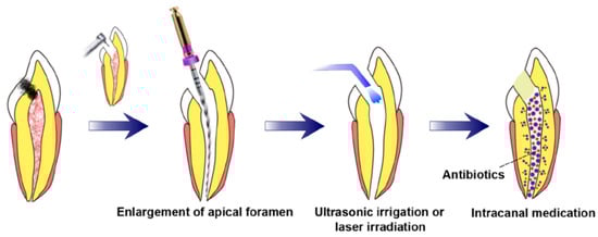
3.3. Prerequisites for Revascularization Approach (Figure 1)

Revascularization studies have established the following prerequisites:
  • There should be open apices and necrotic pulp secondary to trauma.
  • In addition, open apex should be less than 1.5 mm.
  • The following agents can be incorporated to remove microorganisms from the canal.
    Antibiotic paste
    Calcium hydroxide [68]
    Formocresol [69]
  • The coronal seal should be effective.
  • There should be a matrix or the growth of new tissues.
  • When trying to induce bleeding, anaesthesia should be used without a vasoconstrictor [70].
  • Canals should not be instrumented.
  • Sodium hypochlorite should be used as the irrigant.
  • There should be blood-clot formation.
Figure 1. Requisite preconditions for pulp regeneration (root canal disinfection and enlargement of the apical foramen) [71].

4. Postnatal Stem Cell Therapy

Bone, buccal mucosa, fat, and skin are the common sources of postnatal-stem cells. After the apex is opened, the disinfected root-canal system is injected with postnatal-stem cells. This treatment is considered the simplest technique [72]. There are numerous benefits of this type of tissue-engineering technique. Postnatal-stem cells are rationally easy to harvest, and these cells can persuade the regeneration of the pulp. Moreover, these cells are easy to deliver by syringe. In addition, application of these stem-cell therapy is used in regenerative medicine since past many years, for example, bone-marrow replacement and endodontic applications [73]. However, low survival rates are one of the major disadvantages of this technique. Moreover, these cells can migrate into different locations of the body, which presents peculiar forms of mineralization [74]. For the development of dental tissues by the differentiation of stem cells, bioactive-signalling molecules, growth factors, and scaffolds are required [75]. Consequently, with only stem cells that exclude the growth factors or scaffolds, the chance of pulpal regeneration of new tissues is very low. In this approach, the chief identification of a postnatal-stem-cell source that must be able to differentiate into the diverse cell population can be obtained [74]. However, this technique is not approved yet. 

4.1. Pulp Implantation

In this procedure, after cleaning and shaping the root canal, the substituted pulp tissue is transplanted. Purified pulp-stem-cells line is among one the sources of the pulp tissue. This pulp tissue can also grow in the laboratory by cell biopsy. For this invitro technique, pulp tissues can be cultured by biodegradable-polymer nanofibers. Moreover, these tissues can be obtained from collagen I or fibronectin-extracellular-matrix proteins [76]. It has been found that further investigations are required for the proteins, such as vitronectin and laminin. However, it has been proved that for growing pulpal cells, collagens I and III are not fruitful [77]. In the root-canal system, the localization of postnatal-stem cells is a major advantage of pulp implantation. However, there are several disadvantages to this technique. It is a restriction of this technique that the apical portion of the root canal should be harvested with pulp cells. The reason behind this concept is that the sheets of the extracellular matrix are very thin, fragile, and they lack vascularity. Therefore, a scaffold that must have cellular proliferation is required for coronal delivery. If the cells are located 200 μm from a capillary- blood supply which is the maximum oxygen-diffusion distance, these cells are in danger of anoxia and necrosis.

4.2. Scaffold Implantation

For vascularization and cell organization, pulp-stem cells must be systematized into a three-dimensional assembly. This objective can be achieved by seeding pulp-stem cells with a porous-polymer scaffold [79]. Distribution of therapeutic medicines to precise tissues can successfully be accomplished by these nano scaffolds [80]. Moreover, the biological and mechanical properties needed for proper functioning are also provided by these scaffolds [81]. In teeth that have pulp exposure, dentin chips have been introduced which accelerate dentin-bridge formation [82]. These dentin chips aid in the reservoir of growth factors and they offer a matrix for the attachment of pulp-stem cells [83,84]. In reaction to the dentine chip and the use of scaffolds, the regeneration of the pulp-dentin complex occurs. To provide structural support to the tooth it is not necessary to have a tissue-engineered pulp in the root-canal systems [85]. Polymer hydrogel, a soft three-dimensional injectable scaffold matrix, will be administrated by syringe in tissue-engineered pulp tissues [86]. They are easy to deliver into the root-canal systems and are non-invasive.

4.3. Three-Dimensional Cell Printing

The three-dimensional cell printing technique is considered the final approach for the replacement of pulp tissues [90]. This approach can be used to position cells precisely [91]. This technique mimics the natural pulp-tissue structure. In tissue-engineering technique, to maintain and repair dentine, odontoblastoid cells should be positioned around the periphery of the pulp.

4.4. Gene Therapy

To promote tissue mineralization, mineralizing genes would be delivered into the pulp tissues. However, Rutherford worked on this specific field of gene delivery into the pulp tissues, although there is a dearth of literature in this context [93]. He suggested further research to improve the possible gene therapy inside the pulp after he failed in his work when he transduced pulps of ferret animals with cDNA-transfected mouse BMP-7. Researchers used the electroporation method to insert mineralizing genes into the pulp space by culturing of pulpal-stem cells. Initially, the FDA approved the gene therapy research on terminally ill humans; however, after the development of numerous tumours in a nine-year-old boy, the FDA withdrew this decision in 2003. Gene therapy arising from the use of vector systems is posing serious health hazards in contrast to gene expressions [94,95].

4.5. Nitric Oxide

Among many wound healing and pathological processes, angiogenesis is considered an important process. The most potent and critical inducer of angiogenesis is the vascular endothelial growth factor (VEGF). A variety of stimuli take part in the regulation of gene expression of VEGF. The transcription factor is a key factor for hypoxia-mediated VEGF- gene upregulation, which is achieved by hypoxia-inducible factor 1 (HIF-1). Nitric oxide (NO) is a potent vasodilator. Nitric oxide (NO) can simply pervade natural membrane obstacles because it is lipophilic in nature. This VEGF regulates the amount of nitric oxide [97]. Hypoxia as well as nitric oxide upregulate the VEGF genes by enhancing HIF-1 activity. Moreover, dendrimers are released by nitric oxide which acts as antibacterial agents [98,99].

4.6. Platelet-Rich Plasma (PRP)

Special challenges are faced by clinicians for the treatment of an immature tooth with necrotic pulp and open apex. One of the strategies for its treatment is the traditional apexification procedure. This treatment process requires the formation of the apical barrier by multiple applications of calcium hydroxide. This apical barrier can also be formed by placing mineral trioxide aggregate (MTA) into the canal, which is followed by the conventional root-canal procedure [33]. Due to incomplete root formation with these procedures, the chances of root fracture are very common [19,33]. Platelet-rich plasma (PRP) has been suggested as probably the greatest platform for RET that will overcome all these problems [33,101]. Platelet-derived growth factor, transforming growth factor b, and insulin-like growth factor form an integral part of the PRP [5]. PRP can be utilized as a scaffold as it can form a three-dimensional fibrin matrix. It is easily prepared from the patient’s autologous whole blood [33,102,103,104]. Growth factors and cytokines are 4-fold higher in platelets than found in whole blood [105]. Mandibular-continuity defects, for the first time, were healed by the PRP and the placement of cancellous-bone grafts by the dental community [106]. Human dental pulp stem cells (DPSCs), when treated with PRP, resulted in an increase in the differentiation and proliferation of these cells [107].
There are numerous advantages of PRP treatment. During the preparation of the PRP, erythrocytes that would be responsible for necrosis after clot formation was removed [33]. For cell migration, fibrin, fibronectin, and vitronectin are required which is obtained from the formation of PRP clots [104]. Moreover, in regenerative-endodontic procedures, the optimal level of MTA placement is mandatory which can be done by the collagen matrix present in the PRP [70,108]. Before clot formation, PRP does not release growth factor until it is activated. As soon as it is activated either endogenously or through the exogenous, such as by incorporation of calcium chloride or thrombin that acts as a clotting factor, PRP will start secreting growth factors that contribute to the repair and regeneration of the tissues [104,112].

4.7. Cell Homing

In tissue regeneration, the first concept of cell homing was presented in Lancet in 2010. The concept was based on the delivery of transforming growth factor-b3 (TGFb3) without cell transplantation. This approach was first used for the regeneration of the articular cartilage [114]. However, for dental-tissue regeneration, the idea of cell homing was introduced in 2010 [115]. During cell homing, root canal of the extracted human teeth was shaped and cleaned followed by the delivery of the growth factors, scaffold, and stem cells. Residual proteins in the root canal or dentinal tubules were deactivated in the first phase. This can be done by sterilization of extracted teeth in an autoclave. This was followed by the infusion of collagen gel into a shaped and cleaned root canal that might be with or without basic fibroblast growth factors (bFGFs), vascular endothelial growth factors (VEGFs), platelet-derived growth factors (PDGFs), nerve growth factors (NGFs), or bone morphogenetic proteins (BMPs).

The prime difference between the cell homing and cell transplantation approaches is that, in the latter case, for dentine/pulp regeneration, the isolated cells (stem/progenitor) from the host are transplanted into the root canal of the host. Dental pulp-like cells have been differentiated in the cell-homing approach when growth factors are recruited into the root-canal system. Cell-homing-technique-dental-organ regeneration presents a harmonizing and/or balancing approach to cell-transplantation technique and, at the same time, this strategy has shown auspicious results in animal models [23,115,116]. Hematopoietic-stem cells were militarized and transferred to different tissues or organs using active navigation in the cell-homing approach. The ultimate outcome of this process is pulp-dentin re-cellularization and revascularization. Numerous growth factors along with cell homing will result in pulp-dentin regeneration. Tissue revascularization and regeneration-cell homing consist of two distinctive cellular processes. They are differentiation and recruitment [117,118].

 Migration of the cells to the defective site is referred to as recruitment. However, the presence of a mesenchymal stem/progenitor having the ability to differentiate into cells that form pulp and dentine is mandatory [117,118]. When stem/progenitor cells are transformed into mature cells, this process is known as differentiation. During pulp and dentine regeneration, odontoblasts and pulp fibroblasts are formed by the differentiation of the stem/progenitor cells. These processes need to persuade the budding of endothelial cells and neural-fibril cells for angiogenesis. However, more literature is needed to establish the fact that endothelial cells are formed directly by the differentiation of the dental pulp stem/progenitor [117,118].
Extracellular matrix with disconnected cells is found on neo-pulp tissues. This new pulp tissue appears to be dense and contains erythrocyte-filled blood vessels having endothelial-like cell lining. When bFGF, VEGF, PDGF, NGF, and BMP7 are delivered and the entire root canal is filled with dental pulp-like tissues found in microscopic images. Von Willebrand factor, dentin sialoprotein, and NGF are expanded in the ELISA after the combinatory delivery of bFGF, VEGF, or PDGF, with basal NGF and BMP7.

5. Biomimetic Materials in Endodontics

5.1. Biointeractive Materials

5.1.1. Calcium Hydroxide

Regarding regenerative endodontics, disinfecting the root canal with Ca(OH)2 promotes the proliferation of stem cells of the apical papilla (SCAP) [128,129] and increases the release of growth factors from dentine [130,131]. An in vitro study found that the Ca(OH)2 at a concentration of 1 mg/mL in the culture medium promotes the survival and proliferation of SCAPs [132]. Controlled-release calcium hydroxide-loaded microcapsules based on polylactic and ethylcellulose have been developed to improve their biological performance [132]. Such systems ensure the slow and sustained release of calcium and hydroxide over an extended period. The calcified barrier formed by the Ca (OH)2 is permeable and weak, and multiple soft-tissue inclusions create tunnel defects. The high dissolvability disintegrates material over time, leaving voids that can be the potential pathway for bacterial infiltration [133,134,135,136]. Highly alkaline pH also reduces the fracture resistance of dentine and negates its application for a prolonged time [137].

5.1.2. Calcium Sulfate

Chen et al. [143] used CS to repair large-bone defects in dogs’ tibiae, and the treated area showed new-bone formation without foreign-body reaction [143]. Peltier and Jones [144] also found the same results when they used it in patients to fill bony cavities formed by the removal of the unicameral-bone cyst [144]. Yoshikawa et al. [145] obtained favorable results when CS was used to treat osseous defects formed after apicectomy in beagle dogs [145]. Pecora et al. [146] also successfully used calcium sulfate as a bone graft in the surgical treatment of periradicular lesions [146]. In vivo studies have proved that calcium sulfate can induce new-bone formation when placed in bony defects. However, it lacks osteoinductivity, and the proteins present in blood and tissue fluids prolong its setting time. Rapid resorption sometimes hinders its use in large-bone defects [147], and thus, limiting its use in endodontics. To overcome these constraints, biphasic calcium sulfate and composites of calcium sulfate with other bio-materials are also available [148,149,150].

5.2. Bioactive Materials

“A bioactive material is one that elicits a specific biological response at the interface of the material which results in the formation of a bond between the tissues and the material” [151].

5.2.1. Calcium Silicate Based-Cements

Mineral Trioxide Aggregate

Tora Binejad developed MTA in 1993 at Loma Linda University [152], which is composed of purified Portland cement (75%), Bismuth oxide (20%), and Gypsum (5%). Tricalcium silicate, dicalcium silicate, and tricalcium aluminate are the main components of Portland cement [153]. It can set in the presence of moisture, which is omnipresent in the oral cavity. MTA is mixed with water or saline, forming calcium-silicate-hydrate gel and calcium hydroxide. The pH of the set material is 12.5, which is comparable with that of calcium hydroxide [154,155]. It was primarily developed for root-end filling and perforations repair [156], but its immense clinical success has extended its application in various endodontic procedures [157,158].
The properties which favor its application in endodontic procedures are: high biocompatibility, bioactivity, excellent sealing ability, low solubility, and hydrophilicity [159]. MTA releases calcium ions in contact with human tissues and promotes osteoblast proliferation. It causes cytokine production from osteoblast, which favors the migration and differentiation of bone tissue-forming cells, indicating its remineralization potential [160]. MTA encourages reparative-dentine formation, which is rapid and thicker with good structural integrity [161], and a milder degree of pulpal inflammation while maintaining the integrity of the pulp [162,163].

Biodentine

Biodentine was introduced in 2009 as a dentine-replacement material. It is formulated using MTA-based technology to improve the physical, setting, and handling properties while providing the same range of clinical applications of MTA [171]. It is a tricalcium-silicate-based-cement, having two additional components in the liquid: calcium chloride as a setting accelerator and hydrosoluble polymer as a water-reducing agent. It sets in around 12 min [172]. Set material contains Ca, OH, and silicate ions, responsible for their antibacterial and regenerative property. Micromechanical adhesion of biodentine crystals with underlying dentine provides favorable mechanical properties [173]. Wattanapakkavong and Srisuwan [167] demonstrated that BD has a better mineralizing potential than MTA as it can release a greater concentration of TGF-b1 from dentine. [167].

Calcium Aluminate Cement

Calcium-aluminate cement was created at the Federal University of Sao Carlos, Brazil [186]. It is mainly composed of calcium aluminate and calcium dialuminate, which is responsible for its hydraulic-setting reaction [187]. Upon mixing with water, it forms calcium-aluminate hydrate and aluminium hydroxide. The further decomposition of calcium-aluminate hydrate releases Ca and OH ions at a slower rate, producing an alkaline medium and providing therapeutic properties [187].
Pandolfelli et al. and Jacobovitz et al. [170,171,172,173,174,175,176,177,178,179,180,181,182,183,184,185,186,187,188] showed that the calcium-aluminate cement presents adequate biological and antimicrobial properties [170,188]. Garcia et al. [189] assessed the mechanical properties of the cement and found that it possesses higher compressive strength, diametral tensile strength, and microhardness value than MTA [189]. Oliveira et al. [190] evaluated the physical, chemical, and mechanical properties of the cement by incorporating different additives i.e., dispersant, plasticizer, and radiopacifier. They found that by adding these components, it resulted in a cement that sets more rapidly, has better fluidity and handling characteristics, better mechanical strength, and lower porosity than MTA. Lithium bicarbonate also reduced the setting time to 10 min from 60 min [190].

Theracal

Theracal is a new light-cured resin-modified calcium silicate-based biomaterial containing 45% mineral (Portland cement), 10% radiopaque agent, 5% thickening agent, and 45% resin [193]. It was specially designed for direct/indirect pulp-capping procedures, combining the excellent biological properties of calcium silicates, superior handling characteristics, and setting properties of the resin [194,195].
It releases calcium ions that favor the formation of apatite layer and mineralized tissues. Ca releases are in the concentration range of exerting a potential stimulating effect on dental pulp and odontoblast [196,197,198]. It has the ability to alkalinize the surrounding environment to approximately ph 10-11 and has lower solubility than MTA and Dycal. It can be cured to a depth of 1.7 mm [194]. Command setting facilitates the placement of final restoration with no delay. These attributes are of great help in the pulp-capping procedure.

5.2.2. Calcium Phosphate Based Cements

Hydroxyapatite

Hydroxyapatite can be used in bulk form or as a coating for many biomaterials [205,206,207]. Its biocompatibility, osteoconduction, and osseointegration characteristics are well known. Due to its positive features, this material has remained the material of choice in the fields of dentistry as well as medicine for a long time [208,209,210]. The composition of synthetic hydroxyapatite is identical to the calcified part of teeth and bone [211]; as a result, this material is frequently used for dental and medical applications [212]. Despite the favourable bioactive and osteoconductive properties [213], inferior mechanical strength and toughness of hydroxyapatite prevent its applications under greater masticatory load areas [214].

Bioactive Glass

Since the introduction of bioactive glass (BG) by Larry L. Hench, it has gained wide acceptance in the fields of medicine and dentistry [221]. BG has a noncrystalline structure and it shows relatively better bioactivity, compared to the other types of bioceramics with a crystalline structure. The BG is predominantly consisted of CaO, SiO2, and Na2O, and has the ability to proliferate, differentiate, and mineralize the human-dental-pulp cells [222,223,224]. The BG has commonly used to repair periodontal and bony defects. After its placement into the defects, it leads to different biological reactions which ultimately cause the remodelling and transformation of the living matrix and replace the same with fresh osseous tissues [225,226].
BG-based root-canal sealer, namely GuttaFlow Bioseal (GFB), has been made available to clinicians by (Coltène/Whaledent AG, Altstätten, The Switzerland). It exhibits low porosity, biocompatibility, and dentin penetrability. The other sealer, namely Nishika Canal Sealer BG (CS-BG) that is based on BG, has also shown sealing ability, biocompatibility, and better chemo-physical properties. Both sealers are gaining recognition among endodontists for the management of various endodontic issues [227,228,229]. 

5.2.3. Mixture of Calcium Silicate and Phosphate Based-Cements

Bioaggregate

Bioaggregate is recently introduced as a calcium-silicate-based endodontic cement, claiming to present an improved performance than MTA [239]. It is developed using the science of nano-technology and consists of nanosized hydrophilic particles of tricalcium silicate, dicalcium silicate, and tantalum oxide as a radiopacifying agent [240]. It is an insoluble, radiopaque, and aluminum-free material [241], which takes approximately 4 h to set completely [14]. The biocompatibility and sealing ability of the material are comparable to MTA [242,243,244,245,246]. Its ability to promote cementogenesis [247], coupled with bioactive nature, promotes apatite formation at the material/dentine interface, forming an impermeable seal [246]. Tuloglu and Bayrak [248] evaluated the clinical success of MTA and Bioaggregate as an apical barrier material and concluded that Bioaggregate could be an alternative to MTA [248].

Endosequence Root Repair Material

Root-canal perforations are either mechanical or pathologic communications between the external tooth surface, the root-canal system and their etiological factors, including caries, resorption, or iatrogenic [253,254]. To prevent continuous exposure to a contaminating environment and the occurrence of inflammatory reactions in the adjacent tissues [253], a material with good sealing ability should be employed [255]. MTA was introduced by Torabinejad and is considered a good material for creating an effective seal between root canals and outer dental surfaces [256,257,258]. Recently, biodentine has been marketed to address the deficiencies of MTA which include its difficult manipulation and extended setting time [171]. A newer premixed bioceramic material, namely ‘EndoSequence root repair material’, has been investigated for the management of apical surgery, perforation repair, pulp capping, and apical plug [259].

5.2.4. Sealer

Endosequence BC Sealer

Calcium-silicate cements are now widely considered for pulp or periapical regeneration owing to their biocompatibility, bioactivity, antimicrobial properties, and sealing capability [265,266]. The biomineralization and biocompatibility characteristics of calcium silicate-based materials rendered them appropriate for a variety of applications, namely direct pulp capping [267], retrograde filling, and perforation repair [268]. These materials assist in pulpal healing by promoting the proliferation of stem cells of the dental pulp and the resultant formation of a dentine bridge [269,270,271]. In addition, a biological seal of the apical-root canal is also probable with these types of cement [272]. Based on the promising clinical as the well biological performance of these materials, new relevant endodontic sealers are being introduced with a couple of suitable bioactive and sealing properties.
Endosequence BC Sealer (BCS, Brasseler USA, Savannah, GA, USA) is an injectable calcium silicate-based material and it was introduced as a root-canal filling and sealing material [273]. It possesses suitable physicochemical properties and hardens in the presence of moist conditions [274]. To make this sealer suitable for use in the warm-canal-obturating techniques, the composition of Endosequence BC Sealer has been altered into Endosequence BC Sealer HiFlow (Brasseler, Savannah, GA, USA). This new sealer exhibits a lower viscosity on heating and is relatively more radiopaque than its predecessor. Moreover, the results of both BCHiF and BCS are comparable in terms of cell migration, cell adhesion, cytocompatibility, and bioactivity [275].

5.2.5. Gutta-Percha

Bioceramic Coated Gutta-Percha

Gutta-percha points are extensively used as root-canal filling material [278]. However, their cytotoxicity has not been comprehensively evaluated. Some studies provide satisfactory reports regarding biocompatibility of gutta-percha [279] whereas others highlight a delayed healing and persistent periapical radiolucency owing to extruded gutta-percha [280]. To deal with the biocompatibility issue, alterations have been made in the composition of obturation points. EndoSequence BC points (Brasseler USA, Savannah, GA, USA) (BC) are bioactive substances coated on gutta-percha cones with bioceramic nanoparticles [281]. The bioceramic particles present in the EndoSequence Sealer bind to the bioceramic nanoparticles in the BC and form a gap-free filling in accordance with the reports of the manufacturer.

5.3. Remineralizing Agents

5.3.1. Enamel Matrix Derivative (Emdogain) Remineralizing Agent

Enamel matrix derivative (EMD) is extracted from the buds of porcine teeth which mainly comprise amelogenins (about 90%) and smaller quantities of tuftelin, ameloblastin, enamelin, and other nonamelogenin proteins [285]. Its common clinical uses are stimulation of regeneration of periodontal attachment, and some clinicians have also suggested its potential for periodontal regeneration [286,287,288,289]. Several case reports related to the field of endodontics using EMD for perforation repair or root-end resection along with guided tissue regeneration are well evident [290,291,292]. The potential of EMD in endodontic regeneration is not fully agreed upon; however, its important role in odontogenesis via up-regulation of Osterix and Runx2 transcription factors is well-documented [289].

5.3.2. Dentine Matrix Derivative/Demineralized Dentin Matrix

The organic matrix of dentin consistsof a total of 233 proteins which include various collagenous and non-collagenous proteins [299]. Demineralized dentin matrix (DDM) is mechanically better, nonimmunogenic, and biocompatible, and has the potential for osteoinduction and osteoconduction [301,302,303]. It also allows the differentiation of odontoblast-like cells [304]. DDM is considered a complex of type I collagen (COL-I) and a growth factor, and it has significant osteoinductive and osteoconductive biological effects [305]. Various bone injuries and bone defects have been treated using autologous and xenogenous DDM [306,307,308]; however, the influence of DDM on DPSCs is not well documented.

5.4. Miscellaneous

Calcium Phosphate Cements

Calcium phosphate-based biomaterials have gotten increased attention due to their excellent biocompatibility, non-cytotoxicity, and chemical composition similar to teeth and bone [311,312,313,314]. Bioactive nature coupled with osteoinductive potential makes it a suitable material for endodontic applications [315]. It can chemically adhere to bone and teeth [316]. Brown and Chow first introduced the calcium-phosphate cement, a self-hardening cement consisting of a mixture of calcium phosphate which, when mixed with water, hardened into a less soluble calcium phosphate [317]. It has been widely used in orthopaedics and dentistry to repair bone defects, owing to its osteoconductive potential [318]. In-vitro studies have proposed its use for furcation repair, root-apex sealing, root- canal filling, and root-surface desensitization [319,320].

6. Conclusions

It appears that in-vitro and animal experiments regarding biomimetic approaches in regenerative endodontics are widely performed, but they are still at the inception stage. The findings related to stem-cell therapy, pulp implant, scaffold implant, 3D-cell printing, and gene therapy are quite promising as positive features with regard to pulp regeneration and tissue mineralization have been observed. Despite the aforementioned advantages, future developments in pulp-dentin tissue regeneration are needed to demonstrate the functional tissue regeneration and the ultimate favorable clinical benefits. In addition, some bioactive materials seem to be favorable as they promote osteoconduction and osseointegration, and are capable to proliferate, differentiate, and mineralize the human-dental-pulp cells. However, physico-mechanical characteristics of some materials are not satisfactory and warrant further investigations. Enamel matrix and dentine matrix derivatives have also been researched and their role in the dentine regeneration is encouraging, but there is a lack of scientifically validated data.

This entry is adapted from the peer-reviewed paper 10.3390/biomimetics7040229

This entry is offline, you can click here to edit this entry!
Video Production Service