Cell Therapies in Acute Kidney Injury: History
Please note this is an old version of this entry, which may differ significantly from the current revision.
Contributor: , ,

The incidence of renal disease is gradually increasing worldwide, and this condition has become a major public health problem because it is a trigger for many other chronic diseases. Cell therapies using multipotent mesenchymal stromal cells, hematopoietic stem cells, macrophages, and other cell types have been used to induce regeneration and provide a cure for acute and chronic kidney disease in experimental models. 

  • cell therapies
  • tissue repair
  • kidney disease
  • acute kidney injury (AKI)

1. Introduction

As acute kidney injury (AKI) involves inflammatory processes in the kidney that can lead to a complete loss of kidney function and no therapies are available to treat them, cell therapy has proved to be a promising clinical approach and might represent a novel therapeutic strategy to slow the progression of kidney disease [1].
A cell-based regenerative therapy has been studied in animal models of AKI and there have been a few reports of beneficial effects. The cells investigated so far include granulocyte colony-stimulating factor-mobilized peripheral blood CD34 cells [2] and mesenchymal stem cells (MSCs) [3]. In addition, renal progenitor cells generated from human-induced pluripotent stem (iPS) cells have been found to ameliorate an acute kidney injury induced by an ischemia/reperfusion injury (IRI) in mice [4]. The pluripotent nature of iPSs raises concerns of a high risk of tumor development when these cells are administered without pre-differentiation. Although the differentiation of iPSs has been achieved and a renal recovery observed after an injection in AKI models, this occurred without being integrated into the host kidney tissues, indicating that the paracrine effects of the renotrophic factors secreted from the hiPS-derived renal progenitors were the primary cause of the therapeutic benefits. Thus, the iPSCs, although capable of differentiating into almost any cell type, acted by indirect mechanisms and not by substituting specific cells in a direct manner. Other authors [5] have found improvements in renal injuries after the administration of human-induced pluripotent stem cell-derived mesenchymal stromal cells (hiPS-MSCs), and the effect was mediated by extracellular vesicles.
It has been found that MSC and mononuclear cell therapies have a potent immunomodulatory effect. During an ischemia-reperfusion injury, T-regulatory cells exhibit a protective role in ischemia and reperfusion by secreting IL-10 to reduce the ischemia-reperfusion injury [6]. On top of that, plenty of innate immune cells—including mast cells, neutrophils, macrophages, myeloid-derived suppressor cells, dendritic cells, and natural killer cells—are engaged in an ischemia-reperfusion injury [7][8]. These cell therapies have been shown to gradually ameliorate the renal function in animals with AKI. However, no human clinical studies based on a regenerative therapy have succeeded in counteracting the damage caused by AKI. When conducting translational research to apply these novel clinical treatments, we must consider certain aspects such as the accessibility to the cell source, protocol complexity, and cost.
The first problem to be addressed when developing a cell therapy against AKI in clinics relates to the exact timing of the cell administration. Ideally, the administration of a cell therapy should be conducted soon after the renal ischemia when AKI presumably occurs. Unfortunately, an acute kidney dysfunction does not cause any typical symptoms, nor is there any marker molecule available that would allow the rapid and early detection of AKI. From a clinical point of view, it is impossible to define the exact moment at which AKI evolves. Even if it was possible to predict the timing, the cells for the therapeutic administration should be available as soon as possible. Obtaining proangiogenic cells, for example, usually requires 5–7 days. Therefore, AKI should be diagnosed almost a week in advance for this reason. The ideal cell therapy would be one of a rapid preparation to be administered immediately when renal failure is detected. In Figure 1, the different candidates for cell therapies for kidney disease treatments are summarized.
Figure 1. Top candidates to develop a cellular therapy against renal diseases and advances. Advantages of mesenchymal stem cells, iPS, macrophages, peripheral blood mononuclear cells, and extracellular vesicles as a therapeutic use and novel strategies for renal treatment. MSCs: mesenchymal stem cells; MACs: macrophages; iPS: induced pluripotent stem cells; PBMNCs: peripheral blood mononuclear cells; EVs: extracellular vesicles.

2. Multipotent Mesenchymal Stromal Stem Cell Therapies

Multipotent mesenchymal stromal stem cells (MSCs) have been widely investigated for use as a cell therapy. They have shown promise for several diseases, with the goal of restoring homeostasis to inflamed or injured organs [9]. Human mesenchymal stem cells isolated from certain types of tissues, including adipose and bone marrow, have important features such as multilineage differentiation, self-renewal, and a proliferative potential [10][11].
In general, MSCs differ from other cell therapies as their therapeutic effect is not only dictated by cell–cell contact, but may also include the so-called “hit-and-run” mechanism. This process is accompanied by a set of hormones, growth factors, or soluble cytokines that are transferred to the target cells (damaged tissue) through secretion, phagocytosis, or vesicle uptake [12][13]. MSCs migrate to the injury site through the circulation (blood and lymphatics) or through the tissue stroma as a response to suppress the inflammatory process caused by a tissue injury. Such a response also participates in tissue repair and regeneration by secreting local factors that modulate the host immune responses by promoting angiogenesis and regulating both the extracellular matrix and connective tissue deposition [14][15]. Therefore, novel preclinical studies using MSCs have been developed with the aim of ameliorating a kidney injury.
In a preclinical study with the use of an intravenous MSC administration as a treatment for AKI, a reduction in the reactive oxygen species through the signaling of the antioxidant response element/factor 2 related to nuclear factor E2 was detected. In addition, the upregulation of antioxidant enzymes, the decreased expression of proinflammatory cytokines, and reduced evidence of renal apoptosis have been detected [16][17]. Therefore, these studies demonstrated beneficial effects by reducing tissue injuries in AKI. In an in vivo canine acute kidney injury model, MSCs were also shown to improve the renal function, decreasing blood urea nitrogen (BUN) and creatinine as well as recovering renal lesions [18].
In another study, Rodrigues et al., suggested that an MSC therapy improved the glomerular filtration rate and decreased oxidative stress-induced cell senescence and inflammation, promoting cell proliferation after IRI [19]. Thus, MSCs protected against AKI in animal models.
Despite these potential therapeutic effects, the engraftment of cells onto injured tissues has not been systematically demonstrated. Therefore, the protective effects have been attributed only to paracrine mechanisms [20].
On the other hand, clinical studies with MSCs have been reported. Of the three clinical trials of MSC therapies conducted on AKI patients since 2008, only one study (NCT00733876, phase 1) was completed, showing the protective effect of MSC administration on an acute kidney injury. The other two trials (NCT01275612, phase 1; NCT01602328, phase 2) were withdrawn and terminated, respectively. In the full study (NCT00733876), bone marrow-derived mesenchymal stem cells (BM-MSCs) were administered intra-arterially through the adrenal aorta to avoid lung entrapment. The results indicated that the therapy prevented a postoperative and late deterioration of the renal function. In contrast, in the completed ACT-AKI multicenter trial (NCT01602328) in postcardiac surgery AKI patients, the intra-aortic administration of MSCs was not successful. It also did not find a significant difference in the renal function measures (30 day all-cause mortality; the need for dialysis) and, therefore, the trial was terminated due to its uselessness [1][21]. Swaminathan et al., used allogeneic mesenchymal stem cells to treat 156 patients with AKI after cardiac surgery in a multicenter study. The results were not positive, probably because the patients already had established AKI; the aim of the therapy was to shorten the time to recover the baseline renal function, which the cell therapy did not demonstrate [22].
In addition, MSCs have been investigated as a treatment for kidney disorders such as renal transplantations, which started in 2008 (NCT00658073), or kidney/liver failure, which started in 2011 (NCT01429038); both clinical trials used autologous and allogeneic bone marrow, respectively, such as the cell source. In 2013, a treatment for diabetic nephropathy (NCT01843387) began; the cell source was allogeneic mesenchymal precursor cells and bone marrow. All of these were completed [12]; although there were no side effects and the safety of therapy was demonstrated, no conclusive results were reported.

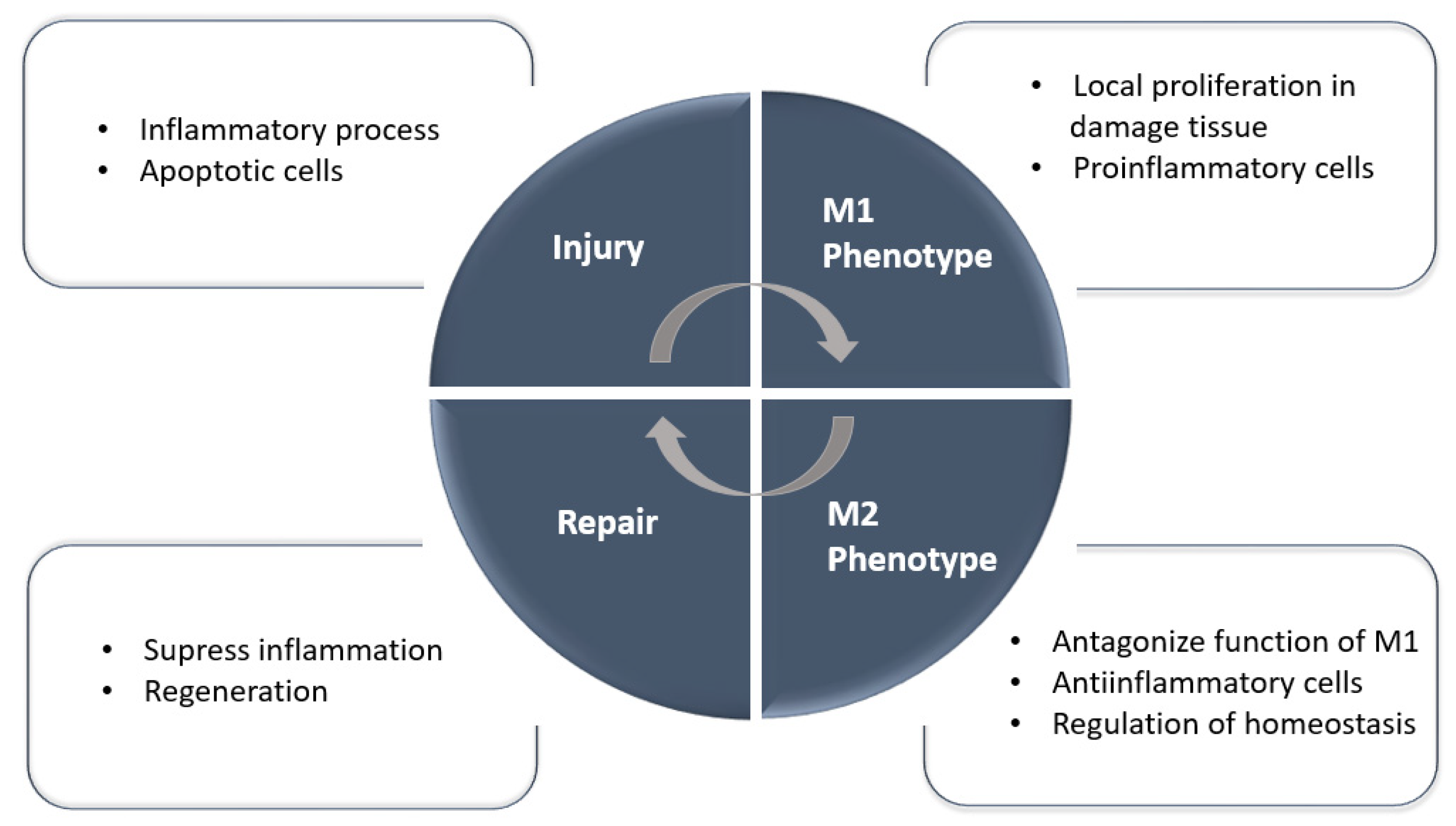
3. Mononuclear and Macrophage Cell Therapies

Unlike MSCs, which require in vitro expansion prior to use (due to their low frequency in the tissue of origin) and a substantial volume of MSCs, peripheral blood mononuclear cells (PBMNCs) can easily be fractionated by apheresis and density centrifugation. Furthermore, after isolation, mononuclear cells (MNCs) can also easily be purified to obtain specific cell types. Studies have also reported on their ability to differentiate into other cell types as well as their extensive involvement in the regeneration and repair of damaged tissue [23]. Thus, PBMNCs have been used in clinical studies for the treatment of different diseases, showing the effectiveness and safety for the patient (NCT00524784 [24]; NCT01503749 [25]; NCT01833585 [26][27]).
Other studies have indicated that human PBMNC cultures in a vasculogenic conditioning medium dramatically improved IRI induced in an AKI mouse model [28]. Although there is much scientific evidence, there have been no completed clinical trials of mononuclear cells for the treatment of AKI.
Recently, researchers described a new autologous cell therapy with polarized PBMNCs administrated intravenously that protected against AKI and AKI-derived fibrosis [29] by reducing inflammation and enhancing kidney regeneration. In this case, the PBMNCs were subjected to a repetitive anoxia/reoxygenation process to promote the anti-inflammatory-specific phenotype of the cells. Cell isolation and the production of a desired phenotype are effective, easy to prepare, and do not require genetic manipulation because PBMNCs subjected to an anoxia/reoxygenation protocol promote a healing phenotype of the cells. Thus, researchers obtained a safer regenerative product to be applied in a clinical setting. The relevance of macrophages is due to their broad participation in the immune system [30][31]; when activated, macrophages tend to polarize into different phenotypes. Researchers highlight M1 as a proinflammatory and M2 as a promoter of tissue repair in Figure 2 [32].
Figure 2. Macrophages in homeostasis, injury, and repair. During the repair phase, M2 macrophages predominate and may originate from in situ proliferation, differentiation from infiltrating monocytes, or phenotype changes from M1 macrophages. M1 macrophages specifically predominate during the injury and inflammation phase.
When IRI occurs, there is an abundance of immune cells—including mast cells, neutrophils, macrophages, myeloid-derived suppressor cells, dendritic cells, and natural killer cells—that are regulated by MSCs. MSCs [6] can secrete prostaglandin E2 [33], quinurenic acid [34], and TNF-stimulated gene-6 [35] that promote macrophage polarization from the M1 phenotype to the M2 phenotype to alleviate inflammation [7]. Distinct macrophage subtypes are involved across different stages of AKI, and, as M2 macrophages have been found to be protective against AKI, there is growing interest in using M2 macrophages and macrophage-modulating agents as therapeutics tools to treat patients with AKI [36]. Interestingly, in a mouse AKI-induced model, the protective role of M2 phenotype peritoneal macrophage transplantation and its possible mechanism of action were evaluated. For this, C57BL/6 mouse macrophages were taken and M2 polarization was induced by IL-4 and IL-13 and injected into the renal cortex of the mouse. A relief of the kidney damage and inflammatory response was observed and the treatment promoted the proliferation of proximal tubular epithelial cells [37].
Resident macrophages in renal tissues are composed of a range of different cells. A few are derived from the yolk sac and others are derived from monocytes [38], and have been shown to actively participate in the resolution of infections and the progression to fibrosis [39][40].
When the kidney is injured or inflamed, macrophages differentiated from monocytes migrate and infiltrate the injured area, eliciting a proinflammatory response. Recently, in a single RNA-seq study, Yao et al., identified a specific inflammatory monocyte-derived infiltrated macrophage as an early responder to AKI and proposed it as a potential therapy. The infiltrated S100A8/A9 macrophage was identified as a mediator of kidney inflammation in an animal model and human AKI. Silencing these macrophages improved the renal function in a bilateral IRI model and decreased the inflammatory response, converting it into a feasible therapy for human AKI [41].
Macrophages can be engineered into an M2 phenotype for the treatment of kidney disease. A few methods have used an ex vivo modification followed by an in vivo modification (the administration of modified macrophages); other methods only used in vivo modifications with genetically modified models. These are explained in Table 1. One of the main concerns about the use of these manipulated M2 macrophages is the possibility of their phenotype changing to M1 during the disease in vivo [36]. Thus, one of the main requirements in macrophage therapies is the maintenance of the healing phenotype and the time needed for tissue recovery. In this sense, the results showed that when researchers infused cells with a specific M2 gene expression profile, isolated renal macrophages maintained the anti-inflammatory and proliferative phenotype during the time needed for tissue recovery [29], confirming again its feasibility to be used in a clinical setting.
Table 1. Cell therapies with M2-induced macrophages.
In addition to understanding the molecular mechanism of therapies using macrophages (which induce renal repairs), new therapeutic strategies have been developed. Several studies have focused on trying to enhance certain healing functions of endogenous macrophages; one of which is based on stimulation by pharmacological agents, as shown in Table 1.
Autophagy has been shown to be closely related to immunity and inflammation. It contributes to the regulation and function of human immunological responses [50].
Macrophages are part of the innate leukocytes that accumulate in the kidney and promote inflammation in acute kidney inflammations [51]. Several studies have shown that a treatment with ursolic acid increases macrophage autophagy. In addition, to enhance macrophage autophagy, it alters the macrophage function and inhibits the secretion of inflammatory factors such as tumor necrosis factor-alpha (TNF-α), interleukin-6 (IL-6), and interleukin-1-beta (IL-1β). This indicates the vital role of autophagy in the regulation of kidney inflammation [52][53] and the possibility of using ursolic acid as an alternative to a cell therapy.
Rapamycin induces autophagy by inhibiting the mTOR signaling pathway, reducing the levels of proinflammatory cytokines such as TNF-α, IL-1β, monocyte chemoattractant protein-1 (MCP-1), and gamma interferon (IFN-γ) as well as enhancing the expansion of renal regulatory T cells (Tregs). It has been found that the adoptive transfer of Tregs with a rapamycin treatment can transform endogenous renal macrophages from M1 to M2 phenotypes and inhibit the expression of proinflammatory cytokines on integrin alpha-M (CD11b+) cells in the kidney whilst increasing the expression of anti-inflammatory cytokines from the kidney [54].
Thus, novel therapeutic interventions designed to enhance autophagy could represent a new approach to overcome the inadequacies of autophagy associated with inflammatory dysregulation [55][56].

4. Conclusions

Cellular therapies are among the most exciting innovations in medicine over the last decade and have the potential to offer curative solutions to kidney disease. Overall, there are various preclinical studies that demonstrate the efficacy of different cell therapies, but fewer clinical trials have demonstrated the efficacy of the different cell therapies. The greatest challenge is to understand how to adapt the experimental innovations to a clinical setting and to use appropriate models that link the preclinical assays with the clinical reality in order to apply these therapies to AKI patients. Future directions point to clinical tests with cellular therapies previously proved in preclinical assays and in models near to clinics with no side effects such the described PBMNC therapy [29].

This entry is adapted from the peer-reviewed paper 10.3390/ijms232415943

References

  1. Tögel, F.E.; Westenfelder, C. Kidney Protection and Regeneration Following Acute Injury: Progress Through Stem Cell Therapy. Am. J. Kidney Dis. 2012, 60, 1012–1022.
  2. Li, B.; Cohen, A.; Hudson, T.E.; Motlagh, D.; Amrani, D.L.; Duffield, J.S. Mobilized Human Hematopoietic Stem/Progenitor Cells Promote Kidney Repair After Ischemia/Reperfusion Injury. Circulation 2010, 121, 2211–2220.
  3. de Almeida, D.C.; Donizetti-Oliveira, C.; Barbosa-Costa, P.; Origassa, C.S.; Câmara, N.O. In Search of Mechanisms Associated with Mesenchymal Stem Cell-Based Therapies for Acute Kidney Injury. Clin. Biochem. Rev. 2013, 34, 131.
  4. Toyohara, T.; Mae, S.-I.; Sueta, S.-I.; Inoue, T.; Yamagishi, Y.; Kawamoto, T.; Kasahara, T.; Hoshina, A.; Toyoda, T.; Tanaka, H.; et al. Cell Therapy Using Human Induced Pluripotent Stem Cell-Derived Renal Progenitors Ameliorates Acute Kidney Injury in Mice. Stem Cells Transl. Med. 2015, 4, 980–992.
  5. Yuan, X.; Li, D.; Chen, X.; Han, C.; Xu, L.; Huang, T.; Dong, Z.; Zhang, M. Extracellular vesicles from human-induced pluripotent stem cell-derived mesenchymal stromal cells (hiPSC-MSCS) protect against renal ischemia/reperfusion injury via delivering specifity propteis (SP1) and trasxcriptional activating of sphingosine kinase 1 and inhibiting necroptosis. Cell Death Dis. 2017, 8, 3200.
  6. Wei, X.; Zhang, J.; Gu, Q.; Huang, M.; Zhang, W.; Guo, J.; Zhou, X. Reciprocal Expression of IL-35 and IL-10 Defines Two Distinct Effector Treg Subsets that Are Required for Maintenance of Immune Tolerance. Cell Rep. 2017, 21, 1853–1869.
  7. Shi, Y.; Wang, Y.; Li, Q.; Liu, K.; Hou, J.; Shao, C.; Wang, Y. Immunoregulatory mechanisms of mesenchymal stem and stromal cells in inflammatory diseases. Nat. Rev. Nephrol. 2018, 14, 493–507.
  8. Pan, B.; Fan, G. Stem cell-based treatment of kidney diseases. Exp. Biol. Med. 2020, 245, 902–910.
  9. Yun, C.; Lee, S. Potential and Therapeutic Efficacy of Cell-based Therapy Using Mesenchymal Stem Cells for Acute/chronic Kidney Disease. Int. J. Mol. Sci. 2019, 20, 1619.
  10. Choi, J.R.; Yong, K.W.; Choi, J.Y. Effects of mechanical loading on human mesenchymal stem cells for cartilage tissue engineering. J. Cell Physiol. 2018, 233, 1913–1928.
  11. Safwani, W.K.Z.W.; Choi, J.R.; Yong, K.W.; Ting, I.; Adenan, N.A.M.; Pingguan-Murphy, B. Hypoxia enhances the viability, growth and chondrogenic potential of cryopreserved human adipose-derived stem cells. Cryobiology 2017, 75, 91–99.
  12. Prockop, D.J. Concise Review: Two negative feedback loops place mesenchymal stem/stromal cells at the center of early regulators of inflammation. Stem Cells 2013, 31, 2042–2046.
  13. Levy, O.; Kuai, R.; Siren, E.M.J.; Bhere, D.; Milton, Y.; Nissar, N.; De Biasio, M.; Heinelt, M.; Reeve, B.; Abdi, R.; et al. Shattering barriers toward clinically meaningful MSC therapies. Sci. Adv. 2020, 6, eaba6884.
  14. Eggenhofer, E.; Luk, F.; Dahlke, M.H.; Hoogduijn, M.J. The Life and Fate of Mesenchymal Stem Cells. Front. Immunol. 2014, 5, 148.
  15. Tsuchiya, A.; Kojima, Y.; Ikarashi, S.; Seino, S.; Watanabe, Y.; Kawata, Y.; Terai, S. Clinical trials using mesenchymal stem cells in liver diseases and inflammatory bowel diseases. Inflamm. Regen. 2017, 37, 16.
  16. Zhuo, W.; Liao, L.; Xu, T.; Wu, W.; Yang, S.; Tan, J. Mesenchymal Stem Cells Ameliorate Ischemia-Reperfusion-Induced Renal Dysfunction by Improving the Antioxidant/Oxidant Balance in the Ischemic Kidney. Urol. Int. 2011, 86, 191–196.
  17. Zhang, G.; Zou, X.; Huang, Y.; Wang, F.; Miao, S.; Liu, G.; Chen, M.; Zhu, Y. Mesenchymal Stromal Cell-Derived Extracellular Vesicles Protect Against Acute Kidney Injury Through Anti-Oxidation by Enhancing Nrf2/ARE Activation in Rats. Kidney Blood Press. Res. 2016, 41, 119–128.
  18. Lee, S.-J.; Ryu, M.-O.; Seo, M.-S.; Park, S.-B.; Ahn, J.-O.; Han, S.-M.; Kang, K.-S.; Bhang, D.-H.; Youn, H.-Y. Mesenchymal Stem Cells Contribute to Improvement of Renal Function in a Canine Kidney Injury Model. In Vivo 2017, 31, 1115–1124.
  19. Rodrigues, C.E.; Capcha, J.M.C.; de Bragança, A.C.; Sanches, T.R.; Gouveia, P.Q.; de Oliveira, P.A.F.; Malheiros, D.M.A.C.; Volpini, R.A.; Santinho, M.A.R.; Santana, B.A.A.; et al. Human umbilical cord-derived mesenchymal stromal cells protect against premature renal senescence resulting from oxidative stress in rats with acute kidney injury. Stem Cell Res. Ther. 2017, 8, 19.
  20. Bi, B.; Schmitt, R.; Israilova, M.; Nishio, H.; Cantley, L.G. Stromal Cells Protect against Acute Tubular Injury via an Endocrine Effect. JASN 2007, 18, 2486–2496.
  21. Perico, N.; Casiraghi, F.; Remuzzi, G. Mesenchymal Stromal Cells for AKI after Cardiac Surgery. JASN 2018, 29, 7–9.
  22. Swaminathan, M.; Stafford-Smith, M.; Chertow, G.M.; Warnock, D.G.; Paragamian, V.; Brenner, R.M.; Lellouche, F.; Fox-Robichaud, A.; Atta, M.G.; Melby, S.; et al. Allogeneic Mesenchymal Stem Cells for Treatment of AKI after Cardiac Surgery. JASN 2018, 29, 260–267.
  23. Zhang, M.; Huang, B. The multi-differentiation potential of peripheral blood mononuclear cells. Stem Cell Res. Ther. 2012, 3, 48.
  24. Mevorach, D.; Zuckerman, T.; Reiner, I.; Shimoni, A.; Samuel, S.; Nagler, A.; Rowe, J.M.; Or, R. Single Infusion of Donor Mononuclear Early Apoptotic Cells as Prophylaxis for Graft-versus-Host Disease in Myeloablative HLA-Matched Allogeneic Bone Marrow Transplantation: A Phase I/IIa Clinical Trial. Biol. Blood Marrow Transplant. 2014, 20, 58–65.
  25. Yu, S.J.; Yoon, J.-H.; Kim, W.; Lee, J.M.; Bin Lee, Y.; Cho, Y.; Lee, D.H.; Lee, M.; Yoo, J.-J.; Cho, E.J.; et al. Ultrasound-guided percutaneous portal transplantation of peripheral blood monocytes in patients with liver cirrhosis. Korean J. Int. Med. 2017, 32, 261–268.
  26. Wahid, F.S.A.; Ismail, N.A.; Jamaludin, W.F.W.; Muhamad, N.A.; Idris, M.A.M.; Lai, N.M. Efficacy and Safety of Autologous Cell-based Therapy in Patients with No-option Critical Limb Ischaemia: A Meta-Analysis. Curr. Stem Cell Res. Ther. 2018, 13, 265–283.
  27. Sermsathanasawadi, N.; Pruekprasert, K.; Chruewkamlow, N.; Kittisares, K.; Warinpong, T.; Chinsakchai, K.; Wongwanit, C.; Ruangsetakit, C.; Mutirangura, P. Peripheral blood mononuclear cell transplantation to treat no-option critical limb ischaemia: Effectiveness and safety. J. Wound Care 2021, 30, 562–567.
  28. Ohtake, T.; Kobayashi, S.; Slavin, S.; Mochida, Y.; Ishioka, K.; Moriya, H.; Hidaka, S.; Matsuura, R.; Sumida, M.; Katagiri, D.; et al. Human Peripheral Blood Mononuclear Cells Incubated in Vasculogenic Conditioning Medium Dramatically Improve Ischemia/Reperfusion Acute Kidney Injury in Mice. Cell Transpl. 2018, 27, 520–530.
  29. Játiva, S.; Torrico, S.; Calle, P.; Muñoz, Á.; García, M.; Larque, A.B.; Poch, E.; Hotter, G. NGAL release from peripheral blood mononuclear cells protects against acute kidney injury and prevents AKI induced fibrosis. Biomed. Pharmacother. 2022, 153, 113415.
  30. Okabe, Y.; Medzhitov, R. Tissue biology perspective on macrophages. Nat. Immunol. 2016, 17, 9–17.
  31. Gosselin, D.; Link, V.M.; Romanoski, C.E.; Fonseca, G.J.; Eichenfield, D.Z.; Spann, N.J.; Stender, J.D.; Chun, H.B.; Garner, H.; Geissmann, F.; et al. Environment Drives Selection and Function of Enhancers Controlling Tissue-Specific Macrophage Identities. Cell 2014, 159, 1327–1340.
  32. Italiani, P.; Boraschi, D. From Monocytes to M1/M2 Macrophages: Phenotypical vs.Functional Differentiation. Front. Immunol. 2014, 5, 514.
  33. Vasandan, A.B.; Jahnavi, S.; Shashank, C.; Prasad, P.; Kumar, A.; Prasanna, S.J. Human Mesenchymal stem cells program macrophage plasticity by altering their metabolic status via a PGE2-dependent mechanism. Sci. Rep. 2016, 6, 38308.
  34. Wang, G.; Cao, K.; Liu, K.; Xue, Y.; Roberts, A.I.; Li, F.; Han, Y.; Rabson, A.B.; Wang, Y.; Shi, Y. Kynurenic acid, an IDO metabolite, controls TSG-6-mediated immunosuppression of human mesenchymal stem cells. Cell Death Differ. 2018, 25, 1209–1223.
  35. Mittal, M.; Tiruppathi, C.; Nepal, S.; Zhao, Y.-Y.; Grzych, D.; Soni, D.; Prockop, D.J.; Malik, A.B. TNFα-stimulated gene-6 (TSG6) activates macrophage phenotype transition to prevent inflammatory lung injury. Proc. Natl. Acad. Sci. USA 2016, 113, E8151–E8158.
  36. Chen, T.; Cao, Q.; Wang, Y.; Harris, D.C.H. M2 macrophages in kidney disease: Biology, therapies, and perspectives. Kidney Int. 2019, 95, 760–773.
  37. Mao, R.; Wang, C.; Zhang, F.; Zhao, M.; Liu, S.; Liao, G.; Li, L.; Chen, Y.; Cheng, J.; Liu, J.; et al. Peritoneal M2 macrophage transplantation as a potential cell therapy for enhancing renal repair in acute kidney injury. J. Cell Mol. Med. 2020, 24, 3314–3327.
  38. Sheng, J.; Ruedl, C.; Karjalainen, K. Most Tissue-Resident Macrophages Except Microglia Are Derived from Fetal Hematopoietic Stem Cells. Immunity 2015, 43, 382–393.
  39. Jang, H.-S.; Kim, J.I.; Jung, K.-J.; Kim, J.; Han, K.-H.; Park, K.M. Bone marrow-derived cells play a major role in kidney fibrosis via proliferation and differentiation in the infiltrated site. Biochim. Biophys. Acta Mol. Basis Dis. 2013, 1832, 817–825.
  40. Malone, A.F. Monocytes and Macrophages in Kidney Transplantation and Insights from Single Cell RNA-Seq Studies. Kidney360 2021, 2, 1654–1659.
  41. Yao, W.; Chen, Y.; Li, Z.; Ji, J.; You, A.; Jin, S.; Ma, Y.; Zhao, Y.; Wang, J.; Qu, L.; et al. Single Cell RNA Sequencing Identifies a Unique Inflammatory Macrophage Subset as a Druggable Target for Alleviating Acute Kidney Injury. Adv. Sci. 2022, 9, 2103675.
  42. Cao, Q.; Wang, Y.; Zheng, D.; Sun, Y.; Wang, Y.; Lee, V.; Zheng, G.; Tan, T.K.; Ince, J.; Alexander, S.I.; et al. IL-10/TGF-β–Modified Macrophages Induce Regulatory T Cells and Protect against Adriamycin Nephrosis. JASN 2010, 21, 933–942.
  43. Ferenbach, D.A.; Ramdas, V.; Spencer, N.; Marson, L.; Anegon, I.; Hughes, J.; Kluth, D.C. Macrophages Expressing Heme Oxygenase-1 Improve Renal Function in Ischemia/Reperfusion Injury. Mol. Ther. 2010, 18, 1706–1713.
  44. Jung, M.; Sola, A.; Hughes, J.; Kluth, D.C.; Vinuesa, E.; Viñas, J.L.; Pérez-Ladaga, A.; Hotter, G. Infusion of IL-10–expressing cells protects against renal ischemia through induction of lipocalin-2. Kidney Int. 2012, 81, 969–982.
  45. Ranganathan, P.V.; Jayakumar, C.; Ramesh, G. Netrin-1-treated macrophages protect the kidney against ischemia-reperfusion injury and suppress inflammation by inducing M2 polarization. Am. J. Physiol. Ren. Physiol. 2013, 304, F948–F957.
  46. Geng, Y.; Zhang, L.; Fu, B.; Zhang, J.; Hong, Q.; Hu, J.; Li, D.; Luo, C.; Cui, S.; Zhu, F.; et al. Mesenchymal stem cells ameliorate rhabdomyolysis-induced acute kidney injury via the activation of M2 macrophages. Stem Cell Res. Ther. 2014, 5, 80.
  47. Du, Q.; Tsuboi, N.; Shi, Y.; Ito, S.; Sugiyama, Y.; Furuhashi, K.; Endo, N.; Kim, H.; Katsuno, T.; Akiyama, S.; et al. Transfusion of CD206+ M2 Macrophages Ameliorates Antibody-Mediated Glomerulonephritis in Mice. Am. J. Pathol. 2016, 186, 3176–3188.
  48. Taguchi, K.; Okada, A.; Hamamoto, S.; Unno, R.; Moritoki, Y.; Ando, R.; Mizuno, K.; Tozawa, K.; Kohri, K.; Yasui, T. M1/M2-macrophage phenotypes regulate renal calcium oxalate crystal development. Sci. Rep. 2016, 6, 35167.
  49. Jung, M.; Brüne, B.; Hotter, G.; Sola, A. Macrophage-derived Lipocalin-2 contributes to ischemic resistance mechanisms by protecting from renal injury. Sci. Rep. 2016, 6, 21950.
  50. Singbartl, K.; Formeck, C.L.; Kellum, J.A. Kidney-Immune System Crosstalk in AKI. Semin. Nephrol. 2019, 39, 96–106.
  51. Lech, M.; Gröbmayr, R.; Ryu, M.; Lorenz, G.; Hartter, I.; Mulay, S.R.; Susanti, E.; Kobayashi, K.S.; Flavell, R.A.; Anders, H.-J. Macrophage Phenotype Controls Long-Term AKI Outcomes—Kidney Regeneration versus Atrophy. JASN 2014, 25, 292–304.
  52. Sun, Q.; He, M.; Zhang, M.; Zeng, S.; Chen, L.; Zhou, L.; Xu, H. Ursolic acid: A systematic review of its pharmacology, toxicity and rethink on its pharmacokinetics based on PK-PD model. Fitoterapia 2020, 147, 104735.
  53. Gong, L.; Pan, Q.; Yang, N. Autophagy and Inflammation Regulation in Acute Kidney Injury. Front. Physiol. 2020, 11, 576463.
  54. Ramanathan, C.; Kathale, N.D.; Liu, D.; Lee, C.; Freeman, D.A.; HogenEsch, J.B.; Cao, R.; Liu, A.C. mTOR signaling regulates central and peripheral circadian clock function. PLoS Genet. 2018, 14, e1007369.
  55. Radi, Z.A. Immunopathogenesis of Acute Kidney Injury. Toxicol. Pathol. 2018, 46, 930–943.
  56. Jia, H.; Yan, Y.; Liang, Z.; Tandra, N.; Zhang, B.; Wang, J.; Xu, W.; Qian, H. Autophagy: A new treatment strategy for MSC-based therapy in acute kidney injury (Review). Mol. Med. Rep. 2017, 17, 3439–3447.
More
This entry is offline, you can click here to edit this entry!
Video Production Service