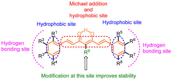
1. Anti-Breast Cancer Properties of Curcumin Analogs
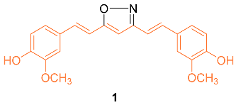
The isoxazole curcumin analog
1 was synthesized and the anti-cancer properties against the MCF-7 breast cancer cell line and its multidrug-resistant (MDR) version, MCF-7R, were compared with curcumin. After 72 h of treatment, the IC
50 of curcumin was calculated from four separate experiments to be 29.3 ± 1.7 μM in MCF-7 and 26.2 ± 1.6 μM in MCF-7R, indicating that the cytotoxic activity of curcumin in the MDR breast cancer cell line is at least equivalent to, and perhaps slightly stronger than, its parental variant. In both the parental and MDR cell line, derivative
1 was more effective than curcumin with an IC
50 of 13.1 ± 1.6 μM in MCF-7 and 12.0 ± 2.0 μM in MCF-7R. An MDR form of HL-60 leukemia also showed comparable outcomes. RT-PCR analyses in MCF-7 and MCF-7R cell lines revealed that curcumin and
1 caused early changes in the quantities of important gene transcripts, which were, nevertheless, primarily varied between the two cell lines. Overall, these results show that the expression of P-gp or the absence of ER in breast cancer cells does not impede the anti-cancer activities of either curcumin or
1. Remarkably, the agents seemed to adjust their molecular actions in response to the different patterns of gene expression found in the MDR and the parental MCF-7 [
18].
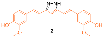
According to Wang et al., research was conducted on hydrazinocurcumin
2 (HC) to investigate its effectiveness against breast cancer cells, specifically in the cell lines MDA-MB-231 and MCF-7. After 72 h of treatment, dose-dependent suppression of tumor cell survival and proliferation was seen for the MDA-MB-231 and MCF-7 cell lines. The IC
50 values for
2 were 3.37 μM and 2.57 μM, respectively, which were both significantly lower than those for curcumin (26.9 μM and 21.22 μM). Compared to curcumin, the results demonstrated that
2 was significantly more effective in suppressing cell viability in both cell lines tested. Apoptosis was induced in MDA-MB-231 and MCF-7 cells using FCM, and the influence of
2 and curcumin on this process was analyzed. At 10 µM,
2 significantly induced cells apoptosis (14% in MDA-MB-231 cells and 26% in MCF-7 cells), whereas at the same concentration, curcumin only induced 9% and 20% cell apoptosis in MDA-MB-231 and MCF-7 cells, respectively. The results showed that
2 caused an increase in the apoptotic rate of cells in a dose-dependent manner after a treatment period of 48 h. In addition, the Western blot analysis demonstrated that
2 was much more effective than curcumin in suppressing the production of STAT3 protein in MDA-MB-231 and MCF-7 cells at the same concentration (10–20 μM). The data showed that
2 was more effective than curcumin at suppressing cell proliferation, losing colony formation, depressing cell migration and invasion, and inducing cell death via inhibiting STAT3 phosphorylation and downregulating an array of STAT3 downstream targets [
19].
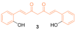
Mohankumar et al. studied the apoptotic mechanism of
3, an
ortho-hydroxy substituted analog of curcumin using an in vitro and in silico approach. In the study, it was found that
3 exhibited a greater potency in the modulation of selective apoptotic markers and inhibited MCF-7 at a dose level of 30 µM (equivalent dosage level to curcumin), and significantly regulated PI3k/Akt, both intrinsic and extrinsic apoptotic pathways, by inhibiting Bcl-2 and inducing p53, Bax, cytochrome c, Apaf-1, FasL, caspases-8, 9, 3, and PARP cleavage. mRNA expression studies for Bcl-2/Bax indicated increased efficiency with
3 compared to curcumin, while an in silico molecular docking study utilizing PI3K revealed that the docking of
8 was more potent than curcumin. Cells treated with
3 effectively induced apoptosis through ROS intermediates, as measured by 2′,7′-dichlorodihydrofluorescein diacetate (DCFH-DA). Results showed
3 induced apoptosis more effectively than curcumin, and this activity can be attributed to the presence of the hydroxyl group in the
ortho position in the structure [
20]. In addition, Western blotting indicated that compound
3 significantly downregulated the expression levels of NF-κB, p65, and c-Rel. In addition, src levels were significantly reduced in comparison to cells treated with curcumin. In silico docking studies were performed with the derivative and curcumin with NF-κB (PDB ID: 1NFK). The results indicated that the derivative displayed a stronger interaction with NF-κB compared to curcumin, with a Lidblock score of 109.814 while curcumin’s was 95.696 [
21].
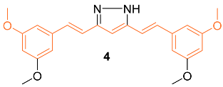
Lien et al. synthesized over 30 curcumin derivatives and published findings that a novel curcumin derivative (
4) inhibits cell proliferation and drug resistance of HER2-overexpressing cancer cells. The mimic was tested in vitro on both the MCF-7 and MDA-MB-435 cell lines transfected with pSV2-
erbB2. Results indicated that the derivative preferentially suppresses the growth of HER2-overexpressing cancer cells. Studies were also carried out to investigate if the derivative would sensitize HER2-overexpressing cancer cells to clinical drugs and it was found that overexpressing cells showed greater cytotoxic activity when the derivative was administered in combination with doxorubicin (DOX), etoposide, or taxol [
22].
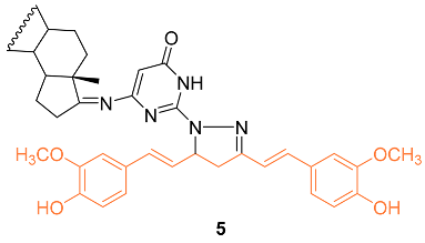
To understand the molecular hybridization impact and the integration of two drugs with different modes of action, affecting the same target, a variety of heterocyclic steroids and curcumin moieties were considered for the synthesis of hybrid conjugates and to determine their anti-cancer activity. The authors synthesized the hetero-steroid compounds and conducted in vitro studies of the cytotoxic effects against the MCF-7 breast cancer cell line. Of all compounds,
5 had the best cytotoxic activity against the MCF-7 cell line with an IC
50 value of 18 μM. This compound is also promising as an anti-cancer compound having pro-apoptotic effects resulting in desired cell growth inhibition [
23].
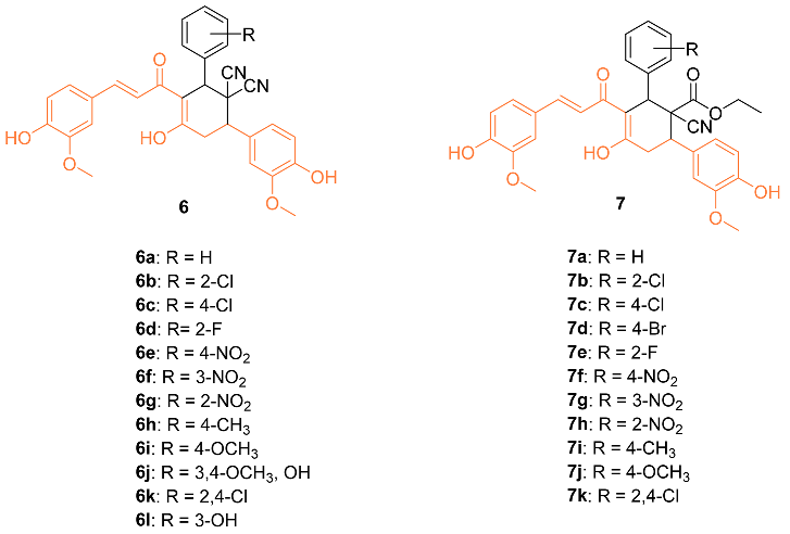
Bhuvaneswari et al. reported the biological evaluation and molecular docking of novel curcumin derivatives
6a–
l and
7a–
k. Firstly, in vitro cytotoxicity was tested against the MCF-7 breast cancer cell line. The IC
50 for
6j and
7i were 15 µM and 10 µM, respectively. When the compounds were tested against normal HBL-100 cells, the cells were resistant to the compounds up to 50 µM doses, showing the compounds are selective and dose-dependent. Molecular docking studies were also conducted in PatchDock and suggested that these two compounds could be the starting point for designing new potent Bcl-2 anti-apoptotic protein inhibitors, with
7 having a geometrical score of 6028 and
6 with a score of 5962 compared to 4190 for curcumin (PDB: 1GJH) [
24].
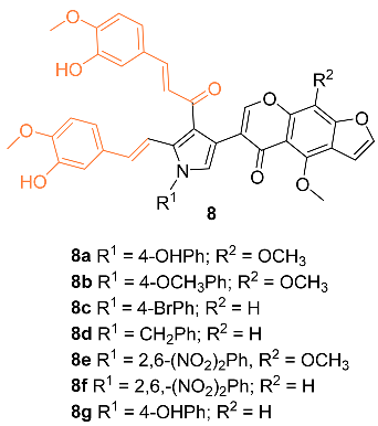
Nagwa et al. synthesized a set of curcumin derivatives
8a–
g and then experimented to determine the efficacy of the derivatives against breast cancer. Preliminary tests were conducted with normal MCF-10A cells and it was found that all derivatives had little cytotoxicity, with more than 85% cell viability. An MTT assay was performed with the derivatives against an MCF-7 breast cancer cell line. Compounds
8a and
8c were the most potent against the breast cancer cells, with an IC
50 of 20 and 22 μg/mL, respectively. Pharmacokinetic (ADME) studies confirm that compounds
8a and
8c have good intestinal absorption and are non-carcinogenic [
25].
Hong et al. reported the synthesis of and anti-cancer studies on the novel curcumin mimic (1
E,4
E)-1,7-bis(4-hydroxyphenyl) (hepta-1,4-dien-3-one)
9 isolated from mistletoe. It was first tested for in vitro cytotoxicity in which it showed activity in the micromolar range. Additionally,
9 showed a higher potency than cis-platinum against four human breast cancer cell lines (SKBR3, MDA-MB231, MCF-7, and MDA-MB453). The IC
50 values for the breast cancer cell lines were significantly lower with
9 compared to cis-platinum. The cytotoxicity of
9 with normal cells was investigated with LO2 human liver cells, GES-1 human gastric epithelial cells, and BEAS-2B human lung epithelial cells. The results indicated that
9 had a little inhibitory effect on normal cells, with each group having a less than 5% inhibition rate, which is much lower than the rate on cancer cells at the same concentration, indicating
9 has a selectivity for the toxic effects of cancer cells rather than normal cells. In addition, in vivo data on the MCF-7 breast cancer model in mice suggest that
9 is more effective than cisplatin. The groups administered
9 had a stable weight for up to 9 days, while a clear weight loss was observed in the positive control group [
26].

Shen et al. tested the efficacy of a curcumin analog
10 in breast cancer cells. The breast cancer cell lines MCF-7 and MDA-MB-231 were used to study the cell viability, cell migration, cell cycle, and apoptosis of this analog. It was shown that when the concentration of
10 was increased, there was a decrease in cell viability. In addition,
10 had an IC
50 of 8.84 μM compared to curcumin with an IC
50 of 16.85 μM against MCF-7 breast cancer cells. It was shown that
10 is a compound that activates the mitochondrial apoptosis pathway in breast cancer cells [
27].

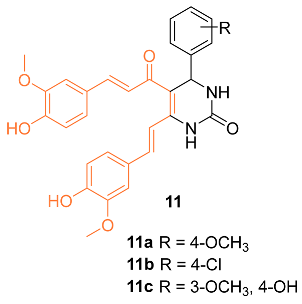
Sharma et al. synthesized 3,4-Dihydropyrimidin-2(1H)-one/thione curcumin analogs and, among them, compounds
11a–
c were submitted to the National Cancer Institute (NCI) to investigate activity against various cell lines, including the breast cancer cell lines MDA-MB-231 and HS 578T. At a concentration of 100 µM, compounds
11a–
c all displayed moderate activity, with compound
11a being the most active. This is supported by a growth percent value (GP) of 55.45 for compound
11a on MDA-MB-231 cells and a GP of 73.39 on HS 578T cells, while activities with a GP of 73.63 and 67.70 on MDA-MB-231 cells were found for compounds
11b and
11c, respectively. The authors believe compounds
11a–
c should be further studied to increase the moderate anti-cancer activity [
28].
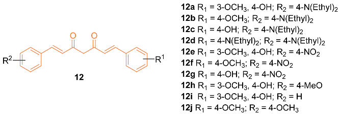
Zhang et al. reported on the synthesis and anti-cancer activity of ten curcumin mimics
12a–
j. Compound
12b exhibited the best anti-cancer activity, with an IC
50 value of 4.99 µM against MDA-MB-231 breast cancer cells compared to the 6.18 µM of cisplatin. In vivo data were obtained and were promising. However, in vivo testing was only carried out on H22 hepatic cells. Further testing is needed to evaluate if compound
12b is a promising anti-breast cancer drug [
29].
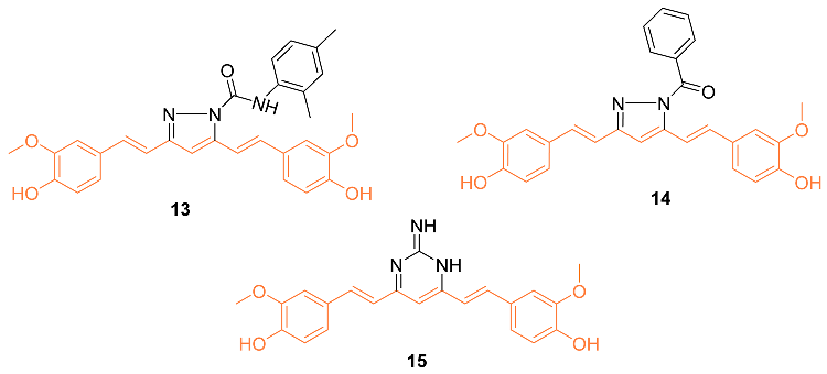
Considering the importance of pyrazole moiety, Ahsan et al. synthesized various curcumin analogs
13–
15 containing a pyrazole or pyrimidine ring to target the epidermal growth factor receptor (EGFR) tyrosine kinase. Fourteen curcumin analogs with pyrazole or pyrimidine moieties were synthesized, with ten being evaluated amongst 60 different cell lines to observe anti-cancer effects. The activity was observed from various compounds, however,
13–
15 displayed anti-cancer activity against various cell lines including MDA-MB-468. Compound
13 showed a cell promotion of −30.34%, compound
14 showed −31.86%, and compound
15 showed −35.04%. Ahsan et al. claim their curcumin analogs are promising and can be a therapeutic intervention in cancer treatment [
30].
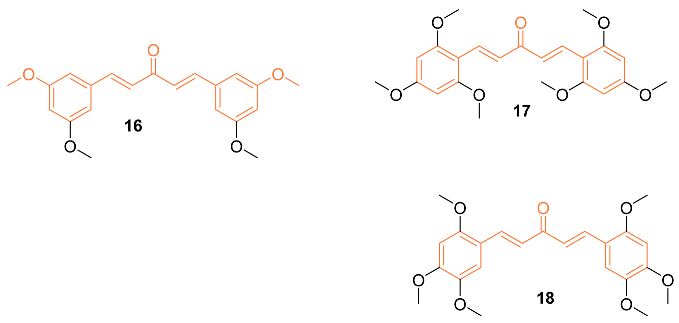
A set of twenty-four different analogs of curcumin containing pentadienone moiety were synthesized and examined for their anti-cancer properties against breast cancer cells (MCF-7 and MDA-MB-231). A dose-dependent suppression of tumor cell survival and proliferation was observed after 72 h of treatment with compounds
16–
18. The IC
50 values for compound
16 were 2.7 ± 0.5 μM and 1.5 ± 0.1 μM for the cell lines MCF-7 and MDA-MB-231, respectively, which were 5-8 times lower than those for curcumin (21.5 ± 4.7 μM and 25.6 ± 4.8 μM). Furthermore, the IC
50 values of compounds
17 (0.4 ± 0.1 μM and 0.6 ± 0.1 μM) and
18 (2.4 ± 1.0 μM and 2.4 ± 0.4 μM) were favorable for the MCF-7 and MDA-MB-231 cell lines, respectively. The non-malignant mammary epithelial cell line (MCF-10) demonstrated no toxicity from any of the three compounds. In comparison to curcumin, which did not exhibit any selectivity against cancer cell lines, it was discovered that compounds
16–
18 displayed a selectivity ratio of at least fivefold or greater. Compound
17, having IC
50 values in the sub-micromolar range and a selectivity ratio greater than 25, was found to be the most potent analog. All three compounds, however, show promise as possible anti-tumor drug candidates for breast cancer [
31].
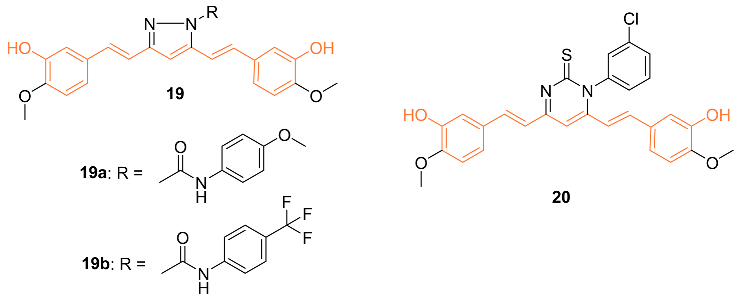
Ali et al. synthesized curcumin analogs
19a,
19b, and
20 and tested them against several breast cancer lines to determine their anti-cancer effects. The compounds were docked against the epidermal growth factor receptor, which allowed for the determination of binding efficiency. All derivatives showed moderate inhibition of epidermal growth factor receptors. Compounds
19b and
20 showed the most anti-cancer activity against BT-549 with GI
50 values of 2.98 μM for 2 and 1.51 μM for 3 [
32].

