Primary Cilia: History
Please note this is an old version of this entry, which may differ significantly from the current revision.
Contributor: , ,

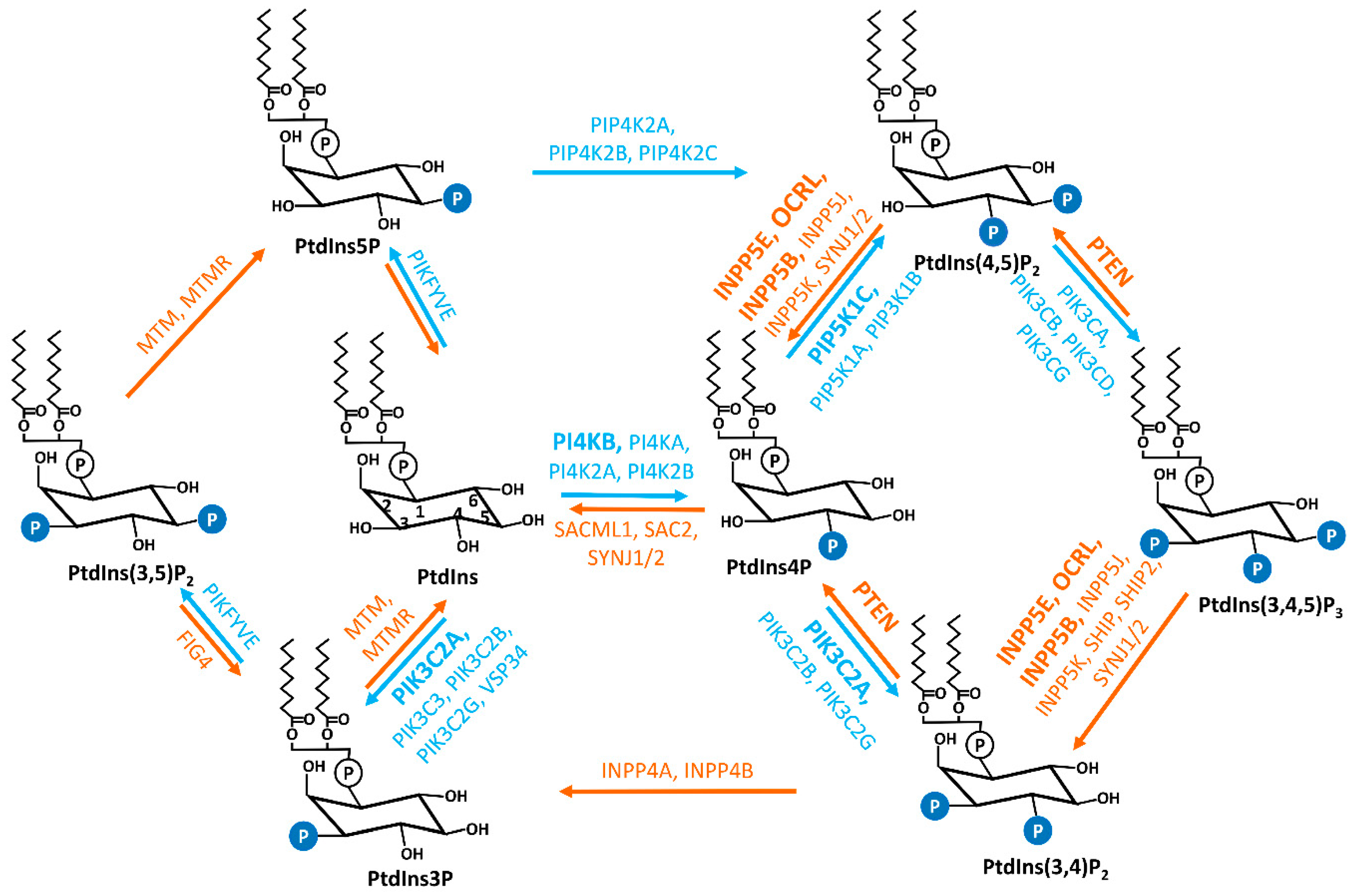
Primary cilia are microtube-based organelles that extend from the cell surface and function as biochemical and mechanical extracellular signal sensors. Primary cilia coordinate a series of signaling pathways during development. Cilia dysfunction leads to a pleiotropic group of developmental disorders, termed ciliopathy. Phosphoinositides (PIs), a group of signaling phospholipids, play a crucial role in development and tissue homeostasis by regulating membrane trafficking, cytoskeleton reorganization, and organelle identity. Accumulating evidence implicates the involvement of PI species in ciliary defects and ciliopathies. The abundance and localization of PIs in the cell are tightly regulated by the opposing actions of kinases and phosphatases, some of which are recently discovered in the context of primary cilia. 

  • phosphoinositide kinases and phosphatases
  • primary cilia

1. Introduction

Phosphoinositides (PIs) are reversibly phosphorylated derivatives of the membrane phosphatidylinositol (PtdIns, Figure 1) [1][2]. PIs consist of two fatty acid chains, a glycerol moiety, and a D-myo-inositol-1-phosphate head group that is decorated by phosphate moiety at the 3-, 4-, and/or 5-hydroxyl position to produce seven phosphorylated derivatives (PtdIns3P, PtdIns4P, PtdIns5P, PtdIns(3,4)P2, PtdIns(3,5)P2, PtdIns(4,5)P2, and PtdIns(3,4,5)P3) [1]. The exact composition of each species varies in different subcellular locations and different cell types. Compared to other phospholipids in cellular membrane compartments, PIs are relatively minor constituents and PtdIns4P and PtdIns(4,5)P2, which contribute the largest amounts, are less than 1% of the total cellular phospholipid pool [1]. Despite the low abundance, PIs are indispensable to the integrity of cells. PIs play essential roles in various cellular events including, but not limited to, cytoskeletal dynamics, cell adhesion and migration, vesicular trafficking, assembly of cargo proteins at membranes, cell proliferation, and survival [1][2][3][4]. PIs are key identity determinants of various cellular membrane compartments and act as spatiotemporal cues to direct signaling events [2][3][5][6][7].
Figure 1. Interconversions of seven PIs. The reversible production of the PIs is mediated by the combined activities of specific phosphatases and kinases, which are labeled in orange and blue, respectively. The kinases and phosphatases that have been reported to be associated with cilia are marked in bold. PtdIns, phosphatidylinositol; PtdIns3P, phosphatidylinositol 3-phosphate; PtdIns4P, phosphatidylinositol 4-phosphate; PtdIns5P, phosphatidylinositol 5-phosphate; PtdIns(3,4)P2, phosphatidylinositol 3,4-phosphate; PtdIns(3,5)P2, phosphatidylinositol 3,5-phosphate; PtdIns(4,5)P2, phosphatidylinositol 4,5-phosphate; PtdIns(3,4,5)P3, phosphatidylinositol 3,4,5-trisphosphate; PI4KA, phosphatidylinositol 4-kinase alpha; PI4KB, phosphatidylinositol 4-kinase beta; PI4K2A, phosphatidylinositol 4-kinase type 2 alpha; PI4K2B, phosphatidylinositol 4-kinase type 2 beta; PIK3C3, phosphatidylinositol 3-kinase catalytic subunit type 3; PIK3C2A, phosphatidylinositol-4-phosphate 3-kinase catalytic subunit type 2 alpha; PIK3C2B, phosphatidylinositol-4-phosphate 3-kinase catalytic subunit type 2 beta; PIK3C2G, phosphatidylinositol-4-phosphate 3-kinase catalytic subunit type 2 gamma; PIP5K1A, phosphatidylinositol-4-phosphate 5-kinase type 1 alpha; PIP5K1B, phosphatidylinositol-4-phosphate 5-kinase type 1 beta; PIP5K1C, phosphatidylinositol-4-phosphate 5-kinase type 1 gamma; PIP4K2A, phosphatidylinositol-5-phosphate 4-kinase type 2 alpha; PIP4K2B, phosphatidylinositol-5-phosphate 4-kinase type 2 beta; PIP4K2C, phosphatidylinositol-5-phosphate 4-kinase type 2 gamma; PIKFYVE, phosphoinositide kinase, FYVE-type zinc finger containing; PIK3CA, phosphatidylinositol-4,5-bisphosphate 3-kinase catalytic subunit alpha; PIK3CB, phosphatidylinositol-4,5-bisphosphate 3-kinase catalytic subunit beta; PIK3CD, phosphatidylinositol-4,5-bisphosphate 3-kinase catalytic subunit delta; PIK3CG, phosphatidylinositol-4,5-bisphosphate 3-kinase catalytic subunit gamma; MTM, myotubularin; MTMR, myotubularin-related; SAC2, Sac domain-containing inositol phosphatase 2; SACML1, SAC1 like phosphatidylinositide phosphatase; SYNJ1/2, synaptojanin 1/2; INPP5E, inositol polyphosphate-5-phosphatase E; INPP5J, inositol polyphosphate-5-phosphatase J; OCRL, oculocerebrorenal syndrome of Lowe; INPP5B, inositol polyphosphate-5-phosphatase B; INPP5K, inositol polyphosphate-5-phosphatase K; PTEN, phosphatase and tensin homolog; INPP4A, inositol polyphosphate-4-phosphatase type I A; INPP4B, inositol polyphosphate 4-phosphatase type II; FIG4, factor-induced gene 4; SHIP, Src homology 2 (SH2) domain containing inositol polyphosphate 5-phosphatase.
Each PI species exhibits unique distribution across the different plasma membrane (PM) and organelle membrane compartments [2]. In general, the lipid tail of PIs inserts into membrane compartments, whereas the phosphorylated inositol head of individual PIs recognizes and binds to unique PI effector proteins [2][8][9]. Current reported PI-binding domains include the Pleckstrin Homology (PH), FYVE, Phox (PX), C2, PROPPIN, PTB, Tubby, TRAF, ANTH/ENTH, FERM, and PDZ domains [9][10][11]. The affinity and specificity vary between different PIs and binding domains [9]. Individual PI species induce the conformational change of effectors and/or recruits them to specific subcellular locales to meet their signaling partners [8][9][12][13]. This leads to the activation/inhibition of PI effectors at unique subcellular locales [8]. Thus, the precisely controlled spatiotemporal availability of PI species in the cell is critical. PIs metabolism and homeostasis are maintained and regulated by specific PI kinases and phosphatases that are conserved in evolution [3][5][8]. Many reports have discovered that the dysfunction of components of the PI signaling pathway is responsible for various human diseases, from rare genetic disorders to the most common conditions such as cancer, obesity, neurological disorders, cardiovascular disease, and diabetes [7][14][15][16][17][18].
The primary cilium is a sensory organelle presenting on the surface of most mammalian cell types [19]. Although the ciliary membrane is reported to be continuous with the plasma membrane, the lipid and protein composition in the ciliary membrane is maintained unique and distinguishable from the plasma membrane [20][21]. To fulfill its function as a signaling hub and cellular antenna, the trafficking of protein and membrane components in and out of cilia is highly active and tightly controlled, suggesting the potential involvement of PIs in cilia. Indeed, it was reported in 2009 that mutations in an inositol polyphosphate 5-phosphatase, INPP5E, are causal for multiple ciliopathies [22][23]. Subsequently, more studies implicated the connection between PIs and primary cilia. PIs are reported to regulate the stability/disassembly of primary cilia [24][25], ciliogenesis [26], and ciliary trafficking [27][28]. The dysfunction of multiple PI enzymes, both phosphatases, and kinases, is connected to ciliary defects and ciliopathies [6]. The localization and function of PIs in cilia have been reviewed in several excellent recent reviews [6][29][30].

2. Primary Cilia

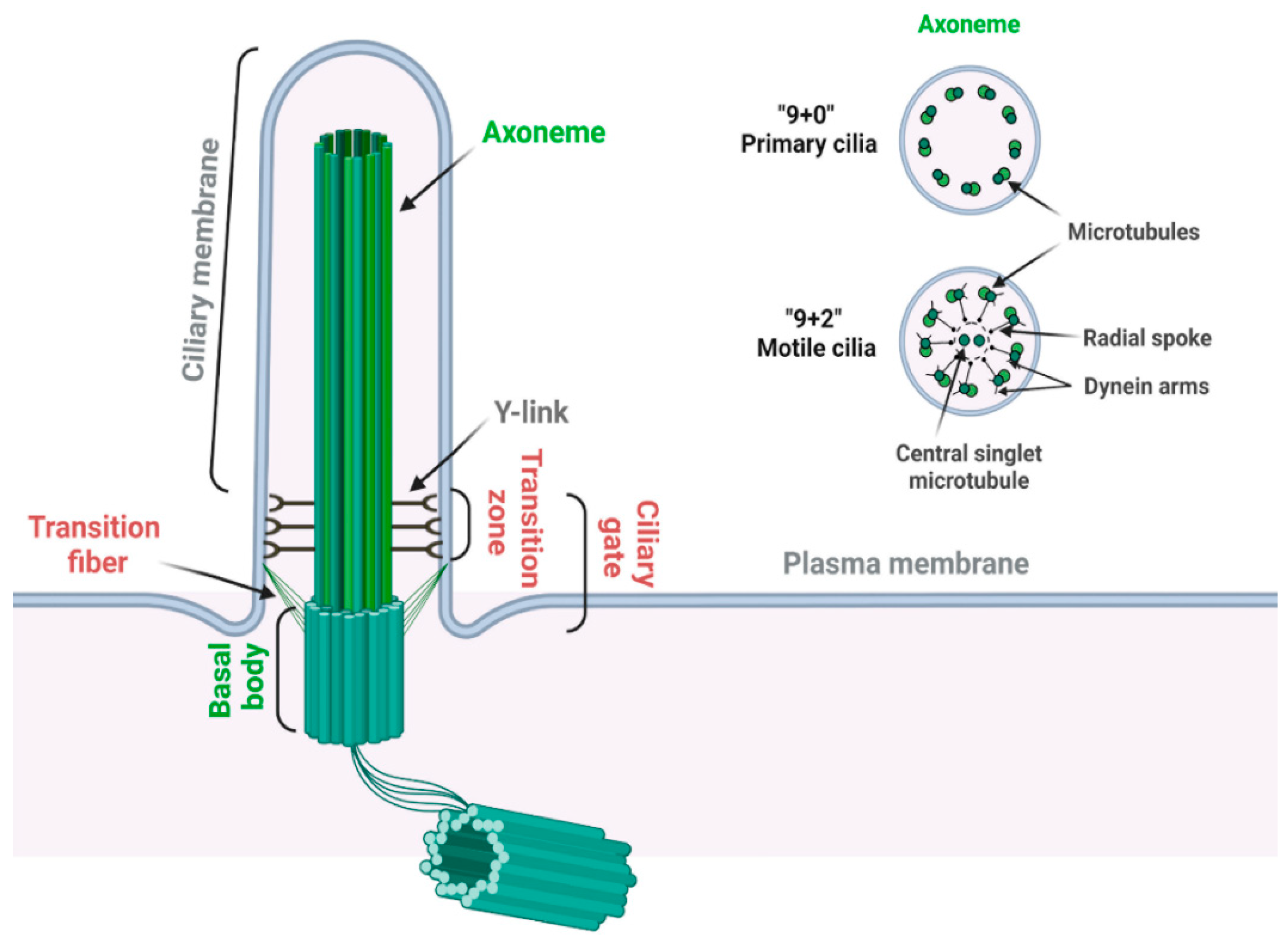
2.1. Structure of Primary Cilia

The primary cilium is a sensory organelle presenting on the surface of most quiescent cells in vertebrates [19][31]. The primary cilium is composed of a microtubule-based core structure called the axoneme, which is surrounded by a ciliary membrane that is continuously extended from the plasma membrane (Figure 2) [32]. Different from motile cilia, which can generate a fluid flow on cells such as respiratory epithelial and ependymal cells, primary cilia only contain nine microtubule doublets without two centrally located singlet microtubules (Figure 2) and are non-motile for lacking the axonemal dynein arms on the outer microtubules [33].
Figure 2. Structure of the primary cilium. A primary cilium is composed of a “9 + 0” microtubule axoneme and basal body complex (labeled in green). A motile cilium has two extra central singlet microtubules, generating a “9 + 2” arrangement (labeled in green). A motile cilium also contains dynein arms for ciliary movement and radial spoke to regulate the motility and motion pattern of motile cilia (labeled in black). Transition zone is characterized by the Y-links, which connect the proximal axoneme to the ciliary membrane. Transition fibers connect the distal end of the basal body to the ciliary membrane. Transition zone and transition fibers together coordinate the ciliary gate function (labeled in red) (created with BioRender.com, accessed on 18 November 2022).
The basal body, transformed from the mother centriole upon serum starvation, nucleates the outgrowth of the axoneme and forms the base of primary cilia. The region between the axoneme and basal body is identified as the transition zone, which is characterized by Y-links connecting the axonemal microtubules to the ciliary membrane [34]. Transition fibers, which are transformed from distal appendages of the mother centriole, are located below the transition zone on the distal end of the basal body [35]. In addition to anchoring the basal body to the plasma membrane, transition fibers function together with the transition zone as the ciliary gate to regulate ciliogenesis and signaling by controlling the selective transport of specific ciliary proteins between the cell body and the cilium [35][36]. Intriguingly, many transition zone and transition fiber proteins are reported to contain phosphoinositide binding domains; however, the physiological significance of their PI binding potential remains unclear [37].

2.2. Ciliopathy

Although largely considered a vestige before 1999, the primary cilium is now demonstrated as a signaling center to interpret both chemical and mechanical signals. Major components of multiple signaling pathways specifically localize in primary cilia [38]. These include pathways that are essential for the biogenesis and homeostasis of tissues and organs, such as the Hedgehog signaling [19], canonical Wnt signaling [39], Planar cell polarity [40], G protein-coupled receptor signaling [41], as well as tyrosine kinase receptor signaling [42] pathways. The activation and regulation of these pathways occur in cilia and depend on the normal structure and functionality of primary cilia [19].
Consistently, defects in primary cilia lead to a panel of genetic human diseases, collectively termed ciliopathies, including numerous seemingly unrelated syndromes, with involvement of the brain, eye, kidney, liver, pancreas, skeletal system, and some others in a growing list [43][44][45][46][47][48]. Common ciliopathies range from organ-specific disorders such as autosomal dominant polycystic kidney disease (ADPKD) and autosomal recessive PKD (ARPKD) to pleiotropic disorders such as Joubert syndrome (JBST), nephronophthisis (NPHP), Bardet–Biedl syndrome (BBS), and Meckel syndrome (MKS) [49][50]. Currently, more than 190 established and 250 candidate cilium-associated proteins are identified as ciliopathy proteins, and over 35 ciliopathies are reported, emphasizing the significance of primary cilia in development and human disease [51][52]. Given the physiological significance of the primary cilium, the molecular mechanisms underlying its biogenesis, function, and related disease pathogenesis have just started to be explored in the recent two decades.

This entry is adapted from the peer-reviewed paper 10.3390/jdb10040051

References

  1. Balla, T. Phosphoinositides: Tiny lipids with giant impact on cell regulation. Physiol. Rev. 2013, 93, 1019–1137.
  2. Schink, K.O.; Tan, K.W.; Stenmark, H. Phosphoinositides in Control of Membrane Dynamics. Annu. Rev. Cell Dev. Biol. 2016, 32, 143–171.
  3. Dickson, E.J.; Hille, B. Understanding phosphoinositides: Rare, dynamic, and essential membrane phospholipids. Biochem. J. 2019, 476, 1–23.
  4. De Craene, J.O.; Bertazzi, D.L.; Bar, S.; Friant, S. Phosphoinositides, Major Actors in Membrane Trafficking and Lipid Signaling Pathways. Int. J. Mol. Sci. 2017, 18, 634.
  5. Posor, Y.; Jang, W.; Haucke, V. Phosphoinositides as membrane organizers. Nat. Rev. Mol. Cell Biol. 2022, 23, 797–816.
  6. Conduit, S.E.; Vanhaesebroeck, B. Phosphoinositide lipids in primary cilia biology. Biochem. J. 2020, 477, 3541–3565.
  7. Hammond, G.R.V.; Burke, J.E. Novel roles of phosphoinositides in signaling, lipid transport, and disease. Curr. Opin. Cell Biol. 2020, 63, 57–67.
  8. Kutateladze, T.G. Translation of the phosphoinositide code by PI effectors. Nat. Chem. Biol. 2010, 6, 507–513.
  9. Hammond, G.R.; Balla, T. Polyphosphoinositide binding domains: Key to inositol lipid biology. Biochim. Biophys. Acta 2015, 1851, 746–758.
  10. Cullen, P.J.; Cozier, G.E.; Banting, G.; Mellor, H. Modular phosphoinositide-binding domains—Their role in signalling and membrane trafficking. Curr. Biol. 2001, 11, R882–R893.
  11. Lemmon, M.A. Phosphoinositide recognition domains. Traffic 2003, 4, 201–213.
  12. Rusten, T.E.; Stenmark, H. Analyzing phosphoinositides and their interacting proteins. Nat. Methods 2006, 3, 251–258.
  13. Jacobsen, R.G.; Mazloumi Gavgani, F.; Edson, A.J.; Goris, M.; Altankhuyag, A.; Lewis, A.E. Polyphosphoinositides in the nucleus: Roadmap of their effectors and mechanisms of interaction. Adv. Biol. Regul. 2019, 72, 7–21.
  14. Staiano, L.; De Leo, M.G.; Persico, M.; De Matteis, M.A. Mendelian disorders of PI metabolizing enzymes. Biochim. Biophys. Acta 2015, 1851, 867–881.
  15. Liu, Y.; Bankaitis, V.A. Phosphoinositide phosphatases in cell biology and disease. Prog. Lipid Res. 2010, 49, 201–217.
  16. Fruman, D.A.; Rommel, C. PI3K and cancer: Lessons, challenges and opportunities. Nat. Rev. Drug Discov. 2014, 13, 140–156.
  17. Dickson, E.J. Recent advances in understanding phosphoinositide signaling in the nervous system. F1000Res 2019, 8, F1000 Faculty Rev-278.
  18. Krajnik, A.; Brazzo, J.A., 3rd; Vaidyanathan, K.; Das, T.; Redondo-Munoz, J.; Bae, Y. Phosphoinositide Signaling and Mechanotransduction in Cardiovascular Biology and Disease. Front. Cell Dev. Biol. 2020, 8, 595849.
  19. Bangs, F.; Anderson, K.V. Primary Cilia and Mammalian Hedgehog Signaling. Cold Spring Harb. Perspect. Biol. 2017, 9, a028175.
  20. Plotnikova, O.V.; Pugacheva, E.N.; Golemis, E.A. Primary cilia and the cell cycle. Methods Cell Biol. 2009, 94, 137–160.
  21. Dutta, P.; Ray, K. Ciliary membrane, localised lipid modification and cilia function. J. Cell Physiol. 2022, 237, 2613–2631.
  22. Bielas, S.L.; Silhavy, J.L.; Brancati, F.; Kisseleva, M.V.; Al-Gazali, L.; Sztriha, L.; Bayoumi, R.A.; Zaki, M.S.; Abdel-Aleem, A.; Rosti, R.O.; et al. Mutations in INPP5E, encoding inositol polyphosphate-5-phosphatase E, link phosphatidyl inositol signaling to the ciliopathies. Nat. Genet. 2009, 41, 1032–1036.
  23. Jacoby, M.; Cox, J.J.; Gayral, S.; Hampshire, D.J.; Ayub, M.; Blockmans, M.; Pernot, E.; Kisseleva, M.V.; Compere, P.; Schiffmann, S.N.; et al. INPP5E mutations cause primary cilium signaling defects, ciliary instability and ciliopathies in human and mouse. Nat. Genet. 2009, 41, 1027–1031.
  24. Dyson, J.M.; Conduit, S.E.; Feeney, S.J.; Hakim, S.; DiTommaso, T.; Fulcher, A.J.; Sriratana, A.; Ramm, G.; Horan, K.A.; Gurung, R.; et al. INPP5E regulates phosphoinositide-dependent cilia transition zone function. J. Cell Biol. 2017, 216, 247–263.
  25. Conduit, S.E.; Ramaswamy, V.; Remke, M.; Watkins, D.N.; Wainwright, B.J.; Taylor, M.D.; Mitchell, C.A.; Dyson, J.M. A compartmentalized phosphoinositide signaling axis at cilia is regulated by INPP5E to maintain cilia and promote Sonic Hedgehog medulloblastoma. Oncogene 2017, 36, 5969–5984.
  26. Xu, Q.; Zhang, Y.; Wei, Q.; Huang, Y.; Hu, J.; Ling, K. Phosphatidylinositol phosphate kinase PIPKIgamma and phosphatase INPP5E coordinate initiation of ciliogenesis. Nat. Commun. 2016, 7, 10777.
  27. Chavez, M.; Ena, S.; Van Sande, J.; de Kerchove d’Exaerde, A.; Schurmans, S.; Schiffmann, S.N. Modulation of Ciliary Phosphoinositide Content Regulates Trafficking and Sonic Hedgehog Signaling Output. Dev. Cell 2015, 34, 338–350.
  28. Garcia-Gonzalo, F.R.; Phua, S.C.; Roberson, E.C.; Garcia, G., 3rd; Abedin, M.; Schurmans, S.; Inoue, T.; Reiter, J.F. Phosphoinositides Regulate Ciliary Protein Trafficking to Modulate Hedgehog Signaling. Dev. Cell 2015, 34, 400–409.
  29. Zhang, R.; Tang, J.; Li, T.; Zhou, J.; Pan, W. INPP5E and Coordination of Signaling Networks in Cilia. Front. Mol. BioSci. 2022, 9, 885592.
  30. Nakatsu, F. A Phosphoinositide Code for Primary Cilia. Dev. Cell 2015, 34, 379–380.
  31. Goetz, S.C.; Anderson, K.V. The primary cilium: A signalling centre during vertebrate development. Nat. Rev. Genet. 2010, 11, 331–344.
  32. Linck, R.W.; Chemes, H.; Albertini, D.F. The axoneme: The propulsive engine of spermatozoa and cilia and associated ciliopathies leading to infertility. J. Assist. Reprod. Genet. 2016, 33, 141–156.
  33. Satir, P. CILIA: Before and after. Cilia 2017, 6, 1.
  34. Goncalves, J.; Pelletier, L. The Ciliary Transition Zone: Finding the Pieces and Assembling the Gate. Mol. Cells 2017, 40, 243–253.
  35. Wei, Q.; Ling, K.; Hu, J. The essential roles of transition fibers in the context of cilia. Curr. Opin. Cell Biol. 2015, 35, 98–105.
  36. Garcia-Gonzalo, F.R.; Reiter, J.F. Open Sesame: How Transition Fibers and the Transition Zone Control Ciliary Composition. Csh Perspect. Biol. 2017, 9, a028134.
  37. Garcia-Gonzalo, F.R.; Reiter, J.F. Scoring a backstage pass: Mechanisms of ciliogenesis and ciliary access. J. Cell Biol. 2012, 197, 697–709.
  38. Nachury, M.V. How do cilia organize signalling cascades? Philos. Trans. R. Soc. Lond B Biol. Sci. 2014, 369, 20130465.
  39. Lee, K.H. Involvement of Wnt signaling in primary cilia assembly and disassembly. Febs. J. 2020, 287, 5027–5038.
  40. Wallingford, J.B. Planar cell polarity signaling, cilia and polarized ciliary beating. Curr. Opin. Cell Biol. 2010, 22, 597–604.
  41. Schou, K.B.; Pedersen, L.B.; Christensen, S.T. Ins and outs of GPCR signaling in primary cilia. Embo. Rep. 2015, 16, 1099–1113.
  42. Christensen, S.T.; Clement, C.A.; Satir, P.; Pedersen, L.B. Primary cilia and coordination of receptor tyrosine kinase (RTK) signalling. J. Pathol. 2012, 226, 172–184.
  43. Sreekumar, V.; Norris, D.P. Cilia and development. Curr. Opin. Genet. Dev. 2019, 56, 15–21.
  44. Anvarian, Z.; Mykytyn, K.; Mukhopadhyay, S.; Pedersen, L.B.; Christensen, S.T. Cellular signalling by primary cilia in development, organ function and disease. Nat. Rev. Nephrol. 2019, 15, 199–219.
  45. Ferkol, T.W.; Leigh, M.W. Ciliopathies: The central role of cilia in a spectrum of pediatric disorders. J. Pediatr. 2012, 160, 366–371.
  46. Quinlan, R.J.; Tobin, J.L.; Beales, P.L. Modeling ciliopathies: Primary cilia in development and disease. Curr. Top Dev. Biol. 2008, 84, 249–310.
  47. Fliegauf, M.; Benzing, T.; Omran, H. When cilia go bad: Cilia defects and ciliopathies. Nat. Rev. Mol. Cell Biol. 2007, 8, 880–893.
  48. Focsa, I.O.; Budisteanu, M.; Balgradean, M. Clinical and genetic heterogeneity of primary ciliopathies (Review). Int. J. Mol. Med. 2021, 48, 176.
  49. Powles-Glover, N. Cilia and ciliopathies: Classic examples linking phenotype and genotype-an overview. Reprod. Toxicol. 2014, 48, 98–105.
  50. Braun, D.A.; Hildebrandt, F. Ciliopathies. Csh Perspect. Biol. 2017, 9, a028191.
  51. Reiter, J.F.; Leroux, M.R. Genes and molecular pathways underpinning ciliopathies. Nat. Rev. Mol. Cell Biol. 2017, 18, 533–547.
  52. Wheway, G.; Mitchison, H.M.; Genomics England Research, C. Opportunities and Challenges for Molecular Understanding of Ciliopathies-The 100,000 Genomes Project. Front. Genet. 2019, 10, 127.
More
This entry is offline, you can click here to edit this entry!
Video Production Service