Monkeypox Infection 2022 in Adults and Children: History
Please note this is an old version of this entry, which may differ significantly from the current revision.

Contributors : Francesca Gaeta  Francesco De Caro  Gianluigi Franci  Pasquale Pagliano  Pietro Vajro  Claudia Mandato

Monkeypox disease has been endemic in sub-Saharan Africa for a long time, attracting remarkable attention only in 2022 through the occurrence of a multi-country outbreak. The latter has raised serious public health concerns and is considered a public health emergency by the World Health Organization. Although the disease is usually self-limiting, it can cause severe illness in individuals with compromised immune systems, in children, and/or the pregnant woman–fetus dyad. Patients generally present with fever, lymphadenopathy, and a vesicular rash suggestive of mild smallpox. Serious eye, lung and brain complications, and sepsis can occur.

https://doi.org/10.3390/children9121832

  • antivirals
  • breastfeeding
  • children
  • monkeypox
  • newborn
  • outbreak
  • pregnancy
  • prevention
  • testing
  • vaccination

1. Introduction

Monkeypox is a sylvatic viral zoonotic (i.e., spreading between animals and humans) infection, originating from the homonymous virus (MPV), first identified by Copenhagen’s Statens Serum Institute in a cluster of monkeys imported from Singapore to Denmark in 1958. The first cases of human infection were identified in children in the year 1970 [1][2]. MPV infections were endemic in sub-Saharan Africa until its complete eradication using first-generation smallpox vaccines in 1980. In western countries, a notable epidemic of MPV infection occurred in the United States in 2003, when infected rodents from Africa, imported as pets, spread the virus to domestic prairie dogs, who later infected human subjects in the Midwest. That epidemic involved 37 confirmed cases and 10 likely cases in six states, but there were no deaths [3]. Thereafter, except in cases associated with the pet trade and travel, monkeypox has recently regained attention from health authorities, due to infections reported in the UK in May 2021 and in the US in November 2021. A few months later, (May 2022), the first outbreak was reported in continental Europe [4]. Since January 2022 and as of 17 November 2022, there have been 29,055 confirmed cases of monkeypox and 11 deaths in the United States [5]. In the same period, 25,465 confirmed cases of monkeypox and four deaths have been reported from 29 European Union countries [6][7]. Currently this global outbreak (80,221 confirmed cases and 52 deaths reported to WHO) is occurring in 110 Member States. A high proportion of these infections are from countries without previously documented monkeypox transmission and are caused by the Clade II of monkeypox, formerly known as the West African clade [7][8]. Presently, except for the South Pole, cases of monkeypox have been reported in every continent, declaring on 23 July a Public Health Emergency of International Concern (PHEIC) by World Health Organization (WHO) [9]. Despite its recent mildly declining trend [7], the current outbreak of MPV gives cause for concern also due to its long incubation period which may mask the culprit contagion and for its quite frequent subtle clinical presentation as well. Overall, epidemiological data show that the ratio M:F is 97:3, and that the population seemingly most affected (approximately 80%) is male, aged between 18 and 44 years old, mostly homosexual men having unprotected sex with other men. However, adolescents, children and babies of both sexes can be affected as well (1.2% reported cases were aged 0–17) [7][8].
Although the infection now is not yet an emergency in Western countries, the persisting outbreak of cases in recent times makes the need for comprehensive information on diagnosis, management, and therapy for all age groups seemingly reasonable. As happened for the COVID-19 pandemic, the most important world health organizations have fielded information and tools on the subject of pediatric monkeypox, including the procedures to contain contagion in pregnancy and lactation which involve the pediatric population too, in a secondary way.

2. Monkeypox Virus: Generalities

Monkeypox virus is a large virus (200–250 nm), part of the group of Poxviridae (subfamily: chordopoxvirinae-genus: orthopoxvirus/species: monkeypox virus). MPV is an enveloped virus, containing a linear genome with double stranded DNA [10]. It is characterized by two phylogenetic clades: Central Africa (native to Congo Basin) and West Africa (native to Nigeria). The first clade appears more pathogenetic causing severe disease with a mortality rate of up to about 10% [11].
WHO experts reached consensus to now refer to the former Congo Basin (Central African) clade as Clade one (I) and the former West African clade as Clade two (II). Additionally, it was agreed that Clade II consists of two subclades IIa and IIb, the latter referring primarily to the group of variants largely circulating in the 2022 global outbreak [12].
In a recent study, the immunological signature of infected patients showed an early expansion of activated CD4+ and CD8+ T-cells which persisted after recovery without distinctions between HIV-positive and HIV-negative patients. Cases with mild clinical pictures showed a less perturbed immune profile early [13]. These data agree with previous studies which showed an overproduction of interleukin [IL]-2R, IL-10, and granulocyte macrophage-colony stimulating factor in patients with serious disease [14]. Cytokine response appears to be turned off in subjects who have already contracted the virus [4].
Although in the past MPV underwent few mutations, overtime, as happens for other DNA viruses, [15][16], more mutations are now increasingly being reported. These recent mutations are different from those already known previously, likely reflecting the fact that the virus is now able to survive in the population more easily [4][16].
A recent bioinformatic analysis study consistently found in the 2022 genomes two specific variation units in repeat located in intergenic regions of the viral DNA helicase and transcription factors. These factors might affect the virulence of the virus during the current outbreak [17], although the monkeypox virus continues to evolve, no mutations have affected the part of its genome that encodes a protein targeted by tecovirimat, an antiviral drug being tested for use against monkeypox in humans [18].

3. Clinical Presentation, Transmission and Prevention
The MPV final reservoir has not yet been clearly identified. Several evidences report that some rodents, originating in Africa, can transmit the virus between members of the same and different species. Additional known zoonotic reservoirs for MPV are the Gambian marsupial, tree squirrel, rope squirrel and sooty mangabey monkey, and possibly others [10]. Body fluids, droplets, scratch, bush meat preparation, lesion material wounds are some of the modes of animal trasmission [19]. Immunopathogenetic studies show that human monkeypox virus (MPV) can enter the body via the the skin or respiratory tract and starts replicating in inoculation sites. Recent data showing high viral loads on the skin and mucosa of infected patients suggest that in humans the transmission most likely occurs mainly through direct contact rather than through contact with bodily fluids or respiratory transmission [20][21].
In both the above scenarios, it is hypothesized that infected Antigen-presenting cells (APC) migrate to nearby draining lymph nodes and facilitate their spread through the lymphatic system with frequent swelling of lymph nodes. After propagating through lymphoid tissue, MPV viremia can target organs such as the spleen and liver and spread further to distant organs like the skin and gonads. MPV has recently been isolated from the semen of infected individuals, underscoring the possibility of sexual transmission. MPV locates in tissues with an incubation period that lasts 7–14 days, up to a maximum of 21 days. In this step, first non-specific symptoms appear, such as fever, maxillary, cervical or inguinal lymphadenopathy, chills, headache and backache. Lymphadenopathy when present is characterized by solid consistency, sometimes painful, and tender lymphnodes. This clinical aspect could allow making differential diagnosis with smallpox as the latter does not cause lymphadenopathy [1]. At this time, patients may already be contagious. Infection of the skin and mucous membranes leads to the appearance of infectious pustules and ulcers. The latter release large amounts of virus into the saliva, possibly leading to aerosolized transmission of MPV [22][23]. MPV can also be transmitted through the placenta in vertical transmission (congenital Monkeypox), which is associated with a high risk of either perinatal loss or vertical infection, and through close contact during and after birth [24][25]. The contagion from humans to pets has been documented as well [26].
To estimate the MPV infection score, it has been used a mathematical model showing a number of reproduction (R) > 1, indicative of the epidemic potential. Quarantine can be a good strategy to prevent infection; based on the knowledge of the potential contagion, it would be useful to identify and isolate affected or potentially affected subjects up to 3 weeks [4][27].
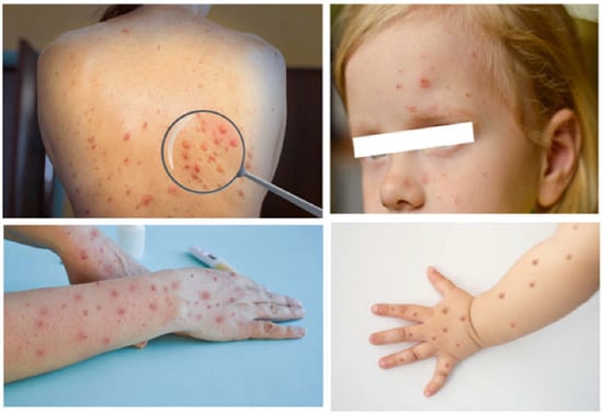
After the initial contagion, and 1–2 days after the end of incubation period described above, the patient develops a rash, mainly to the face, which spreads throughout the body progressively. Typically, the lesions are initially visible within the oropharynx; afterwards they spread centrifugally to other areas of the body, including palms and soles. The rash can also be localized, and skin lesions can be from few to thousands. In a period between 2 and 4 weeks, the skin lesions change in appearance, and become macular, papular, vesicular, and pustular, most frequently all at the same time and are painful (Figure 1).
Figure 1. Diverse phases of the evolution of the monkeypox rash in adults (left) and in children (right) Photo credit: Shutterstock.
The size of the lesions is between 2 and 10 mm, remaining in the pustular phase for 5–7 days. At this point one can observe crusts that tend to desquamate in 7–14 days. In 3–4 weeks, the clinical pathological situation is resolved, and patients are no longer contagious at the fall of all the crusts [1]. In the current outbreak, affected people have however been reported with fewer lesions than in previous outbreaks in Africa, with a higher proportion of (likely more uncomfortable) hidden mucosal lesions [28].
Noticeably, in the current 2022 outbreak, the febrile prodrome may be absent and skin/mucosal lesions may be isolated to the genital and anal regions [29][30].
In addition to classical symptoms, MPV can present with rare symptoms and complications that are observed with the highest rate in immunocompromised patients. Vomiting and diarrhea can appear from the second week after infection and can cause secondary dehydration. Corneal infections are among the most complex and dangerous complications, with the possibility of vision loss. Patients affected by monkeypox can report neurological complications with rare cases of encephalitis, which need intensive supportive treatments [25][31].
Bacterial superinfection, bronchopulmonary infections, and respiratory distress can occur in immunocompromised and co-infected with influenza virus patients. Sepsis and septic shock due to bacterial over-infections can occur when the general state of the patient is impaired [32]. In view of the above complications, the MPV patients need careful monitoring.
Currently, the mortality rate is estimated between 1 and 10% in relation to the clade involved and the proper health care [33]. Quantification of environmental contamination is not well known. A study published in 2022 [34] showed that hospital environmental contamination is possible. Biological samples, collected on contaminated room surfaces examined by real-time polymerase chain reaction (RT-PCR) after a 4-day hospital stay of MPV patients, showed up to 105 viral copies/cm2. In view of these data, it appears important to decontaminate the hospital rooms and to protect healthcare professionals from exposure to the contagion. Currently, the following measures appear to be good practices to be implemented: (i) avoiding direct contact with wild animals or pets suspected of hosting MPV; (ii) using negative pressure rooms to isolate affected patients in hospital; (iii) killing of affected animals; (iv) avoiding contact surfaces possibly contaminated by infected persons or animals. All persons and operators who meet affected subjects or animals must wear protective equipment, such as waterproof clothing over the whole body, a double layer of gloves, and an N95 mask [4][35].

4. Diagnosis

MPV diagnosis is based on the patient’s medical history and epidemiological data. Suspicion of MPV infection should arise in the case of travel or possible contact with wild animals from endemic areas and evidence of suggestive symptoms. When collecting the history of possible trips in endemic areas, details such as the destination and the dates of arrival and departure to assess potential incubation period and any activities carried out abroad and any exposure to people or animals potentially infected should be recorded [19]. Due to the difficulty of a differential diagnosis between smallpox and human monkeypox infection, the CDC created a specific protocol, named the “Acute, Generalized Vesicular or Pustular Rash Illness Protocol” [36]. Lymphadenopathy was used as the primary criteria to discriminate which patients are sent to second-level tests, because, as mentioned above, this was an MPV characteristic feature  [37]. However, in the present outbreak, this symptom may be absent [38].
Although history and symptoms can be indicative, it is crucial to use laboratory diagnostics, represented by the detection of viral DNA isolated from patient lesions. According to the WHO, RT-PCR specific to the MPV genome used alone, or in combination with sequencing is considered the gold standard for diagnosis in adults and children. Moreover, at all ages, the recommended specimen type for laboratory confirmation of monkeypox is skin lesion material, including swabs of the lesion surface and/or exudate, roofs from more than one lesion, or lesion crusts [39][40].
MPV infection may also be confirmed by several other newer molecular techniques, or electron microscope vision and immunohistochemistry staining for Orthopoxvirus which are reliable tests for diagnosis but are expensive or not commercially available.
Serology (IgG and IgM) with enzyme-linked immunosorbent assay (ELISA) early (after 5 and 8 days of infection for detection of IgM and IgG, respectively), is particularly important for the reconstruction of the timing of infection [1]. Antibody assay detects its presence more clearly after about two weeks of infection [1][4]. Orthopox biothreat alert © (Tetracore, Rockville, MD, USA) allows Smallpox virus antigens to be detected directly, but unfortunately, it is not able to distinguish between smallpox and monkeypox [41].
The differential diagnosis should be placed with pathologies that manifest themselves on the skin, but that have systemic involvement [42], such as smallpox, disseminated zoster, chickenpox, eczema herpeticum, scabies, and rickettsialpox. Due to MPV sexual transmission, skin manifestations of some sexually transmitted diseases (STD) such as syphilis should be considered in differential diagnosis. Furthermore, bacterial infections of wounds and skin infections can mimic monkeypox. Skin reactions to medications sometimes manifest as ulcerated lesions and monkeypox-like crusts.
Complications of monkeypox can sometimes appear as the first symptom of the disease and delay the diagnosis. A patient who typically presents with fever, characteristic lymphadenopathy, a history of exposure to risk and impaired clinical conditions (for example, pneumonia, gastroenteritis, or neurological symptoms) should arise suspicion in relation to monkeypox. Dehydration, sepsis, encephalitis, and pneumonia deserve even more attention in the pediatric population because dehydration is faster and more severe in the child than in the adult patient. As mentioned, in children lymphadenopathy may be less marked than in adults, but the history and the presence of any cases in the family should make monkeypox suspected [43].
The asymptomatic occurrence of monkeypox remains a vulnus also for the risk of vertical transmission of monkeypox, partly addressed through rigorous assessments of testing facilities in pregnant women [44][45].

5. Treatment

Several antivirals and vaccines which can be used for both smallpox and MPX are presently available. Their effectiveness and best use in patients with MPV disease are still unclear, and they are not widely accessible worldwide [46].

5.1. Antiviral Medicines

Currently, MPV has no specific treatment; symptomatic drugs, such as non-steroidal anti-inflammatory drugs, are mainly used to control non-specific symptoms (i.e., fever and asthenia). Immunocompetent patients usually overcome the disease without treatment. Due to the similarity between monkeypox and smallpox, the CDC [47] has proposed the use of previously approved drugs for smallpox in patients who contract MPV. Treatment with antiviral drugs, according to the CDC, should be reserved for particularly compromised patients. The following three available antiviral drugs used for MPV infection have side effects and a still poorly known clinical value [48]:
(a)
Cidofovir (Vistide) is primarily used as a treatment for retinitis, encephalitis and oesophagitis caused by cytomegalovirus, especially in people with HIV. It is the phosphorylated active metabolite of brincidovir. In-vitro and preclinical studies showed that is effective against poxviruses [4];
(b)
Brincidofovir (Tembexa) is available as oral suspension/tablets, approved byFood and Drug Administration (FDA) for smallpox disease [49]. Both drugs are inhibitors of DNA replication with a broad spectrum of activity against multiple families of double-stranded DNA viruses.
(c)
Tecovirimat (ST-246): is an antiviral medication which impairs the function of the VP37 envelope protein necessary for the formation of the extracellular enveloped virus required for cell-to-cell transmission; it has more specific activity against orthopoxviruses [49]. It has been approved by FDA, used to treat human smallpox disease but can be used against MPV. Tecovirimat is given orally (TPOXX®: 200 mg capsule) or as an injectable formulation [41]. Capsules should be taken within 30 min after a full meal with moderate to high fat. Per CDC guidelines, for those who cannot swallow they can be opened and mixed with liquids/soft food [50] Because it is an inducer of cytochrome P450 (CYP) 3A and CYP2B6, co-administration with this drug may lead to reduced plasma exposures of sensitive substrates of CYP3A4 or CYP2B6, reducing the effects. Because of the presence in its IV formulation of a potentially nephrotoxic substance (hydroxypropyl-β-cyclodextrin), it is advisable to dose creatinine clearance (CrCl) and liver function before starting treatment. Intravenous therapy is safe in mild/moderate renal impairment but is contraindicated in severe nephropathies (CrCl < 30 mL/min), both in adults and children [43]. Dose adjustments for oral therapy instead are not required in the case of mild, moderate, severe nephropathy or even in patients requiring hemodialysis in end-stage renal disease [51]. Although reduced fertility due to testicular toxicity was found in mouse models, no human data are available [51].
The most frequent adverse effects in oral treatment are headache and nausea, followed by abdominal pain and vomiting. The main reactions to IV tecovirimat are pain and redness at the injection site, headache, myalgia, arthritis, back pain, muscle tightness, diarrhoea, photophobia, and generalized pruritus. Recently, the largest safety study concluded was a trial on 549 mostly non hospitalized adults affected by MPV disease with/without HIV, who were prescribed oral tecovirimat under an Expanded Access Investigational New Drug protocol. The median interval was three days from initiation of tecovirimat to subjective improvement, with no difference by HIV infection status. In 3.5% of patients, adverse events were reported, one of which was nonserious. Tecovirimat is generally well tolerated, and these data support continued access to treatment with tecovirimat during the current monkeypox outbreak [52].
The effectiveness of antiviral drugs as post exposure prophylaxis in PEP monkeypox is unknown. Antiviral medications, primarily tecovirimat, can be used as PEP only in exceptional cases, for example when it is not possible to administer vaccines for allergies. To find new therapeutic strategies, studies of the efficiency of some antivirals generally used against Orthopoxvirus species are underway [53].

5.2. Children’s Treatment

Although no study is available in the pediatric age, the use of specific drugs in paediatric patients is strongly recommended in the “severe disease”. According to the AAP, [43] the risk conditions for “severe disease” are infants, children < 8 years of age, children with eczema and other skin conditions, immunocompromise, presence of lesions on the eyes, mouth, genitals, anus (especially in adolescents), with complications from MPV [43][54][55].
Tecovirimat and Brincidofovir are authorized also for the treatment of paediatric MPV, including neonates [4][51][55][56]. Tecovirimat represents the first-line treatment and is being used under an investigational protocol. Recently, the CDC streamlined the process to obtain it. As for adults, it is available in both oral and intravenous forms [43][51]. In paediatric patients with monkeypox, treatment with tecovirimat should follow specific dosages. CDC [51] but not yet the European Medicine Agency [57] has given indications for the administration also to children of less than 13 kg body weight.
In patients where it is necessary to administer the drug via the nasogastric tube (NGT), this way should be preferred to the IV administration. In fact, in children it can be difficult to find effective venous access and syringe pumps. NGT administration is allowed under last updates in terms of therapy, to provide an alternative option in case of limited supply of IV Tecovirimat or if infusion is not feasible. At the moment, however, there are no reliable data on the enteral absorption of Tecovirimat [51].
The adverse reactions during treatment with Tecovirimat remain unknown in the pediatric age [58]. Regarding Brincidovir, pediatric recommendations [59] indicate the following dose: from 10 to 48 Kg = 4 mg/kg, once weekly for two doses; <10 Kg = 6 mg/kg once weekly for two doses.

Further detailed and up-to-date information on: 

  • antiviral drugs treatment during pregnancy and breast-feeding 
  • vaccines for pre- and post-exposure prophylaxis at all ages 
  • population, ethics and risk of discrimination

is available inside the article by Gaeta et al. Monkeypox Infection 2022: An Updated Narrative Review Focusing on the Neonatal and Pediatric Population. Children 2022, 9, 1832. https://doi.org/10.3390/children9121832  

6. Conclusions

MPV is an emerging zoonosis in the Western countries. Generally, severity has been low, with few reported hospitalizations and deaths. However, while the availability of a vaccine, the generally mild presentation of the disease, and its fairly low transmissibility argue for an epidemic situation still under control, the broad host range of monkeypox virus raises concerns about the possible establishment of new reservoirs [60]. Moreover, although MPXV is not as deadly or contagious as the variola virus that causes smallpox, it poses a threat because it could evolve into a more potent human pathogen [61].
Therefore, the need to be prepared to manage the clinic and therapy of a possible increase in the outbreaks is a priority. In all cases, physicians should not only maintain a high level of clinical suspicion for MPV disease, but also follow the available protocol for diagnosing, reporting, and isolating cases, and allaying public fears and misconceptions.
The most recent Technical Report of CDC (October 2022) observed that the outbreak is slowing as the availability of vaccines has increased and people have become more aware of how to avoid infection. Still, it is their opinion that monkeypox is unlikely to be eliminated from the U.S. in the near future [62].
Undertreatment and underdiagnosis availabilities in under-resourced countries remain a relevant issue needing urgent attention. Particularly, due to the low prevalence in the paediatric population, it may happen that a pediatrician may fail to recognize and manage patients with potential MPV infection. Knowing the clinical signs, differential diagnosis with other common pediatric exanthemas, and treatment of children with MPV can help in the infection containment, and its early treatment which is needed especially in the most severe forms.
As with other physicians, pediatricians and obstetricians need to be aware that the clinical manifestations of MPV are wide-ranging. Due to the variety of the first and more apparent signs, all clinical departments could become the first point of contact for MPV cases in the general hospitals as well as in the children’s hospitals [63].
The choice of therapeutic strategies, including the treatment of complications, and the management of side effects must be within the reach of all personnel working in the first intervention, especially with the pediatric population. In conjunction with these challenges, stigma and discrimination also negatively affect case detection and may put some communities at an avoidable increased risk of worse outcomes and continuing transmission.

This entry is adapted from the peer-reviewed paper 10.3390/children9121832

References

  1. Moore, M.J.; Rathish, B.; Zahra, F. Monkeypox; Stat Pearls Publishing LLC: Treasure Island, FL, USA, 2022. Available online: www.ncbi.nlm.nih.gov/books/NBK574519/ (accessed on 18 November 2022).
  2. Ladnyj, I.D.; Ziegler, P.; Kima, E. A human infection caused by monkeypox virus in Basankusu Territory, Democratic Republic of the Congo. Bull. World Health Organ. 1972, 46, 593–597.
  3. Reynolds, M.G.; Yorita, K.L.; Kuehnert, M.J.; Davidson, W.B.; Huhn, G.D.; Holman, R.C.; Damon, I.K. Clinical Manifestations of Human Monkeypox Influenced by Route of Infection. J. Infect. Dis. 2006, 194, 773–780.
  4. Kumar, N.; Acharya, A.; Gendelman, H.E.; Byrareddy, S.N. The 2022 outbreak and the pathobiology of the monkeypox virus. J. Autoimmun. 2022, 131, 102855.
  5. CDC. 2022 Monkeypox Outbreak Global Map. Available online: https://www.cdc.gov/poxvirus/monkeypox/response/2022/world-map.html (accessed on 18 November 2022).
  6. ECDC. 2022 Monkeypox Situation Update. Available online: https://www.ecdc.europa.eu/en/news-events/monkeypox-situation-update (accessed on 18 November 2022).
  7. WHO. Monkeypox Outbreak 2022. Available online: https://www.who.int/emergencies/situation-reports (accessed on 18 November 2022).
  8. Hennessee, I.; Shelus, V.; McArdle, C.E.; Wolf, M.; Schatzman, S.; Carpenter, A.; Minhaj, F.S.; Petras, J.K.; Cash-Goldwasser, S.; Maloney, M.; et al. Epidemiologic and Clinical Features of Children and Adolescents Aged < 18 Years with Monkeypox—United States, 17 May–24 September 2022. MMWR Morb. Mortal. Wkly. Rep. 2022, 71, 1407–1411.
  9. WHO Director-General Declares the Ongoing Monkeypox Outbreak a Public Health Emergency of International Concern. Available online: https://www.who.int/europe/news/item/23-07-2022-who-director-general-declares-the-ongoing-monkeypox-outbreak-a-public-health-event-of-international-concern (accessed on 18 November 2022).
  10. Alakunle, E.; Moens, U.; Nchinda, G.; Okeke, M. Monkeypox Virus in Nigeria: Infection Biology, Epidemiology, and Evolution. Viruses 2020, 12, 1257.
  11. Likos, A.M.; Sammons, S.A.; Olson, V.A.; Frace, A.M.; Li, Y.; Olsen-Rasmussen, M.; Davidson, W.; Galloway, R.; Khristova, M.L.; Reynolds, M.G.; et al. A tale of two clades: Monkeypox viruses. J. Gen. Virol. 2005, 86 Pt 10, 2661–2672.
  12. WHO. Variants Names. Available online: https://www.who.int/news/item/12-08-2022-monkeypox--experts-give-virus-variants-new-names (accessed on 18 November 2022).
  13. Agrati, C.; Cossarizza, A.; Mazzotta, V.; Grassi, G.; Casetti, R.; De Biasi, S.; Pinnetti, C.; Gili, S.; Mondi, A.; Cristofanelli, F.; et al. Immunological Signature in Human Cases of Monkeypox Infection in 2022 Outbreak. Lancet Infect. Dis. 2022.
  14. Johnston, S.C.; Johnson, J.C.; Stonier, S.W.; Lin, K.L.; Kisalu, N.K.; Hensley, L.E.; Rimoin, A.W. Cytokine modulation correlates with severity of monkeypox disease in humans. J. Clin. Virol. 2015, 63, 42–45.
  15. Sanjuán, R.; Domingo-Calap, P. Mechanisms of viral mutation. Cell. Mol. Life Sci. 2016, 73, 4433–4448.
  16. Gigante, C.M.; Plumb, M.; Ruprecht, A.; Zhao, H.; Wicker, V.; Wilkins, K.; Matheny, A. Genomic deletions and rearrangements in monkeypox virus from the 2022 outbreak, USA. bioRxiv 2022.
  17. Wassenaar, T.M.; Wanchai, V.; Ussery, D.W. Comparison of Monkeypox virus genomes from the 2017 Nigeria outbreak and the 2022 outbreak. J. Appl. Microbiol. 2022, 133, 3690–3698.
  18. Kozlov, M. The monkeypox virus is mutating. Are scientists worried? Nature 2022. epub ahead of print.
  19. Angelo, K.M.; Petersen, B.W.; Hamer, D.H.; Schwartz, E.; Brunette, G. Monkeypox transmission among international travellers—Serious monkey business? J. Travel Med. 2019, 26, taz002.
  20. York, A. The bodily distribution of monkeypox virus. Nat. Rev. Genet. 2022, 20, 703.
  21. Palich, R.; Burrel, S.; Monsel, G.; Nouchi, A.; Bleibtreu, A.; Seang, S.; Bérot, V.; Brin, C.; Gavaud, C.; Wakim, Y.; et al. Viral loads in clinical samples of men with monkeypox virus infection: A French case series. Lancet Infect. Dis. 2022.
  22. Lum, F.-M.; Torres-Ruesta, A.; Tay, M.Z.; Lin, R.T.P.; Lye, D.C.; Rénia, L.; Ng, L.F.P. Monkeypox: Disease epidemiology, host immunity and clinical interventions. Nat. Rev. Immunol. 2022, 22, 597–613.
  23. Di Gennaro, F.; Veronese, N.; Marotta, C.; Shin, J.I.; Koyanagi, A.; Silenzi, A.; Antunes, M.; Saracino, A.; Bavaro, D.F.; Soysal, P.; et al. Human Monkeypox: A Comprehensive Narrative Review and Analysis of the Public Health Implications. Microorganisms 2022, 10, 1633.
  24. Vouga, M.; Nielsen-Saines, K.; Dashraath, P.; Baud, D. The monkeypox outbreak: Risks to children and pregnant women. Lancet Child Adolesc. Health 2022, 6, 751–753.
  25. D’Antonio, F.; Pagani, G.; Buca, D.; Khalil, A. Monkeypox infection in pregnancy: A systematic review and metaanalysis. Am. J. Obstet. Gynecol. MFM 2022, 5, 100747.
  26. CDC. Monkeypox in Animals. Available online: https://www.cdc.gov/poxvirus/monkeypox/veterinarian/monkeypox-in-animals.html (accessed on 18 November 2022).
  27. Grant, R.; Nguyen, L.-B.L.; Breban, R. Modelling human-to-human transmission of monkeypox. Bull. World Health Organ. 2020, 98, 638–640.
  28. WHO. Disease Outbreak News. Available online: https://www.who.int/emergencies/disease-outbreak-news/item/2022-DON396 (accessed on 18 November 2022).
  29. Pastula, D.M.; Tyler, K.L. An Overview of Monkeypox Virus and Its Neuroinvasive Potential. Ann. Neurol. 2022, 92, 527–531.
  30. CDC. Clinical Recognition. Available online: https://www.cdc.gov/poxvirus/monkeypox/clinicians/clinical-recognition.html (accessed on 18 November 2022).
  31. Sejvar, J.J.; Chowdary, Y.; Schomogyi, M.; Stevens, J.; Patel, J.; Karem, K.; Fischer, M.; Kuehnert, M.J.; Zaki, S.R.; Paddock, C.D.; et al. Human Monkeypox Infection: A Family Cluster in the Midwestern United States. J. Infect. Dis. 2004, 190, 1833–1840.
  32. Kaler, J.; Hussain, A.; Flores, G.; Kheiri, S.; Desrosiers, D. Monkeypox: A Comprehensive Review of Transmission, Pathogenesis, and Manifestation. Cureus 2022, 14, e26531.
  33. Adalja, A.; Inglesby, T. A Novel International Monkeypox Outbreak. Ann. Intern. Med. 2022, 175, 1175–1176.
  34. Nörz, D.; Pfefferle, S.; Brehm, T.T.; Franke, G.; Grewe, I.; Knobling, B.; Aepfelbacher, M.; Huber, S.; Klupp, E.M.; Jordan, S.; et al. Evidence of surface contamination in hospital rooms occupied by patients infected with monkeypox, Germany, June 2022. Eurosurveillance 2022, 27, 2200477.
  35. CDC. Isolation and Infection Control at Home. Available online: https://www.cdc.gov/poxvirus/monkeypox/clinicians/infection-control-home.html (accessed on 18 November 2022).
  36. Rash Illness Testing Protocol. Acute, Generalized Vesicular or Pustular Rash Illness Testing Protocol in the United States. Available online: https://www.cdc.gov/smallpox/lab-personnel/laboratory-procedures/rash-testing.html (accessed on 15 October 2022).
  37. Mccollum, A.M.; Damon, I.K. Human Monkeypox. Clin. Infect. Dis. 2014, 58, 260–267.
  38. Thornhill, J.P.; Barkati, S.; Walmsley, S.; Rockstroh, J.; Antinori, A.; Harrison, L.B.; Palich, R.; Nori, A.; Reeves, I.; Habibi, M.S.; et al. Monkeypox Virus Infection in Humans across 16 Countries—April–June 2022. N. Engl. J. Med. 2022, 387, 679–691.
  39. Ramnarayan, P.; Mitting, R.; Whittaker, E.; Marcolin, M.; O’Regan, C.; Sinha, R.; Bennett, A.; Moustafa, M.; Tickner, N.; Gilchrist, M.; et al. Neonatal Monkeypox Virus Infection. N. Engl. J. Med. 2022, 387, 1618–1620.
  40. WHO. Laboratory Testing for the Monkeypox Virus: Interim Guidance 23 May 2022. Available online: https://www.who.int/publications/i/item/WHO-MPX-laboratory-2022.1 (accessed on 18 November 2022).
  41. Singhal, T.; Kabra, S.K.; Lodha, R. Monkeypox: A Review. Indian J. Pediatr. 2022, 89, 955–960.
  42. MacNeil, A.; Reynolds, M.; Braden, Z.; Carroll, D.S.; Bostik, V.; Karem, K.; Smith, S.K.; Davidson, W.; Li, Y.; Moundeli, A.; et al. Transmission of Atypical Varicella-Zoster Virus Infections Involving Palm and Sole Manifestations in an Area with Monkeypox Endemicity. Clin. Infect. Dis. 2009, 48, e6–e8.
  43. Monkeypox—AAP Book American Academy of Pediatrics. Available online: https://www.aap.org/en/patient-care/monkeypox/ (accessed on 18 November 2022).
  44. Dashraath, P.; Nielsen-Saines, K.; Mattar, C.; Musso, D.; Tambyah, P.; Baud, D. Guidelines for pregnant individuals with monkeypox virus exposure. Lancet 2022, 400, 21–22.
  45. Baud, D.; Nielsen-Saines, K.; Dashraath, P. Approach to monkeypox in pregnancy: Conjecture is best guided by evidence. Am. J. Obstet. Gynecol. 2022. epub ahead of print.
  46. Webb, E.; Rigby, I.; Michelen, M.; Dagens, A.; Cheng, V.; Rojek, A.M.; Dahmash, D.; Khader, S.; Gedela, K.; Norton, A.; et al. Availability, scope and quality of monkeypox clinical management guidelines globally: A systematic review. BMJ Glob. Health 2022, 7, e009838.
  47. CDC. Monkeypox. Available online: https://www.cdc.gov/poxvirus/monkeypox/if-sick/treatment.html#:~:text=There%20are%20no%20treatments%20specifically,and%20treat%20monkeypox%20virus%20infections (accessed on 18 November 2022).
  48. Adler, H.; Gould, S.; Hine, P.; Snell, L.B.; Wong, W.; Houlihan, C.F.; Osborne, J.C.; Rampling, T.; Beadsworth, M.B.; Duncan, C.J.; et al. NHS England High Consequence Infectious Diseases (Airborne) Network. Clinical features and management of human monkeypox: A retrospective observational study in the UK. Lancet Infect. Dis. 2022, 22, 1153–1162, Erratum in Lancet Infect. Dis. 2022, 22, e177.
  49. Siegrist, A.E.; Sassine, J. Antivirals with Activity Against Monkeypox: A Clinically Oriented Review. Clin. Infect. Dis. 2022. epub ahead of print.
  50. CDC. Guidance for Tecovirimat Use. Available online: https://www.cdc.gov/poxvirus/monkeypox/pdf/tecovirimat-ind-protocol-cdc-irb.pdf (accessed on 18 November 2022).
  51. Expanded Access IND Protocol: Use of Tecovirimat (TPOXX®) for Treatment of Human Non-Variola Orthopoxvirus Infections in Adults and Children IND No. 116,039 CDC IRB No. 6402 Version 6.1 10 August 2022. Available online: https://solanocounty.com/documents/TecovirimatINDProtocol_CDCIRB6402_v5.105.20.22_versionfor2022MPXOutbreak.pdf (accessed on 18 November 2022).
  52. O’Laughlin, K.; Tobolowsky, F.A.; Elmor, R.; Overton, R.; O’Connor, S.M.; Damon, I.K.; Petersen, B.W.; Rao, A.K.; Chatham-Stephens, K.; Yu, P.; et al. Clinical Use of Tecovirimat (Tpoxx) for Treatment of Monkeypox Under an Investigational New Drug Protocol—United States, May–August 2022. MMWR Morb. Mortal. Wkly. Rep. 2022, 71, 1190–1195.
  53. Rabaan, A.A.; Abas, A.H.; Tallei, T.E.; Al-Zaher, M.A.; Al-Sheef, N.M.; Fatimawali; Al-Nass, E.Z.; Al-Ebrahim, E.A.; Effendi, Y.; Idroes, R.; et al. Monkeypox Outbreak 2022: What We Know So Far and Its Potential Drug Targets and Management Strategies. J. Med. Virol. 2022. epub ahead of print.
  54. Clinical Considerations for Monkeypox in Children and Adolescents. Available online: https://www.cdc.gov/poxvirus/monkeypox/clinicians/pediatric.html (accessed on 18 November 2022).
  55. Red Book Online Outbreak: Monkeypox Virus Outbreak. Available online: https://publications.aap.org/redbook/resources/20705?autologincheck=redirected?nfToken=00000000-0000-0000-0000-000000000000 (accessed on 18 November 2022).
  56. Medscape Drug and Disease. Tecovirimat. Available online: https://reference.medscape.com/drug/tpoxx-tecovirimat-1000237 (accessed on 18 November 2022).
  57. EMA European Medicine Agency—Tecovirimat. Available online: www.ema.europa.eu/en/medicines/human/EPAR/tecovirimat-siga (accessed on 18 November 2022).
  58. Grosenbach, D.W.; Jordan, R.; Hruby, D.E. Development of the small-molecule antiviral ST-246® as a smallpox therapeutic. Futur. Virol. 2011, 6, 653–671.
  59. Rizk, J.G.; Lippi, G.; Henry, B.M.; Forthal, D.N.; Rizk, Y. Prevention and Treatment of Monkeypox. Drugs 2022, 82, 957–963.
  60. Forni, D.; Cagliani, R.; Molteni, C.; Clerici, M.; Sironi, M. Monkeypox virus: The changing facets of a zoonotic pathogen. Infect. Genet. Evol. 2022, 105, 105372.
  61. Mukherjee, A.G.; Wanjari, U.R.; Kannampuzha, S.; Das, S.; Murali, R.; Namachivayam, A.; Renu, K.; Ramanathan, G.; Doss, C.; Vellingiri, B.; et al. The pathophysiological and immunological background of the monkeypox virus infection: An update. J. Med. Virol. 2022. epub ahead of print.
  62. Technical Report 3. Available online: https://www.cdc.gov/poxvirus/monkeypox/cases-data/technical-report/report-3.html (accessed on 18 November 2022).
  63. Yao, K. The diversity of clinical manifestations of human monkeypox should be emphasized in practice. Pediatr. Investig. 2022, 6, 224–225.
More
This entry is offline, you can click here to edit this entry!
ScholarVision Creations