Microneedles are micron-sized devices that are used for the transdermal administration of a wide range of active pharmaceutics substances with minimally invasive pain. 3D-printing technologies that have the potential to revolutionize the manufacturing of microneedles. 3D-printed microneedles have applications in various fields, such as drug delivery, vaccine delivery, cosmetics, therapy, tissue engineering, and diagnostic devices. Microneedles are classified into five types, which include solid microneedles, hollow microneedles, coated microneedles, hydrogel-forming microneedles, and dissolving microneedles. This review enumerates the challenges that are posed by the 3D-printing technologies, including the manufacturing cost, which limits its viability for large-scale production, the compatibility of the microneedle-based materials with human cells, and concerns around the efficient administration of large dosages of loaded microneedles.
1. Introduction
Additive manufacturing (AM) is a technology that is evolving very rapidly and currently has applications in advanced manufacturing and in our day-to-day lives [
1,
2,
3,
4,
5,
6]. It is also referred to as three-dimensional (3D)-printing, layered manufacturing, rapid prototyping, or solid free-form fabrication. This manufacturing approach uses computer-aided design (CAD) files to build three-dimensional objects for applications in the biomedical, health care, manufacturing, fashion, food industry, military, automotive, and aerospace sectors. AM was first developed in the 1980s, when Chuck Hull invented the first form of 3D-printer technology called stereolithography. He was the first to build an AM method that utilized CAD files in order to build 3D objects using rapid prototyping systems. AM technologies build objects from the bottom up by adding material one cross-sectional layer at a time [
7,
8]. The layers are built in the X-Y direction and are consolidated in order to generate the third dimension, which is the z-dimension. AM gives engineers the flexibility to collaborate and design customizable, complex products from any location in a timely fashion, which in turn breaks the barriers of localized engineering or manufacturing [
4,
9,
10,
11,
12]. Additive manufacturing mainly consists of the following five basic steps to build 3D objects:
-
A computerized 3D solid model is developed;
-
It is converted into a standard AM file format, such as the standard tessellation language format (STL) [
13];
-
The STL file is sent to a 3D printer where it is modified, e.g., changing the position and orientation of the part or scaling the part;
-
The part is built layer-by-layer on the 3D-printing machine;
-
The cleaning, finishing, and post-processing of the printed parts are conducted.
The advantages and disadvantages of AM processes are illustrated in
Table 1. AM processes build three-dimensional objects in a layer-by-layer fashion, as discussed earlier, and can be utilized to rapidly develop 3D structures with complicated designs that are based on a computer-aided design (CAD) model. AM processes are compatible with various types of materials, such as metals, polymers, biomaterials, ceramics, and composites [
14,
15,
16,
17,
18,
19]. The capability to utilize biomaterials in AM processes enables the fabrication of a wide range of 3D structures for clinical and point-of-care applications, including tissue engineering, stem cell research, wound healing, organ-on-chip technology, cancer research assays, and cosmetics [
20,
21,
22,
23,
24,
25].
Table 1. Advantages and disadvantages of additive manufacturing processes.
AM manufacturing processes enable the fabrication of macro- and micro-scale 3D structures for patients with special requirements and materials. AM is a promising technique that can be used for the fabrication of customizable, complex, and cost-effective microneedle arrays. AM techniques such as stereolithography (SLA), selective laser sintering (SLS), digital light processing (DLP), fused deposition modeling (FDM), two-photon polymerization (2PP), and continuous liquid interface production (CLIP) can manufacture microneedle arrays [
26,
27,
28,
29]. Generally, the microelectromechanical fabrication techniques such as molding, chemical wet etching, dry etching, direct laser micromachining, ultraviolet (UV) lithography, and micro milling are used for the fabrication of microneedle-based devices. The limitations of the traditional manufacturing techniques include advanced manufacturing facilities, time-consuming processes, expensive specialized equipment, limited customizability, and a lack of flexibility over specific parameters, such as array size, height, and aspect ratio [
30]. These above-mentioned limitations can be addressed with the AM processes. Each AM process has a particular set of tradeoffs in terms of biocompatibility, design structure, resolution, cost-effectiveness, material type, and particular application. Other 3D-printing technologies include direct-write inkjet methods that use a combination of different materials and inks [
31,
32,
33,
34,
35,
36,
37,
38]. The emerging applications of 3D-printed microneedles include healthcare systems, tissue engineering, biomedical engineering, and healthcare systems. These applications specifically include drug and vaccine delivery, cosmetics, therapy, diagnosis, sample extraction, and stem cell research.
2. Microneedles
Microneedles (MNs) are minimally invasive, tiny needle devices that can be fabricated from a variety of materials, such as biomaterials, metals, polymers, ceramics, and composites [
39,
40,
41], which are designed to penetrate the skin’s stratum corneum layer for various applications. The aim of microneedles is the delivery of bioactive materials, vaccines, and pharmaceutical agents, and the collection of bio-signals and substances from the body with minimal invasiveness. The administration of drugs through the gastrointestinal passage has not been the most efficient due to the poor absorption of orally ingested drugs and the pharmacokinetic activities of the body, which leaves only a fraction of the drug to achieve its intended therapeutic effect. Patients’ compliance with the conventional use of hypodermic needles has dropped significantly over the years due to the pain, anxiety, and discomfort that accompany their usage. A more appealing approach that offers the possibility of controlled release at the expense of the time of administration is transdermal drug delivery (TDD) using a microneedle patch. However, TDD is severely limited by the inability of most single drug particles to cross the skin at therapeutic rates due to the great barrier that is imposed by the skin’s stratum corneum layer [
42]. In order to increase the skin’s permeability, different approaches have been investigated, including, but not limited to, chemical lipid enhancers, electric field employing iontophoresis, and electroporation to pressure waves that are generated by ultrasound or photoacoustic effects [
43]. An alternative approach involves creating a pathway of micron-scale needles that serves to create microscopic holes on the outermost layer, called the stratum corneum, by inserting microneedles that are made of silicon, metal, or polymeric material. Microneedle arrays are promising devices in transdermal drug delivery applications.
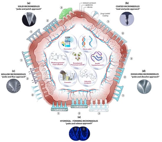
Microneedles are designed to be able to create an easier passage to the rich blood supply in the lower dermal layers, allowing an easy, pain-free delivery of a wide range of medicines across the skin [
44]. The advantages of microneedles include painless administration, faster healing, ease of administration, and more control over the rate of drug delivery. Microneedle patches are categorized into five types, as shown in
Figure 1, which include the following: solid microneedles, coated microneedles, dissolvable microneedles, hollow microneedles, and hydrogel-based microneedles. Each microneedle type has particular fabrication procedures and application areas. The first report of the term “microneedle” dates back to 1921 (Chambers, 1921), as a means of the micro-dissection of echinoderm eggs [
45].
Figure 1. A schematic diagram of microneedle-based drug delivery approaches with a cross-section of the upper layer of the skin. The approaches are (
a) solid microneedles, (
b) coated microneedles, (
c) hollow microneedles, (
d) dissolving microneedles, and (
e) hydrogel-forming microneedles. The step-by-step process of each delivery approach is numbered from 1 to 3 [
46].
The microneedle concept started with using large needles, until it evolved over the years to the current micro-sized needles. In 1905, a German dermatologist used a motor-powered dental brush to treat skin ailments [
47]. In the 1970s, Gerstel et al. introduced the microneedle concept, however, this concept was not demonstrated experimentally until the 1990s [
48]. In 1998, Henry et al. were the first to propose a microneedle to be used for transdermal drug delivery [
49]. The microneedle array in their study was fabricated using silicon as the material, with etching and photolithography as the manufacturing techniques. Initially, the purpose of the microneedle was to increase the skin’s permeability by using a solid microneedle. Another purpose was to fabricate hollow microneedles with advanced functionality compared to the ordinary hypodermic needles [
50]. Eventually, this concept was extended to different applications, such as drug delivery, vaccine delivery, therapeutics, diagnostics, and cosmetic applications. A variety of materials, such as silicon, stainless steel, sugar, and polymers, have been used in order to fabricate solid microneedles, coated microneedles, hollow microneedles, or dissolvable microneedles [
51]. Silicon was the first proposed material for fabricating a solid microneedle [
49]; however, many other materials were studied in order to manufacture microneedles, such as stainless steel [
52], ceramic [
53], glass [
54], and polymer [
55]. Various types of manufacturing methods and techniques have been utilized over the years to fabricate specific and customized microneedle arrays. These manufacturing methods include, but are not limited to, lithography [
56,
57], micro milling, mold-based techniques, injection molding [
58], laser ablation [
59,
60], an elasto-capillarity-driven self-assembly mechanism, and additive manufacturing [
27,
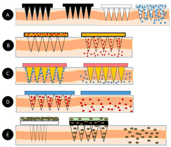
61]. Many of these conventional fabrication methods have some limitations, such as cost-efficiency, requiring manual steps, requiring sophisticated equipment, and being labor-intensive. Hence, accessible and cost-efficient technologies, such as additive manufacturing, are needed in order to produce microneedles. A summary of the fabrication techniques for the different types of microneedles is illustrated in
Table 2.
Table 2. Source: “
Microneedles: A smart approach and increasing potential for transdermal drug delivery system” [
62].
This entry is adapted from the peer-reviewed paper 10.3390/pharmaceutics14122693