Immunotherapy in Gastric Cancer Management: History
Please note this is an old version of this entry, which may differ significantly from the current revision.
Contributor: , , , ,

Gastric cancer (GC) is the fifth most commonly diagnosed cancer worldwide and the fourth most common cause of cancer death. As GC is often diagnosed at an advanced stage, mortality remains very high. GC shows both genetic and environmental risk factors, with Helicobacter pylori (H. pylori) infection being the most well-described risk factor leading to GC. Germline genetic alterations are also involved in 1–3% of cases. Due to the poor survival rates of GC, immunotherapy has been widely explored as a potential treatment. Both active and passive immunotherapies have been examined. Active immunotherapies involve using a patient’s own immune system to treat the disease whereas passive immunotherapies rely on exogenous agents administered to patients such as antibodies in order to treat the tumour. The great efficacy observed in melanoma has propelled immunotherapies to be explored in a variety of other tumours, particularly breast, prostate, and lung cancer].

  • gastric cancer
  • immunotherapy
  • PD-L1
  • H. pylori
  • EBV
  • immune microenvironment

1. Molecular Classification

The earliest classification system of GC is the Lauren classification which separates gastric cancer into intestinal and diffuse subtypes [6]. Intestinal tumours are more adhesive than diffuse, forming tubular or glandular formations. They are more common in older patients and associated with a more favourable prognosis. Intestinal adenocarcinoma is more often associated with H. pylori infection. Diffuse GC cells are less adhesive and typically occur in younger patients [7]. Diffuse type GC is associated with a poorer prognosis than intestinal type [8].
In 2020, the WHO classified GC into four main histological subtypes: tubular, papillary, mucinous, and poorly cohesive (including signet ring cell carcinoma) [9]. Tubular adenocarcinoma is the most common subtype of early GC and is characterised by irregular tubules, frequently found with intraluminal mucous [10]. Papillary cancers more often affect older patients and present as epithelial projections on a fibrovascular core. Mucinous adenocarcinomas are composed of extracellular mucinous pools, comprising greater than 50% of the tumour bulk. Poorly cohesive carcinomas are formed of signet ring cells and other ring cells, often resulting in lymph node invasion [9]. In addition to these subtypes, other less common variants exist which do not fit into the above categories.
A study in 2014 by the Cancer Genome Atlas Research Network classified gastric adenocarcinoma (GAC) into four new categories: Epstein–Barr virus (EBV)-positive, Microsatellite instability (MSI), Genomically stable (GS), and chromosomal instability (CIN) [11] (Figure 1). These novel categories provide a potential method for the stratification of patients to enable the use of targeted therapies.
Figure 1. Main features of GC subtypes. Schematic representation of the molecular characteristics associated with GC molecular subtypes.

2. The Role of Tumour-Infiltrating Immune Cells in the Gastric Cancer Microenvironment

Tumour-infiltrating immune cells are important components of the tumour microenvironment with many described roles. Numerous studies have reported interactions between solid tumours and their immune microenvironment promoting invasion and metastasis. In GC, the main immune cell types in the microenvironment are tumour-associated macrophages (TAMs) and tumour-infiltrating lymphocytes (TILs).
TAMs are an important immune cell subtype in GC. Derived from lymphatic and blood monocytes, TAMs infiltrate the tumour and secrete various chemokines to regulate cell growth, invasion, and metastasis [16]. TAMs are typically described as having an M1 or M2 polarisation, with classical M1 TAMs being anti-tumourigenic and pro-inflammatory, producing IL-1β, IL-1α, IL-12, and TNF-α, whereas alternative M2 TAMs display anti-inflammatory activity and immunoregulation, promoting tumourigenic functions through IL-4, IL-6, and IL-10 [17].
Various properties of TAMs have been proposed as prognostic biomarkers in GC [17]. These include TAM density [18] and M2 infiltration [19] which are associated with poor prognosis and M1 infiltration which correlates with better prognosis [19]. Various secreted factors associated with TAMs are also potential biomarkers, such as Tim-3 which correlates with increased tumour invasion and lymph node metastasis [20] and CCL5/RANTES and NFKB1 where SNPs are associated with altered clinical outcome [21].
Importantly, TAMs induce immune tolerance in GC where programmed cell death protein 1 (PD-1) and cytotoxic T-lymphocyte antigen 4 (CTLA-4) promote an immunosuppressive microenvironment by blocking cytotoxic T cell anti-tumour activity. Macrophages can induce PD-L1 expression in GC cells through TNF-α and IL-6 signalling [22]. These pro-inflammatory cytokines can regulate STAT3 and NF-kB signalling in tumour cells, inducing PD-L1 expression and contributing to immunosuppression in tumours. TAMs are also known to play important roles in angiogenesis where they accumulate in hypoxic regions in tumours [23,24]. TAM infiltration correlates highly with PD-L1 expression, impacting metastasis and survival rate [25].
Natural killer (NK) cells are important players in GC as they can often attack cancer cells after the tumours have escaped detection by CD8+ T cells [26]. NK cells work by releasing granules containing perforin and granzymes to cause cancer cell lysis. They also express TNF-related apoptosis-inducting ligand family (TRAIL) and Fas ligand (FASL) inducing apoptosis in cancer cells [27]. Cytokine production (IFN-γ and TNF-α) also increases the cytotoxic anti-tumour response. In GC, NK cells are also able to target CD133+ cancer stem cells [28]. Despite this, as GC progresses, NK activity decreases through increased apoptosis [29], upregulation of inhibitory receptors and downregulation of activating receptors [30], a decrease in cytotoxic granule production and cytokine release [31], and a reduction in infiltration [32].
Dendritic cells (DCs) are another important cell type in GC. They present cancer antigens to immune cells to regulate the immune response against tumour cells. DC infiltration has been associated with an increased 5-year survival rate in GC [33]; however, CD83+ DCs are associated with a poorer prognosis in GC, both in the primary tumour and in lymph nodes [34]. Clinical studies have explored the use of DCs pulsed with tumour-associated antigens, followed by autologous transplant into patients, for instance HER2 peptide-activated DCs could induce a T cell response against the antigen [35]. Additional DC therapies in GC are reviewed in Tewari et al. [36].
Another key population of immune cells in GC is TILs. These cells have prognostic significance in gastric cancer. TILs are comprised of T cells, B cells and NK cells [37]. The anti-tumour immune response occurs when tumour-specific antigens are processed by DCs and presented to T cells. Stromal TILs have previously been found as predictors of poorer disease-free and recurrence-free survival in GC [38] whereas intratumoural TILs are associated with increased overall survival and cancer-specific survival in EBV-associated GC [39,40].

3. Role of Chronic Infection with Helicobacter pylori and the Association with an Immunosuppressive Microenvironment

Helicobacter pylori (H. pylori) is a facultative, spiral-shaped, Gram-negative bacterium that selectively colonises the gastrointestinal mucosa [45,46,47]. Four decades after its discovery in 1982, H. pylori represents a well-established risk factor for GC, and it is recognised as a type I carcinogen by the International Agency for Research on Cancer (IARC), with 90% of non-cardia gastric cases attributable to this bacterium [47,48,49,50].

3.1. Contribution of H. pylori Virulence Factors to Chronic Inflammation

During H. pylori infection, stimulation of several inflammatory signals is key to the establishment of an inflammatory environment in the gastric epithelium. This marks an important step towards initiation of a more complex inflammatory and immune response which can eventually culminate in the development of peptic ulceration and gastric malignancies.
A range of genes expressed by H. pylori are involved in the infection and remodelling process of the microenvironment [45,54,55]. These include ureases that confer this pathogen the ability to colonise and neutralise the highly acidic environment found within the stomach by converting urea into ammonia, therefore establishing the optimal pH conditions for its growth [54]. The subsequent increase in pH contributes to alter the viscosity of gastric mucus facilitating H. pylori diffusion through the mucosal barrier and allowing the pathogen to gain access to the underlying epithelial cells [56]. This event is crucial for the gastric epithelium colonisation process, which is the basis of the inflammatory reaction induced by H. pylori. Moreover, urease could contribute to gastric carcinogenesis by producing reactive oxygen species and activating the lipoxygenase pathway, resulting in differentiation of endothelial cells [57,58].
H. pylori flagella favour the colonisation of the gastrointestinal mucosa and contribute to bacterial motility [59]. FlaA is one of the main structural proteins of the H. pylori flagellum which can evade the host immune response, as it can escape recognition by the Toll-like receptor 5 (TLR5), a member of the Toll-like receptor family that normally recognises most bacterial flagellins [59,60]. As flagellins are critical for persistent H. pylori colonisation, and given that H. pylori colonisation is the basis of inflammation, flagella can be considered responsible for both inflammation and immune evasion [46,59].
Other players can also act in a cascade of events inducing damage to the gastric mucosa and host inflammatory response. Amongst these, there is the cytotoxin-associated gene CagA which is part of the cag pathogenicity island (cag PAI)—locus of approximately 40 kb containing 31 genes, the majority of which encode for the cag secretion system (T4SS) [46,60]. Importantly, cag Pal+ H. pylori strains increase the risk of gastritis, atrophic gastritis, and GC than strains lacking the cag island [61]. CagA modulates the host cell signalling both in a phosphorylation-dependent and independent manner. Phosphorylation of CagA has been reported to induce sustained activation of the ERK1/2 MAP kinase and NF-kB signalling pathways, and disruption of epithelial cell tight junctions with damage to the gastric mucosa [57]. Activation of proinflammatory responses, mostly through IL-8 pro-inflammatory cytokine production instead, appears to be independent of CagA phosphorylation [46,51,60].

4.2. Host Immune Response to H. pylori Infection

During H. pylori infection, both innate and acquired immune responses are intensely stimulated. Despite the strong immune responses, H. pylori has the remarkable ability to persist for a very long time in the gastric mucosa, actively modulating and evading the host response to establish an immunosuppressive environment that maintains chronic infection [52].
Most of the factors mentioned above are thought to intensify local inflammation with consequent infiltration of inflammatory cells to the gastric mucosa. Innate host defence mechanisms are triggered as a first line of defence and are crucial to increase risk of gastric carcinogenesis and severity of the disease [60]. The innate immune response includes the nucleotide-binding oligomerisation domain protein 1 (Nod1) [64]. This is a pattern recognition receptor (PRR) responsible for the recognition of H. pylori peptidoglycan components secreted by the cag secretion system, and activation of NF-kB-dependent proinflammatory responses. The most studied PRRs are the Toll-like receptors (TLRs), expressed on epithelial and innate immune cells, which interact with diverse H. pylori antigens (including lipoteichoic acid, lipoproteins, lipopolysaccharide, and flagellin) and initiate the adaptive immune responses [65].
Amongst the main players of the innate response to H. pylori, there are macrophages which, along with monocytes and DCs, are responsible for the recruitment of lymphocytes and the stimulation of T-helper (Th) cell-specific responses by releasing factors such as IL-12. In particular, stimulation of Th1 cells is predominant, and it is central in the T-cell response to H. pylori, as it produces cytokines such as IFN-γ and leads to pro-inflammatory cytokine release, for example TNFα, and interleukins [67] (Figure 2).
Figure 2. H. pylori and EBV mechanisms of infection at a glance. H. pylori infection causes a local inflammation state with consequent infiltration of inflammatory cells, and increased risk of gastric carcinogenesis (left panel), EBV infection process, associated with development of EBV-associated GC (right panel).
Upon intense stimulation, CD4+ helper T cells and CD8+ killer T cells are recruited to the gastric mucosa, with activation of CD4+ T cells [65]. In this scenario, other CD4+ T cell subsets, such as Th17 and Tregs also play a role: while Th1 and Th17 cells enhance gastric inflammation, Tregs seem to exhibit a protective role against inflammation and contribute to bacterial persistence [52]. In fact, regulatory T cells are responsible for the secretion of suppressive cytokines (IL-10 and TGFβ), through which, they negatively modulate immune and inflammatory responses, facilitating persistent bacterial colonisation [60].

4.3. H. pylori Infection Promotes an Immunosuppressive Tumour Microenvironment

As discussed above, the establishment of H. pylori-mediated chronic infection has been shown to have profound implications on the host immune system and its functions. In fact, by exerting immunomodulatory effects on a variety of immune cells (through negative modulation of Th1 and Th17 cells, macrophages and dendritic cells, and promotion of Treg cells activity) H. pylori is thought to be responsible for the creation of an immunosuppressive environment [70,71].
Under normal conditions, the immune system retains the ability to generate tumour-specific immune responses to thwart cancer formation and progression, a concept known as anti-tumour immunity [72]. Such anti-tumour immunity can be boosted by cancer immunotherapies, for instance by using immune checkpoint inhibitors (ICIs) that block the natural function of immune checkpoints and prevent attenuation of immune cell activation [71]. These include inhibitors of the cytotoxic T-lymphocyte antigen 4 (CTLA-4), or the programmed cell death protein 1 (PD-1)/programmed cell death ligand 1 (PD-L1) [73].

4. Molecular Mechanisms Underlying the Pathology of Microsatellite Instable Gastric Cancer

Genomic instability is one of the major hallmarks of cancer development [84]. It is believed to be one of the initial steps of gastric carcinogenesis and can be found in all different histological subtypes of GC [85]. Microsatellites (MS) are repetitive and specific DNA sequences characterized by a high mutation rate [86,87]. Microsatellite instability (MSI) is a hyper-mutable phenotype caused by a non-functional DNA mismatch repair (MMR) machinery at MS sites. During DNA replication, the insertion or deletions of nucleotides in MS regions because of germline mutations or epigenetic silencing cause malfunction of MMR system [88,89].
The MMR system includes several proteins: human MutL homolog 1 (hMLH1), human MutL homolog 3 (hMLH3), human MutS homolog 2 (hMSH2), human MutS homolog 3 (hMSH3), human MutS homolog 6 (hMSH6), human post meiotic segregation increased 1 (hPMS1), and human post meiotic segregation increased 2 (hPMS2) [87]. During DNA replication, hMSH2/hMSH6 and hMSH2/hMSH3 complexes are responsible for detecting and binding small DNA mismatch errors while the excision and re-synthesis of the corrected DNA bases in the mismatch site is detected by the heterodimeric complex hMLH1/hPMS2. Defects in one or more MMR machinery elements determine the unsuccessful repair of the DNA [87]. Different processes including promoter methylation, chromosomic rearrangements that lead to loss of heterozygosity or mutations in the coding region are responsible for the inactivation of MMR proteins [90,91]. The main cause of MMR deficiency in both sporadic and familial MSI GCs is represented by the hypermethylation of hMLH1 promoter [92,93]. Conversely, mutations of hMLH1 and hMSH2 are relatively rare (15% and 12%, respectively) [94]. Some reports showed that MSI represents an early molecular event during gastric carcinogenesis [95,96]. However, Ling et al. reported that the promoter methylation of hMLH1 represents a later event during the natural process of tumour growth and the time-dependent acquisition of MSI may be due to the hMLH1 silencing [97]. Both sporadic GC and Lynch syndrome (LS) show MSI [91,98]. LS is mainly caused by autosomal dominant mutations affecting hMLH1 and hMSH2 and less frequently hPMS2 and hMSH6 [91]. Moreover, the epigenetic silencing of hMSH2 by a constitutional 3′ end deletion of EPCAM can also cause LS [99,100]. Patients affected by LS show an increased predisposition to develop GC at a younger age (11.3-fold in the 30s and 5.5-fold in the 40s) [90,91].

5. EBV-Related Gastric Cancer

Epstein–Barr virus (EBV) emerged as an important virulence factor for nasopharyngeal carcinoma. EBV infection also causes the development of T-cell lymphoma and EBV-associated GC (EBVaGC) [106,107]. Immunotherapy drug treatments were successful against EBV-positive and MSI GCs [108]. Once EBV infects the human body, this does not immediately produce GC and EBV-positive GC is not characterized by any evident clinical manifestations [109,
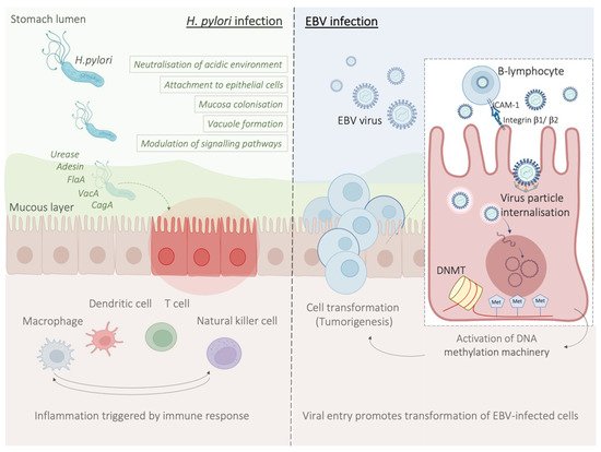
110]. Two theories have been reported about the mechanism of EBV infection. According to the first theory, EBV produces the infection of B-lymphocytes and oral epithelial cells [111]. In particular, since EBV is present in the saliva, this causes the infection of the epithelial cells [111]. The second theory reports EBV reactivation in B-lymphocytes in the stomach and its subsequent release to cause the infection of epithelial cells [111]. EBV infection of lymphocytes results in the interaction of these cells with epithelial cells [112]. This interaction is mediated by integrin β-1/β-2 and the translocation of intracellular adhesion molecule-1 to the cell surface (ICAM-1) produce cell-to cell contact [112]. The virus particles are then internalised by recipient cells through clathrin-mediated endocytosis [112]. EBV-particles inside the host cell nucleus are characterized by a naked, linear DNA genomes and a specific protein capsid protect them [113]. The exposed DNA linear genome is then circularised into a functional chromosome [113]. After circularisation, the chromatinised viral DNA protects it from DNA damage and provides accurate regulation of gene expression [113] (Figure 2). EBV genome is characterised by widely methylated CpG motifs enabling it to establish latent infection [114]. The two types of infection caused by EBV are the lytic and the latent form [114]. The latent infection is the one preferred by EBV and during the long incubation period, EBV causes the methylation of the host DNA and the expansion of GC [113,114]. Latent EBV proteins such as EBERs, BARF-0, EBNA-1, and LMP2A downregulate the miR-200 family causing a reduction in E-cadherin expression [115]. This mechanism is mediated by the upregulation of the E-cadherin repressors, ZEB1 and ZEB2 [115]. Tumour progression involves the loss of cell-to-cell adhesion and this event is also an important step in the carcinogenesis of EBVaGC [115]. EBV is the first human virus expressing many microRNAs [116]. The EBV miRNA BART11 has been shown to downregulate forkhead box protein P1 (FOXP1) transcription factor [117]. FOXP1 downregulation activates the epithelial-mesenchymal transition involving the gastric tumour cells or affecting the tumour microenvironment [117]. This, in turn, accelerates cancer invasion and metastasis, thereby affecting the survival and prognosis of patients [117]. 

6. Potential Role and Relevance of POLE/D Mutations in Gastric Cancer

The immune checkpoint inhibitor (ICI) biomarkers approved by the US Food and Drug Administration (FDA) in the treatment of certain cancers are the use of PD-L1 expression, microsatellite instability (MSI)-H/deficient mismatch repair (dMMR) and tumoural mutation burden (TMB) [123,124,125]. Since some responses seen with ICIs do not fully correlate with any of the biomarkers above, other potential predictive biomarkers of response to ICI can exist in GC [126,127]. POLE and POLD1 genes encode for the DNA polymerases ε and δ [127,128]. TMB and clinical benefits of immunotherapy have been associated with mutations in POLE and POLD1 in different cancer types [129,130]. 

7. HER2 Overexpression in Gastric Cancer

Human epidermal growth factor receptor 2 (HER2) is a receptor tyrosine kinase proto-oncogene that is increasingly understood to be overexpressed in GC. Different studies have found HER2 overexpression to occur in between 4.4% and 53.4% of GCs, with an average of 17.9% [132]. Prognostically, HER2 appears to be correlated with poorer survival and increased recurrence in GC [133,134]. HER2 targeting through the monoclonal antibody trastuzumab together with chemotherapy has in fact been shown to increase survival in HER2+ GC patients; however, the effect is small with a clinical trial only finding survival increasing from 11.1 months to 13.8 months [135].
During treatment, the tumour microenvironment contributes to tumourigenesis and proliferation but is also associated with immune cell infiltration and treatment efficacy [136]. Suh et al. reported that the HER2 pathway modulates the tumour microenvironment and this is correlated with tumour pathological characteristics and patient survival [137].

8. Immunotherapy in the Clinical Management of Gastric Cancer

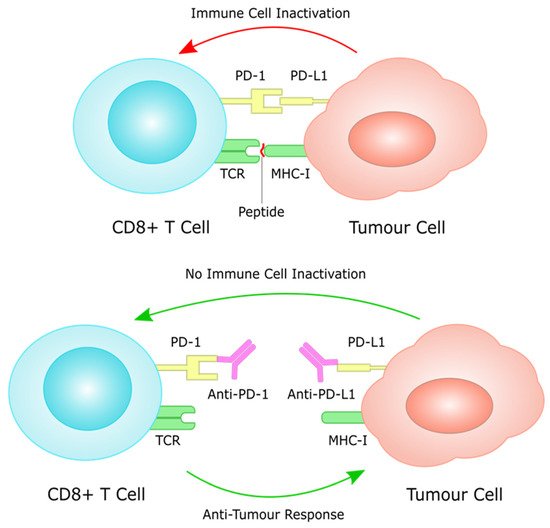
Over the last few years, immunotherapy has been actively incorporated as part of first and later lines of systemic treatment for advanced gastric cancer. PD-1 inhibitors have been shown to significantly improve efficacy in several large phase III trials when added to platinum-based chemotherapy, which has for many years been the standard of care in the first line setting for metastatic GC (Figure 3). 
Figure 3. PD-L1 on tumour cells works through the PD-1 receptor on T cells to induce immune cell inactivation. Treatment with immune checkpoint inhibitors inhibits this interaction, often through monoclonal antibodies against PD-1 and PD-L1, restoring immune cell function against tumour cells.

8.1. PD-L1 as a Prognostic Biomarker

There have been several studies investigating a potential prognostic role for PD-L1 expression in GC, with diverse and controversial results. In a retrospective study by Morihiro, et al., including 283 patients with GC, PD-L1 expression was significantly correlated with a poor prognosis (p  =  0.0025). Multivariate analysis revealed that PD-L1 expression was found to be an independent poor prognostic factor, along with diffuse histological type and lymph node involvement. [140]. Furthermore, Chang et al., using tissue microarrays in 464 GC samples, showed that PD-L1 and PD-1 expression was significantly correlated with several adverse prognostic pathologic features, such as T stage, lymphatic invasion and diffuse Lauren histologic type. In the same study, subgroup analyses in which patients were divided into two groups according to CD8+ TILs expression levels (high and low), it was shown that high PD-L1 expression was a negative prognostic factor only in the high CD8+ TILs subgroup [141].

8.2. Immunotherapy in Chemo-Resistant/Refractory Setting; PD-L1 Expression as a Predictive Biomarker

One of the first phase III trials that investigated the use of PD-1 monotherapy in the chemo-refractory setting in metastatic GC was the ATTRACTION-2 trial, which randomised previously treated patients with metastatic gastric or gastro–oesophageal junction (GOJ) adenocarcinoma with more than two lines of treatment, to nivolumab or placebo. Median overall survival was 5.26 months (95% CI 4.60–6.37) in the nivolumab group and 4.14 months (3.42–4.86) in the placebo group (hazard ratio 0.63, 95% CI 0.51–0.78; p < 0.0001). [147]. A more recent update of the trial with 2-year follow-up data confirmed the long-term benefit of nivolumab over placebo, with a higher OS rate noted in the nivolumab vs. placebo group at 2 years (10.6% vs. 3.2%). The OS benefit was observed regardless of tumour PD-L1 expression, although this must be interpreted with caution because only 192 of 493 patients (39%) had PD-L1 results available for analysis [148]. Despite the fact that the main population of the study was Asian patients, the positive benefit of nivolumab in overall survival established this option as a standard third line and beyond option regardless of PD-L1 expression levels in many countries.

8.3. Immunotherapy in Previously Untreated Patients; PD-L1 Expression as a Predictive Biomarker

The next logical step was to investigate the incorporation of immunotherapy in the first line setting, in previously untreated patients, with advanced gastric cancer. One of the first phase III trials that attempted to answer this specific question was the Keynote-062 trial. In this trial, patients were randomized 1:1:1 to pembrolizumab, pembrolizumab plus chemotherapy (cisplatin plus fluorouracil or capecitabine) or chemotherapy plus placebo. Primary end points were OS and PFS in patients with PD-L1 CPS ≥ 1 or ≥10. This trial demonstrated non-inferiority for pembrolizumab to chemotherapy for OS in patients with CPS ≥ 1 (median, 10.6 vs. 11.1 months; hazard ratio [HR], 0.91; 99.2% CI, 0.69–1.18), however pembrolizumab monotherapy was not found to be superior to chemotherapy in patients with CPS ≥ 1. In patients with CPS ≥ 10, pembrolizumab numerically prolonged OS vs. chemotherapy (median, 17.4 vs. 10.8 months; HR, 0.69; 95% CI, 0.49–0.97); however, this difference was not tested for statistical significance. Pembrolizumab combination with chemotherapy was not found to be superior to chemotherapy for OS in patients with CPS ≥ 1 (12.5 vs. 11.1 months; HR, 0.85; 95% CI, 0.70–1.03; p = 0.05) or CPS ≥ 10 (12.3 vs. 10.8 months; HR, 0.85; 95% CI, 0.62–1.17; p = 0.16) or for PFS in patients with CPS ≥ 1 (6.9 vs. 6.4 months; HR, 0.84; 95% CI, 0.70–1.02; p = 0.04) [152]. In a more recent update of the trial in ASCO GI 2022, with a median follow-up of 54.3 months (range, 46.8–66.1), efficacy was consistent with the initial final analysis data; 24-month OS rates (pembrolizumab vs. chemotherapy) were 26.6% versus 18.8% in the CPS ≥ 1 population and 39.1% versus 21.1% in the CPS ≥ 10 population. Twenty-four-month OS rates (pembrolizumab + chemotherapy vs. chemotherapy) were 24.5% versus 18.8% in the CPS ≥ 1 population and 28.3% versus 21.1% in the CPS ≥ 10 population [153]. Although Keynote-062 was considered a negative trial, it provided further valuable supporting evidence with regards to the potential predictive role of PD-L1 CPS in terms of the selection of patients that would most benefit from immunotherapy, namely patients with PD-L1 CPS ≥ 1, and ideally PD-L1 CPS ≥ 10.

8.4. Immunotherapy in HER2-Positive Gastric Cancer

PD-1 inhibition is thought to work synergistically with HER2 inhibition increasing ADCC (antibody-dependent cellular cytotoxicity), and this concept has been investigated in the Keynote-811 trial, where treatment naive patients with HER2-positive GC or GOJ adenocarcinoma were randomized to receive pembrolizumab or placebo plus trastuzumab and investigator’s choice of fluorouracil/cisplatin or capecitabine/oxaliplatin. Overall, 84% of patients had a PD-L1 CPS ≥ 1. Confirmed ORR (95% CI) was 74.4% (66.2–81.6) for pembrolizumab + chemotherapy vs. 51.9% (43.0–60.7) for placebo + chemotherapy, p = 0.00006); CR (complete response) rate was 11.3% vs. 3.1% and DCR (disease control rate) (95% CI) was 96.2% (91.4–98.8) vs. 89.3 (82.7–94.0). [160]. The very encouraging preliminary results from the Keynote-811 provide a proof of concept for the synergistic effect of PD-1 and HER2 inhibition in HER2-positive gastric cancer, which stands a very good chance to consist of a new standard of care in the first line setting, providing long-term analysis also reveals a survival benefit for pembrolizumab, trastuzumab and chemotherapy in this setting.

8.5. Immunotherapy Efficacy in Rare Subgroups: Role of MSI, EBV and TMB (Tumour Mutational Burden) Status as Predictive Biomarkers

The role of immunotherapy in the management of mismatch-repair deficient (MMR-D) and EBV-associated gastric tumours has long been established through several studies. In stage IV advanced gastric cancer, EBV-positive and MMR-D tumours are identified both in approximately 6% of cases [161]. In the Keynote-158 phase II trial investigating the efficacy of pembrolizumab in MMR-D tumours, 11 of 24 patients with advanced gastric cancer demonstrated an objective response as their best response (ORR, 45.8%), which was associated with a median PFS of 11.0 months and a median OS which was not reached [162]. In a post hoc analysis of the outcomes of MSI-H GC patients included in the phase 2 single-arm trial KEYNOTE-059 (≥3 lines of treatment) and the phase 3 KEYNOTE-061 (second line) and KEYNOTE-062 (first line treatment) trials, the median overall survival was not reached (NR) for pembrolizumab in all three of these trials. The median PFS) for pembrolizumab was NR (95% CI, 1.1 months to NR) in KEYNOTE-059 and 17.8 months (95% CI, 2.7 months to NR) in KEYNOTE-061 (vs. 3.5 months for chemotherapy). In KEYNOTE-062, the median PFS was 11.2 months (95% CI, 1.5 months to NR) for pembrolizumab, NR (95% CI, 3.6 months to NR) for pembrolizumab plus chemotherapy, and only 6.6 months (95% CI, 4.4–8.3 months) for chemotherapy alone. The ORR for pembrolizumab was 57.1% in KEYNOTE-059 and 46.7% (vs 16.7% for chemotherapy alone) in KEYNOTE-061. In KEYNOTE-062, the ORR was 57.1% for pembrolizumab, 64.7% for pembrolizumab plus chemotherapy, and 36.8% for chemotherapy alone [163]. Very few clinical trial data exist in regards to the efficacy of immunotherapy in EBV-positive GC; in a phase II study of pembrolizumab in metastatic GC patients with MSI-H and Epstein–Barr virus-positive tumours, impressive responses to pembrolizumab were noted (overall response rate (ORR) 85.7% in MSI-H metastatic GC and ORR 100% in Epstein–Barr virus-positive tumours) [164]. The results above unequivocally establish MSI-H status and EBV positivity as by far the most robust biomarkers of response to immunotherapy in GC, however it has to be noted that despite the high and durable responses, there is a significant fraction of patients which test positive for the above biomarkers that do not respond to immunotherapy, and therefore there is a great unmet need for further molecular subclassification of these gastric cancer subtypes in order to elucidate mechanisms of primary resistance to immunotherapy.

9. Conclusions

In conclusion, immunotherapy has been incorporated in the clinical management of advanced GC based on recent positive phase III clinical trial results. Most of the benefit is seen in PD-L1 CPS positive, MSI-H/MMR-D, EBV-positive, and possibly TMB-high tumours. Although these biomarkers have been associated with durable responses to immunotherapy, there is still a considerable proportion of patients who fail to respond, while ultimately cancer cells develop immune-evasion strategies and develop resistance. Further clinical combined with translational research is eagerly needed to optimise immunotherapy efficacy and overcome emerging PD-1/PD-L1 resistance in order to improve GC patients’ outcomes.

This entry is adapted from the peer-reviewed paper 10.3390/cancers14184378

This entry is offline, you can click here to edit this entry!
Video Production Service