Human Pluripotent Stem Cell-Based Models for Hirschsprung Disease: History
Please note this is an old version of this entry, which may differ significantly from the current revision.

Hirschsprung disease (HSCR) is a complex congenital disorder caused by defects in the development of the enteric nervous system (ENS). It is attributed to failures of the enteric neural crest stem cells (ENCCs) to proliferate, differentiate and/or migrate, leading to the absence of enteric neurons in the distal colon, resulting in colonic motility dysfunction. Due to the oligogenic nature of the disease, some HSCR conditions could not be phenocopied in animal models. Building the patient-based disease model using human induced pluripotent stem cells (hPSC) has opened up a new opportunity to untangle the unknowns of the disease. The expanding armamentarium of hPSC-based therapies provides needed new tools for developing cell-replacement therapy for HSCR. 

  • Hirschsprung disease
  • induced pluripotent stem cells
  • disease modeling

1. Introduction

Hirschsprung disease (HSCR), which is also known as congenital aganglionic megacolon, is a neurocristopathy caused by the incomplete colonization of the colon by enteric neural crest cells (ENCCs). HSCR is a multigenic congenital disorder with over 80% heritability [1], and its incidence is approximately 1 per 5000 births worldwide, with the highest incidence rate (1.4 per 5000 births) in the Asian population. The incidence rate in males is 3–4 times higher than in females [2][3]. Missing enteric nervous system (ENS) in the colon causes uncoordinated muscular peristalsis and functional bowel obstruction. As a consequence, HSCR patients experience delayed passage of meconium in the first two days after birth and suffer from vomiting, feeding difficulties, abdominal distension, and constipation [4].
HSCR patients are classified into three subtypes based on the extent of aganglionosis (i.e., bowel segment without enteric neurons), namely short-segment HSCR (S-HSCR), long-segment HSCR (L-HSCR) and total colonic aganglionosis (TCA). S-HSCR accounts for around 80% of the cases, while 15% and 5% of the HSCR cases are L-HSCR and TCA, respectively. Different genetic architectures are found in these HSCR subtypes [5]. L-HSCR and TCA mostly are autosomal dominant, while the S-HSCR follows non-Mendelian oligogenic inheritance where patients may carry different constellations of coding and noncoding variants leading to the malformation of the ENS. The disease risk and expressivity are influenced by the underlying genetic background of the individuals [6]. The oligogenic nature of HSCR makes it challenging to build a definitive diagnostic framework for prenatal genetic screening. Till now, surgical removal of the aganglionic segment of the colon is still the only available treatment for HSCR. However, many patients still suffer from constipation and enterocolitis after surgery, particularly short bowel syndrome frequently happens in TCA patients [4]. Therefore, tremendous efforts have been made to develop a new and efficacious treatment for HSCR patients.
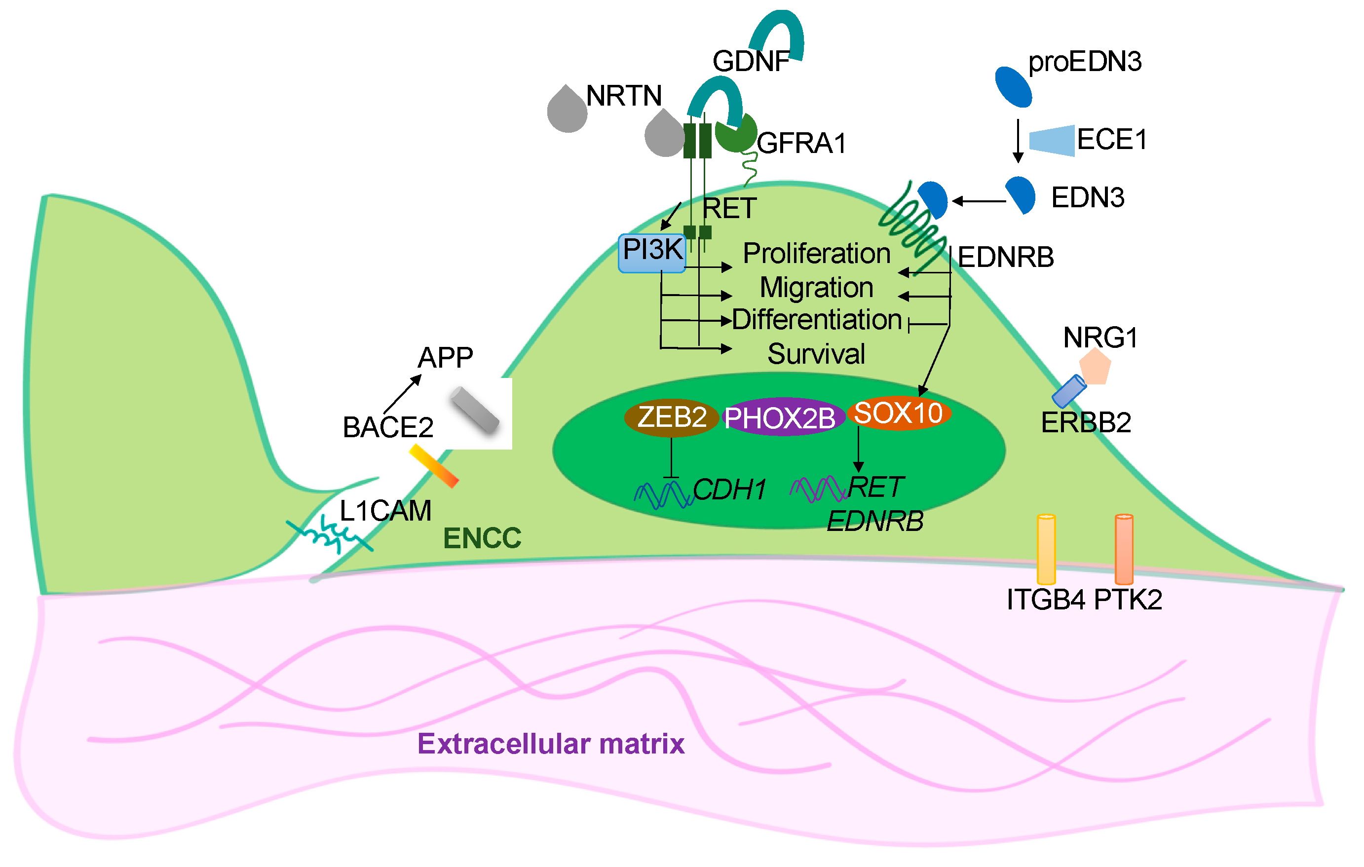
Genetic variants or mutations in RET, ZEB2, EDNRB, SOX10, L1CAM, PHOX2B, GDNF, NRTN, EDN3, ECE1, GFRA1, NRG1 were identified in HSCR patients, imparting up to 50% of the cases [3][7][8][9]. These disease-associated genes encode signaling molecules, cell surface receptors, trophic factors, or transcription factors which are essential for the proper development of the ENS (Figure 1). The individual effects of these disease-associated genes have been studied using the genetic approach through the generation of mutant animal models of the corresponding genes [10]. Currently, the mouse, rat, chick, Xenopus and zebrafish are still the most popular animal models used for studying HSCR and other congenital disorders. Compelling evidence suggests that genetic background influences the disease expressivity of HSCR, especially the epistasis among various coding and noncoding variants, a patient-based model which carries the matched genetic background represents an alternative tool for bridging the gaps between human genetic screens and functional studies performed with animal models. In particular, current advances in stem cell research enable the use of human induced pluripotent stem cells (hPSCs) derived from the patient’s somatic cells to generate two-dimensional (2-D) ENS models and even three-dimensional (3-D) “mini-gut” to recapitulate the human HSCR conditions. In this research, the researchers will summarize the recent findings from the studies of hPSC-based models of HSCR and highlight their potential applications in drug discovery and cell replacement therapy.
Figure 1. Overview of the functions of the disease genes. Simplified illustration showing the functional roles of the HSCR-associated genes in ENS development. These disease-associated genes encode for the cell surface receptors (RET, EDNRB and ERBB2) in the ENCCs and their ligands (GDNF, NRTN, EDN3, ECE1 and NRG1); the transcriptional factors (SOX10, ZEB2 and PHOX2B); or the cell-cell (L1CAM) and cell-extracellular matrix (IGGB4, PTK2) interacting molecules. BACE2 is a protease that acts on APP to prevent the production of toxic Ab peptides from protecting the survival of ENCCs.

2. hPSC-Based Disease Models of HSCR

2.1. In Vitro 2-D ENS Model

By mimicking the endogenous signaling events that happened during the neural crest cell (NCC) formation, human pluripotent stem cells (hPSCs) can form NCCs with sequential activation and inhibition of various developmental cues. To generate ENCCs from hPSC, hPSCs are first directed to the ectoderm lineage using dual SMAD inhibition where both BMP and TGF-β signaling pathways are inhibited by the addition of BMP antagonist (LDN193189) and TGF-β antagonist (SB431542). Subsequent activation of WNT signaling using WNT agonist CHIR99021 further guides the ectoderm cells to NCC lineage [11]. The NCCs are then caudalized to vagal lineage by the treatment with retinoic acid, resembling the in vivo somitic environment. The vagal NCC-like cells can then be enriched by cell sorting using ENCC-specific markers, such as p75NTR, HNK1, and RET. Various types of enteric neurons can be derived from these ENCC-like cells by promoting their neuronal lineage differentiation by adding different neurotrophic factors [11][12][13].
Using this stepwise induction protocol, hPSCs derived from healthy individuals or HSCR patients can generate the disease-relevant cells (i.e., ENCCs and enteric neurons) in a progressive differentiation manner resembling the in vivo developmental processes of the ENS. It is noteworthy that cells derived from patient hPSC lines carrying exactly the same genetic makeup as the HSCR patient, the global and accumulative effects of various genetic mutations or variants on the ENS development can be recapitulated in vitro. The specific developmental process interfered with by the mutations can be further elucidated based on the changes in their cellular phenotypes compared to the isogenic control or the corresponding control group.
With CRISPR-Cas9 genome editing techniques [14][15][16][17], a single or multiple specific HSCR-associated variants or mutations can be introduced into the genome of healthy (control) hPSCs to generate “diseased” ENCCs. Similarly, the disease-associated variant(s) or mutation(s) can be “corrected” in the patient-derived hPSCs to illustrate the primary biological implications of the particular variant(s) or mutation(s) [6][18][19]. A proof-of-concept study was conducted to demonstrate the dose-dependent effect of RET on the differentiation and migration of hPSC-derived ENCCs. A heterozygous or homozygous deletion of RET was introduced into the control hPSC line. ENCCs derived from these mutant hPSC lines exhibited severe defects in making enteric neurons and failed to migrate, as monitored by the in vitro differentiation and stretch assays. In line with these observations, correcting a deletion mutation in RET from a TCA-hPSC line could nicely restore the functions of ENCCs [19]. Moreover, by cross-referencing the transcriptome profiles of the patient-derived ENCCs with the whole exome sequencing data of the corresponding patient, a novel mutation in vinculin has been identified, and the subsequent rescue experiment with a “corrected” hPSC line directly illustrated the functional impact of vinculin in ENCC development [19].
With the rapid development of the whole genome genetic screens, hundreds of genetic variations have been identified in HSCR patients. hPSC-based model of HSCR has been used to complement the whole genome genetic screen to unbiasedly discover the causative mutations, with defined disease mechanisms [6][18]. For instance, the analysis of transcriptomic profiles of HSCR-ENCCs from different subtypes of HSCR revealed that the common and distinctive biological pathways are dysregulated in HSCR patients with different RET-sensitized genetic backgrounds. More importantly, the biological implications of a novel HSCR susceptibility gene, BACE2, which was identified from a high coverage whole-genome sequencing of S-HSCR, could be demonstrated using an hPSC-based model of the ENS and a new disease mechanism involving the BACE1-APP-BACE2 pathway underlying the HSCR pathogenesis has been proposed [6].
Unveiling the noncoding regions of the human genome, particularly defining the disease-relevant functional noncoding variants, remains the most formidable challenge in the field of human genetics. To tackle this issue, the epigenome of hPSC-derived ENCCs was obtained to define the ENS-specific noncoding regulatory regions. With the support of a novel association and prioritization bioinformatic framework that considers convergent effects of different genetic variants in one or more regulatory regions of the same gene and uses transcription factor binding motifs as a functional proxy, additional novel HSCR susceptibility loci have been identified. Functional assays in hPSCs-derived ENCCs further confirmed the regulatory role and HSCR-relevance of an enhancer of Phosphatidylinositol-4-phosphate 3-kinase C2 domain-containing beta polypeptide (PIK3C2B) and a long-range enhancer in RET [18]. This research has clearly demonstrated the power of integrative analysis, where integration of the stem cell and bioinformatic platforms allows the researchers to have a more sophisticated cause-and-effect study of the disease-relevant variants or mutations that facilitates the reconstruction of an HSCR-related regulatory network and to better define the molecular mechanisms underpinning the HSCR pathogenesis.

2.2. 3-D Human Colonic Organoids

Continuous interactions between the ENCCs with the endodermal epithelium and the adjacent gut mesenchyme are essential for the proper development of the ENS, and disruption in these interactions may lead to HSCR. It is conceivable that a three-dimensional (3-D) “mini-gut” composed of the intestinal epithelium and ENCCs derived from the patient hPSCs may provide a close-to-physiological environment and, thus, a better disease model of HSCR. By resembling the embryonic intestinal development with a series of growth factor manipulations, hPSCs can be induced into the endodermal lineage to generate intestinal epithelium, which can later self-organize into a 3-D organ-like structure called intestinal organoids (HIOs) [20][21]. By incorporating hPSC-derived ENCCs into HIOs, a “mini-gut” with the intestinal epithelium and the ENS can be generated. This “mini-gut” can then be engrafted into mouse kidney capsules to develop smooth muscle layers in vivo and thereby become a more mature intestinal tissue with a similar cytoarchitecture as seen in the human intestine. The enteric neurons in the “mini-gut” can also respond to electrical stimulation and trigger the contractile movement in the muscle wall of the “mini-gut” [22][23][24]. With a brief activation of the BMP signaling, the HIOs can be directed to a more posterior fate to generate colonic organoids (HCOs) [24][25][26]. Since the HSCR defects mostly occur in the colon regions, the HCOs can serve as a more relevant disease model for HSCR.
The first attempt to establish an organoid model of enteric neuropathy was reported by Workman et al. using hPSC-derived HIOs in which both wild-type and PHOX2B mutant (PHOX2BY14X/Y14X) ENCCs were recombined with wild-type HIOs. The authors demonstrated the cell-autonomous effect of PHOX2B on the development of ENS in this ex vivo organoid model [22]. More intriguingly, this research also revealed the ENCCs-mesenchyme interaction and showed that ENCCs have non-cell-autonomous effects on the development of smooth muscle cells in HIOs [22]. Similar observations were reported in a more recent study from the same group. The authors showed that ENCCs promote the development of gut mesenchyme and the glandular morphogenesis of antral stomach organoids engineered from three primary germ layers derived separately from hPSCs [27]. These studies highlight the potential use of an organoid model for exploring the potential interactions between ENCCs and their neighboring cells. Following the same idea, the ENCCs derived from “diseased” hPSCs can be incorporated with the colonic organoids derived from the control or “diseased” hPSCs to generate human “diseased” colon-like structures for evaluating the biological impacts of the HSCR-associated mutations, in which the environmental effects can also be taken into considerations. Moreover, the functionality of the diseased enteric neurons and the environmental effects can be assessed through the contractile motions of the “diseased” “mini-gut” to unveil how the HSCR mutations are correlated to neuromuscular transmission.
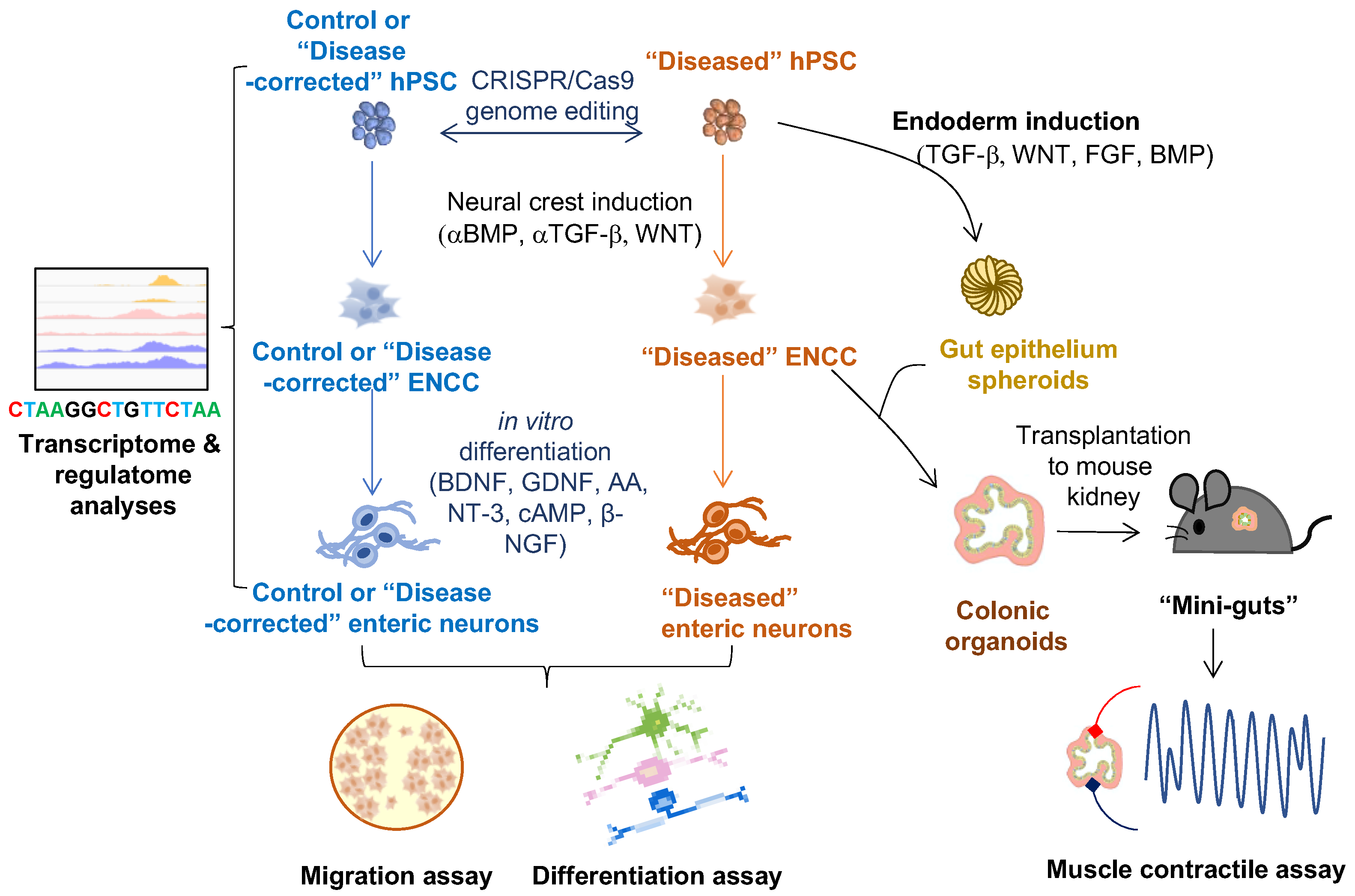
An overview of 2-D and 3-D hPSC-based ENS models in HSCR-related studies is shown in Figure 2.
Figure 2. Applications of hPSC-derived 2-D and 3-D cell models of ENS on studying HSCR disease. ENCC-like cells and intestinal organoids are derived from hPSCs by manipulating different developmental cues (BMP, TGF-b, WNT, and FGF). Innervated “mini-gut” are generated by combining the intestinal organoids with the ENCCs and engrafted to the mouse kidney capsule for further maturation. The 2-D cell models (ENCCs and enteric neurons) can be used for multi-omics studies (e.g., transcriptome and regulatome analyses) and functional assays (e.g., migration and in vitro differentiation assays). The 3-D cell models (colonic organoids and “mini-guts”) can be used for neuromuscular studies such as muscle contractile assay.

This entry is adapted from the peer-reviewed paper 10.3390/cells11213428

References

  1. Tilghman, J.M.; Ling, A.Y.; Turner, T.N.; Sosa, M.X.; Krumm, N.; Chatterjee, S.; Kapoor, A.; Coe, B.P.; Nguyen, K.H.; Gupta, N.; et al. Molecular Genetic Anatomy and Risk Profile of Hirschsprung’s Disease. N. Engl. J. Med. 2019, 380, 1421–1432.
  2. Sergi, C. Hirschsprung’s disease: Historical notes and pathological diagnosis on the occasion of the 100(th) anniversary of Dr. Harald Hirschsprung’s death. World J. Clin. Pediatr. 2015, 4, 120–125.
  3. Heuckeroth, R.O. Hirschsprung disease-integrating basic science and clinical medicine to improve outcomes. Nat. Rev. Gastroenterol. Hepatol. 2018, 15, 152–167.
  4. Goldstein, A.M.; Thapar, N.; Karunaratne, T.B.; De Giorgio, R. Clinical aspects of neurointestinal disease: Pathophysiology, diagnosis, and treatment. Dev. Biol. 2016, 417, 217–228.
  5. Badner, J.A.; Sieber, W.K.; Garver, K.L.; Chakravarti, A. A Genetic-Study of Hirschsprung Disease. Am. J. Hum. Genet. 1990, 46, 568–580.
  6. Tang, C.S.M.; Li, P.; Lai, F.P.L.; Fu, A.X.; Lau, S.T.; So, M.T.; Lui, K.N.C.; Li, Z.X.; Zhuang, X.H.; Yu, M.; et al. Identification of Genes Associated With Hirschsprung Disease, Based on Whole-Genome Sequence Analysis, and Potential Effects on Enteric Nervous System Development. Gastroenterology 2018, 155, 1908−+.
  7. Chow, K.H.-M.; Tam, P.K.-H.; Ngan, E.S.-W. Neural Crest and Hirschsprung’s Disease. In Stem Cells and Human Diseases; Srivastava, R.K., Shankar, S., Eds.; Springer: Dordrecht, The Netherlands, 2012; pp. 353–386.
  8. Obermayr, F.; Hotta, R.; Enomoto, H.; Young, H.M. Development and developmental disorders of the enteric nervous system. Nat. Rev. Gastroenterol. Hepatol. 2013, 10, 43–57.
  9. Heanue, T.A.; Pachnis, V. Enteric nervous system development and Hirschsprung’s disease: Advances in genetic and stem cell studies. Nat. Rev. Neurosci. 2007, 8, 466–479.
  10. Lake, J.I.; Heuckeroth, R.O. Enteric nervous system development: Migration, differentiation, and disease. Am. J. Physiol. Gastrointest. Liver Physiol. 2013, 305, G1–G24.
  11. Lee, G.; Chambers, S.M.; Tomishima, M.J.; Studer, L. Derivation of neural crest cells from human pluripotent stem cells. Nat. Protoc. 2010, 5, 688–701.
  12. Barber, K.; Studer, L.; Fattahi, F. Derivation of enteric neuron lineages from human pluripotent stem cells. Nat. Protoc. 2019, 14, 1261–1279.
  13. Fattahi, F.; Steinbeck, J.A.; Kriks, S.; Tchieu, J.; Zimmer, B.; Kishinevsky, S.; Zeltner, N.; Mica, Y.; El-Nachef, W.; Zhao, H.; et al. Deriving human ENS lineages for cell therapy and drug discovery in Hirschsprung disease. Nature 2016, 531, 105–109.
  14. Kwart, D.; Paquet, D.; Teo, S.; Tessier-Lavigne, M. Precise and efficient scarless genome editing in stem cells using CORRECT. Nat. Protoc. 2017, 12, 329–354.
  15. Mali, P.; Yang, L.; Esvelt, K.M.; Aach, J.; Guell, M.; DiCarlo, J.E.; Norville, J.E.; Church, G.M. RNA-guided human genome engineering via Cas9. Science 2013, 339, 823–826.
  16. Paquet, D.; Kwart, D.; Chen, A.; Sproul, A.; Jacob, S.; Teo, S.; Olsen, K.M.; Gregg, A.; Noggle, S.; Tessier-Lavigne, M. Efficient introduction of specific homozygous and heterozygous mutations using CRISPR/Cas9. Nature 2016, 533, 125–129.
  17. Ran, F.A.; Hsu, P.D.; Wright, J.; Agarwala, V.; Scott, D.A.; Zhang, F. Genome engineering using the CRISPR-Cas9 system. Nat. Protoc. 2013, 8, 2281–2308.
  18. Fu, A.X.; Lui, K.N.; Tang, C.S.; Ng, R.K.; Lai, F.P.; Lau, S.T.; Li, Z.; Garcia-Barcelo, M.M.; Sham, P.C.; Tam, P.K.; et al. Whole-genome analysis of noncoding genetic variations identifies multiscale regulatory element perturbations associated with Hirschsprung disease. Genome Res. 2020, 30, 1618–1632.
  19. Lai, F.P.; Lau, S.T.; Wong, J.K.; Gui, H.; Wang, R.X.; Zhou, T.; Lai, W.H.; Tse, H.F.; Tam, P.K.; Garcia-Barcelo, M.M.; et al. Correction of Hirschsprung-Associated Mutations in Human Induced Pluripotent Stem Cells Via Clustered Regularly Interspaced Short Palindromic Repeats/Cas9, Restores Neural Crest Cell Function. Gastroenterology 2017, 153, 139–153.e8.
  20. McCracken, K.W.; Cata, E.M.; Crawford, C.M.; Sinagoga, K.L.; Schumacher, M.; Rockich, B.E.; Tsai, Y.H.; Mayhew, C.N.; Spence, J.R.; Zavros, Y.; et al. Modelling human development and disease in pluripotent stem-cell-derived gastric organoids. Nature 2014, 516, 400–404.
  21. Spence, J.R.; Mayhew, C.N.; Rankin, S.A.; Kuhar, M.F.; Vallance, J.E.; Tolle, K.; Hoskins, E.E.; Kalinichenko, V.V.; Wells, S.I.; Zorn, A.M.; et al. Directed differentiation of human pluripotent stem cells into intestinal tissue in vitro. Nature 2011, 470, 105–109.
  22. Workman, M.J.; Mahe, M.M.; Trisno, S.; Poling, H.M.; Watson, C.L.; Sundaram, N.; Chang, C.F.; Schiesser, J.; Aubert, P.; Stanley, E.G.; et al. Engineered human pluripotent-stem-cell-derived intestinal tissues with a functional enteric nervous system. Nat. Med. 2017, 23, 49–59.
  23. Schlieve, C.R.; Fowler, K.L.; Thornton, M.; Huang, S.; Hajjali, I.; Hou, X.; Grubbs, B.; Spence, J.R.; Grikscheit, T.C. Neural Crest Cell Implantation Restores Enteric Nervous System Function and Alters the Gastrointestinal Transcriptome in Human Tissue-Engineered Small Intestine. Stem Cell Rep. 2017, 9, 883–896.
  24. Lau, S.T.; Li, Z.; Lai, F.P.L.; Lui, K.N.C.; Li, P.; Múnera, J.O.; Pan, G.; Mahe, M.M.; Hui, C.C.; Wells, J.M.; et al. Activation of Hedgehog Signaling Promotes Development of Mouse and Human Enteric Neural Crest Cells, Based on Single-Cell Transcriptome Analyses. Gastroenterology 2019, 157, 1556–1571.e5.
  25. Múnera, J.O.; Sundaram, N.; Rankin, S.A.; Hill, D.; Watson, C.; Mahe, M.; Vallance, J.E.; Shroyer, N.F.; Sinagoga, K.L.; Zarzoso-Lacoste, A.; et al. Differentiation of Human Pluripotent Stem Cells into Colonic Organoids via Transient Activation of BMP Signaling. Cell Stem Cell 2017, 21, 51–64.e6.
  26. Crespo, M.; Vilar, E.; Tsai, S.Y.; Chang, K.; Amin, S.; Srinivasan, T.; Zhang, T.; Pipalia, N.H.; Chen, H.J.; Witherspoon, M.; et al. Colonic organoids derived from human induced pluripotent stem cells for modeling colorectal cancer and drug testing. Nat. Med. 2017, 23, 878–884.
  27. Eicher, A.K.; Kechele, D.O.; Sundaram, N.; Berns, H.M.; Poling, H.M.; Haines, L.E.; Sanchez, J.G.; Kishimoto, K.; Krishnamurthy, M.; Han, L.; et al. Functional human gastrointestinal organoids can be engineered from three primary germ layers derived separately from pluripotent stem cells. Cell Stem Cell 2022, 29, 36–51.
More
This entry is offline, you can click here to edit this entry!
Video Production Service