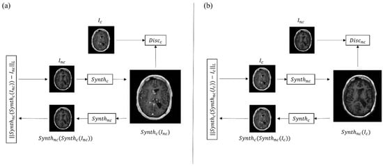
In neuroradiology, the contrast enhancement of a lesion inside the brain tissue reflects a blood–brain barrier rupture. This event occurs in many different brain diseases such as some primary tumors and metastases, neuroinflammatory diseases, infections, and subacute ischemia, for which contrast injection during MRI examination is considered mandatory for accurate assessment, differential diagnosis, and monitoring [
55,
56,
57,
58,
59,
60]. Other uses of contrast include the evaluation of vascular malformation and aneurysms. In this scenario, the implementation of AI algorithms to reduce contrast usage could result in significant benefit for the patients, and reduced scan times and costs [
61]. However, the main drawback of AI analysis, especially through deep learning (DL) methods, reside in the need for a large quantity of data. For this reason, the literature of AI virtual contrast in neuroradiology is focused on MRI examination of relatively common diseases such as tumors and multiple sclerosis (MS) (
Table 2).
In neuro-oncology, contrast enhancement is particularly useful not only as a marker for differential diagnosis and progression, but it is also considered the target for neurosurgical removal of a lesion and an indicator of possible recurrence. Although in recent years some authors suggested expanding the surgical resection of brain tumors beyond the contrast enhancement [
62,
63], injection of gadolinium remains a standard for both first and follow-up MR scans. To avoid the use of gadolinium, Kleesiek et al., applied a Bayesan DL architecture to predict contrast enhancement from non-contrast MR images in patients with gliomas of different grades and healthy controls [
64]. The authors obtained good results in terms of qualitative and quantitative assessment (approximate sensitivity and specificity of 91%). Similarly, other studies applied a DL method to pre-contrast MR images of a large group of glioblastomas and low-grade gliomas with a good structural similarity index between simulated and real postcontrast imaging, and the ability of the neuroradiologist to determine the tumor grade [
32,
65]. These methods can also be applied to sequences different from the T1. Recently, Wang et al., developed a GAN to synthetize 3D isotropic contrast-enhanced FLAIR images from a 2D non-contrast FLAIR image stack in 185 patients with different brain tumors [
66]. Interestingly, the authors went beyond simple contrast synthesis and added super-resolution and anti-aliasing tasks in order to solve MR artifacts and create isotropic 3D images, which give a better visualization of the tumor, with a good structural-similarity index to the source images [
66]. Calabrese et al., obtained good results in synthetize contrast-enhanced T1 images from non-contrast ones in 400 patients with glioblastoma and lower-grade glioma [
65]. In addition, the authors included an external validation analysis, which is always recommended in DL-based studies [
65]. However, the simulated images appeared blurrier than real ones, a problem that could especially affect discriminating progression in follow-up exams [
65]. This shortcoming appears to be a common issue of all ‘simulated imaging’ studies. As stated above, contrast enhancement reflects disruption of the blood–brain barrier, information that is usually inferred from the pharmacokinetics of gadolinium-based contrasts within the brain vasculature; this explains the difficulty of generating information from sequences that may not contain it. Moreover, virtual contrast may hinder interpretation of derived measures, such as perfusion-weighted imaging, which have been proven crucial for differential diagnosis and prognosis prediction in neuro-oncology [
15,
16,
67,
68]. Future directions could make use of ultrahigh field scanners that may have enough resolution to be closer to molecular imaging. In the meantime, another approach has been explored to address the issue. Rather than eliminating contrast injection, different studies used AI algorithms to enhance a reduced dose of gadolinium (10% or 25% of the standard dose), a method defined as ‘augmented contrast’ [
30,
31,
69,
70]. This method, used on images of different brain lesions, including meningiomas and arteriovenous malformations, allows the detection of a rupture of the blood–brain barrier with a significantly lower contrast dose. Another advantage is the better quality of the synthetized images, as perceived by evaluating neuroradiologists [
30,
31]. Such benefits persist with data obtained across different scanners, including both 1.5T and 3T field strengths, a fundamental step for the generalizability of results [
30,
69,
70]. Nevertheless, augmented contrast techniques are not exempt from limitations. Frequently encountered issues with these techniques are the difficulty of detecting small lesions, the presence of false positive enhancement, probably due to flow or motion artifacts, and coregistration mismatch [
31,
70]. Another concern is the lack of time control between the two contrast injections. Most of the studies perform MRI in a single session by acquiring the low-dose sequence first, followed by full-dose imaging after injecting the remaining dose of gadolinium [
30,
31,
69,
70]. The resulting full-dose images are, thus, a combination of the late-delayed pre-dose (10% or 25%) and the standard-delayed contrast, which can result in a slightly different enhancement pattern from the standard full-contrast injection. Future studies could acquire low and standard dose exams on separate days, with controlled postcontrast timing. Lastly, future directions could include prediction of different contrast imaging. In fact, other types of contrast are being developed to give additional information on pathologic processes, such as neuroinflammation [
71]. Another interesting application of AI in neuro-oncology imaging consists of augmenting the contrast signal ratio in standard dose T1 images, in order to better delineate tumors and detect smaller lesions. Increasing contrast signal for better detection of tumors, in fact, has always been a goal when developing new MR sequences, leading recently to a consensus for brain tumor imaging, especially for metastasis [
72]. A recent study by Ye et al., used a multidimensional integration method to increase the signal-to-noise ratio in T1 gradient echo images [
73], also resulting in contrast signal enhancement. By comparison, Bône et al., implemented an AI-based method to increase contrast signal similarly to the ‘augmented contrast’ studies [
74]. The authors trained an artificial neural network with T1 pre-contrast, FLAIR, ADC, and low-dose T1 (0.025 mmol/kg). Once trained, the model was leveraged to amplify the contrast on routine full-dose T1 by processing it into high-contrast T1 images. The hypothesis behind this process was that the neural network learned to amplify the difference in contrast between pre-contrast and low-dose images. Hence, by replacing the low-dose sequence with a full-dose one, the synthesis led to a quadruple-dose contrast [
74]. The results led to a significant improvement in image quality, contrast level, and lesion detection performance, with a sensitivity increase (16%) and similar false detection rate with respect to routine acquisition.
MS is the most common demyelinating chronic disorder, and mostly affects young adults [
75]. MRI is a fundamental tool in both MS diagnosis and follow-up, and gadolinium injection is usually mandatory. In fact, contrast enhancement is essential to establish dissemination in time according to the McDonald’s criteria, and to evaluate ‘active’ lesions in follow-up scans [
76]. Due to the relatively young age at diagnosis, MS patients undergo numerous MRIs throughout the years. For this reason, different research groups are trying to limit contrast administration in selected cases to minimize exposition and costs [
40,
53]. One study tried to develop a DL algorithm to predict enhancing MS lesions from non-enhanced MR sequences in nearly 2000 scans, reporting accuracy between 70 and 75%, leading the way to further research [
77]. The authors used only conventional imaging such as T1, T2, and FLAIR. Future studies could add DWI to the analysis as this sequence has been proven to be useful in identifying active lesions [
78]. Small dimensions of MS plaques, as already seen above for brain tumor studies, could be a reason for the low accuracy of synthetic contrast imaging; future directions could try enhancing low-dose contrast injections by means of AI algorithms.
In conclusion, virtual and augmented contrast imaging have an interesting role in neuroradiology for assessing different diseases for which gadolinium injection is, for now, mandatory.

