Roles of Calcium Ions in Parkinson’s Disease: History
Please note this is an old version of this entry, which may differ significantly from the current revision.
Contributor: , ,

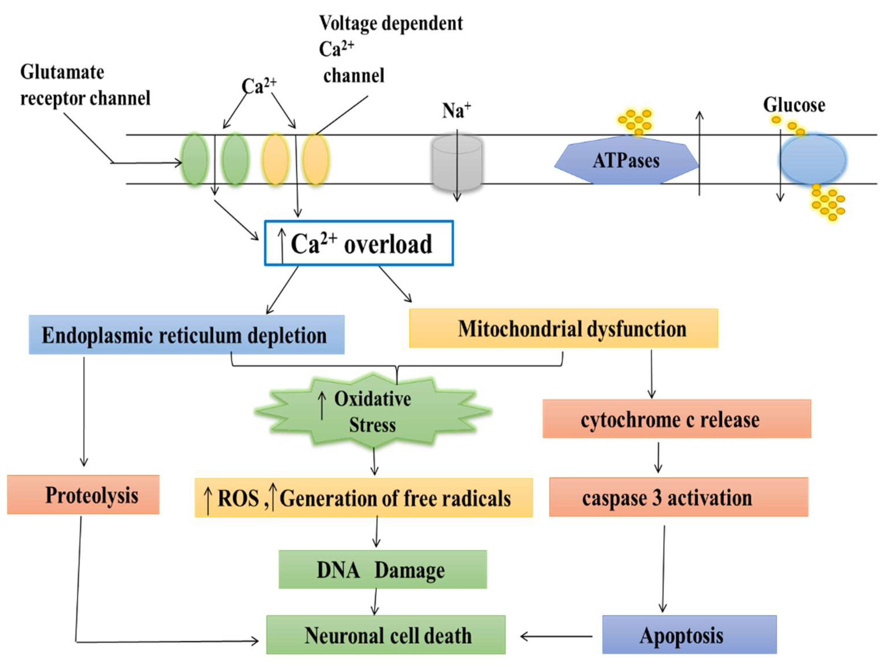
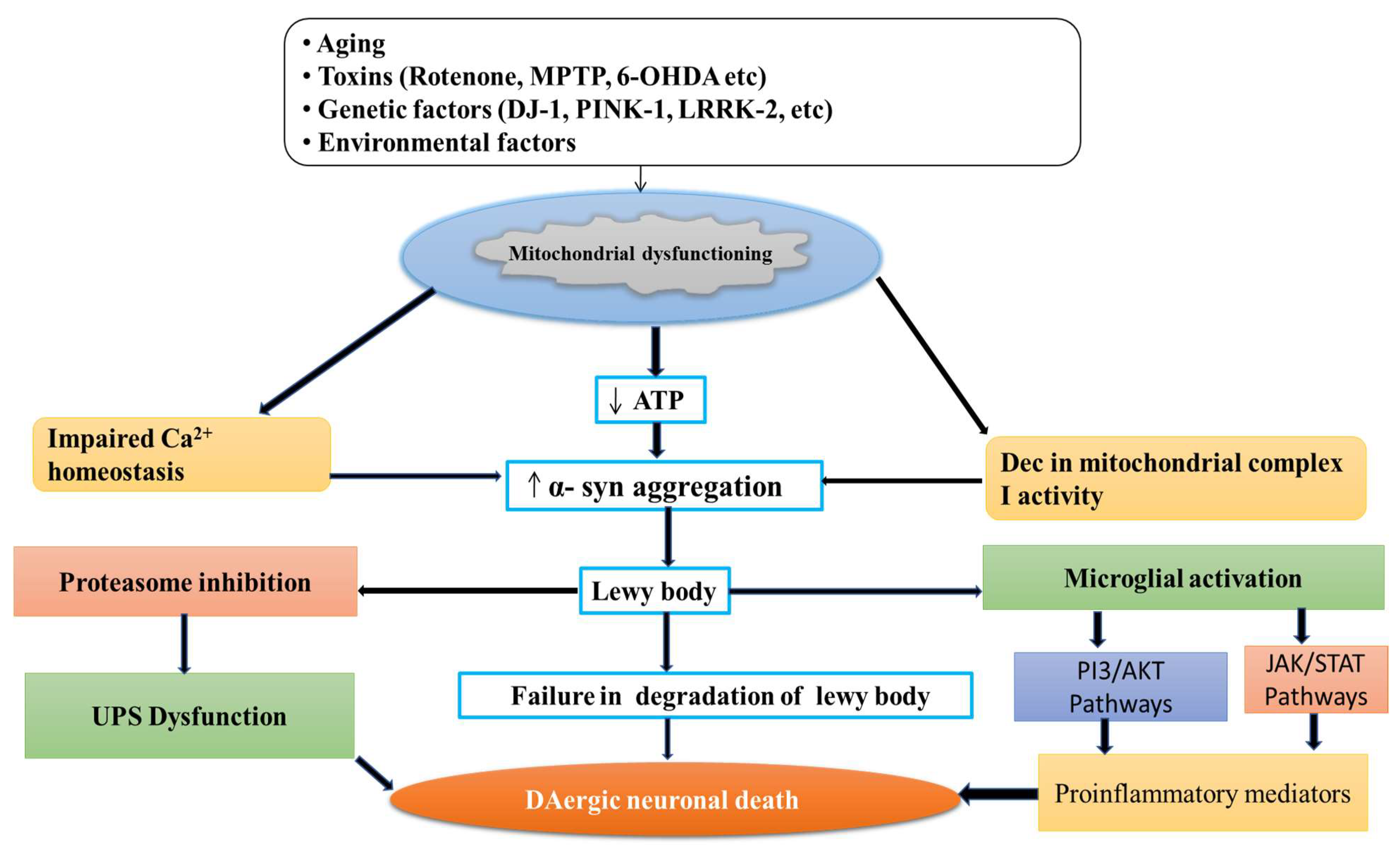
Parkinson’s disease (PD) is a neurodegenerative movement disorder characterized by the loss of dopaminergic neurons, which results in motor impairment.  Ca2+ homeostasis disruption and mitochondrial dysfunction play a vital role in PD aetiology. In addition, the L-type voltage-gated calcium channel is expressed at high levels amongst nigral neurons, and could play a role in the pathogenesis of PD. In the dopaminergic neurons, Ca2+ entry through plasma membrane Cav1 channels drives a sustained feed-forward stimulation of mitochondrial oxidative phosphorylation. The R-type calcium channel is a type of voltage-dependent calcium channel. Available findings suggest that calcium homeostasis in dopaminergic neurons might be a valuable target for developing new drugs for PD patients.

  • Parkinson’s disease
  • dopaminergic neurons
  • mitochondrial
  • calcium channel
  • calcium channel blocker
  • neurodegenerative disorder
  • mitochondrial dysfunction
  • oxidative stress
  • neuroinflammation
  • Lewy bodies
  • dihydropyridine

1. Introduction

Parkinson’s disease (PD) is a neurodegenerative disorder, characterized by cardinal motor symptoms such as bradykinesia, rigidity, and tremor [1]. PD is strongly associated with ageing, increases exponentially in incidence above the age of 65 years, and has no cure [2]. The motor symptoms of PD first appear clinically caused by the degeneration and death of selective dopaminergic (DA) neurons within the substantia nigra pars compacta (SNpc) [3]. The neurological processes underpinning dementia and its accompanying intellectual abnormalities are referred to as PD. Recent neuroscientist findings have begun to untangle the diverse participation of numerous separate neural networks underpinning cognitive impairments in Parkinson’s Disease Dementia (PDD) and their regulation by both dopaminergic and non-dopaminergic transmitter systems in the brain [4]. Cognitive impairment is a typical symptom of PD, which has a high morbidity and fatality rate. The severity of these symptoms ranges from modest executive dysfunction to full-blown dementia affecting numerous areas [5].
The pathological hallmarks of PD are the presence of Lewy bodies (LBs) and the loss of DA neurons containing neuromelanin [6]. LBs are spherical eosinophilic cytoplasmic protein aggregates composed of proteins including α-synuclein, ubiquitin, parkin, and neurofilaments, and these proteins are found in the affected regions of the brain [7][8]. LBs are most commonly found in the brain regions with the greatest neuron loss in PD, such as the SN, locus coeruleus, the dorsal motor nucleus of the vagus, and the nucleus basalis of Meynert, but they have also been found in the neocortex, diencephalon, spinal cord, and even peripheral autonomic ganglia [9]. DJ-1, encoded by the PARK7 gene, causes early-onset autosomal recessive PD and is likely the most thoroughly investigated [10]. DJ-1 is an essential regulator of the pro-inflammatory response, and knocking it out in astrocytes reduces inflammatory-related damage. PARK2 and PINK1 are both expressed at comparable amounts in astrocytes and neurons [11]. Interestingly, astrocytes lacking Parkin, expressed by the PARK2 gene, showed a stress-induced increase in NOD2 expression, a receptor that integrates endoplasmic reticulum stress and inflammation, and these astrocytes displayed increased cytokine release and reduced neurotrophic factor production [12]. Parkin has also been involved in astrocyte responses to inflammatory signals; stimulation with TNF- leads in Parkin overexpression, whereas activation with IL-1 results in Parkin downregulation [13]. PINK1 expression, which encodes the protein PTEN-induced putative kinase 1 (PINK1), is a loss of function mutation related to early-onset PD. Furthermore, neurotoxic kynurenine metabolites in plasma and cerebrospinal fluid (CSF) are related to symptom severity and nigral pathology in PD [14]. The various mechanisms involved in PD are mitochondrial dysfunction, oxidative stress (OS), neuroinflammation, gene mutation, and some environmental toxins [15] (Figure 1).
Figure 1. Different mechanisms involved in Parkinson’s disease (mitochondrial dysfunctioning, oxidative stress (OS), neuroinflammation, gene mutation, and some environmental toxins).
Oxidative stress has long been thought to be one of the pathophysiological mechanisms implicated in PD, which led to the investigation of the antioxidant systems as a promising therapy more than two decades ago. A useful antioxidant must have certain characteristics: it must be capable of interacting with biologically relevant oxidants and free radicals; its reaction by-products must be harmless; and, finally, it must reach a sufficiently high concentration in tissue and cell compartments to ensure quantitatively relevant activity [16]. Patients with Parkinson’s disease (PD) frequently experienced gastrointestinal problems prior to the start of motor symptoms. Parkinson’s disease neuropathology has also been found in the enteric nervous system (ENS). Many studies have found substantial PD-related changes in gut microbiota. The microbiota–gut–brain axis is a dynamic bidirectional communication network that plays a role in the aetiology of PD. The aggregation of misfolded protein alpha-synuclein, the neuropathological characteristic of PD, is thought to start in the stomach and move to the CNS via the vagus nerve and olfactory bulb. Changes in the architecture of the gut microbiota raise the concentrations of short-chain fatty acids (SCFAs) and other metabolites, which act on the neuroendocrine system and modulate the concentrations of GABA, serotonin, and other neurotransmitters. Furthermore, it affects the vagus and intestinal nerve systems, impacting the brain and behavior through the activation of microglia and systemic cytokines. An increasing collection of experimental and clinical evidence suggests that gut dysbiosis and microbiota host interaction play a role in neurodegeneration [17]. Dopaminergic neurodegeneration is directly associated with metal accumulation or elevated inflammatory cytokines such as interleukin-1 (IL-1, IL-6), TNF-α, which causes neuroinflammation, and, ultimately, neuronal death. Metals are the primary natural elements of the earth’s crust and are spread throughout the biosphere by human activities. Metals are commonly found in mining, industrial waste, tailings, agricultural runoff, treated timber, paints, ageing water supply infrastructure, lead-acid batteries, vehicle emissions, fertilizers, and microplastics [18]. Metals’ significance in the aetiology of PD remains a key topic in neurotoxicology and medicinal chemistry. Heavy metals, such as Fe (III) and Mn (II), cause oxidative stress by boosting ROS generation via the Fenton and Haber–Weiss reaction and changing the antioxidant system in cells [19][20].
These metal toxins aggravate the oxidative stress process in the cell, resulting in cell death by producing an imbalance between free radical and antioxidant enzymes [21][22]. In addition, neuroinflammation aggravates oxidative stress pathways, which can lead to protein aggregation via changing the activity of the UPS. Protein aggregates can accumulate due to defective protein breakdown machinery, disrupting cellular activities and causing cell death [23]. In addition, heavy metals inhibit the action of mitochondrial complexes, resulting in a slowed metabolic process, increased ROS generation, and oxidative stress [24][25].
Among these, mitochondrial dysfunction and an increase in oxidative stress play a significant role in the pathogenesis of PD [26]. Although the mechanisms are unclear, the mitochondrial dysfunction in dopaminergic neurons of idiopathic and familial PD is well known. Langston et al. and Burns et al. reported that the accidental administration of 1-methyl-4-phenyl-1,2,3,6-tetrahydropyridine (MPTP) selectively inhibits complex I of the mitochondrial electron transport chain (ETC) and because mitochondria play a significant role in the pathogenesis of PD [27][28]. Rotenone, trichloroethylene, pyridaben are the other complex I inhibitors that induce dopaminergic neurodegeneration in PD [29]. MPTP, a strong inhibitor of mitochondrial complex-1 of the electron transport chain, induces parkinsonian symptoms in rats and mimics dopaminergic degenerations via the nigrostriatal pathway [30][31]. As a result, in animal PD models, MPTP is widely utilized to investigate the molecular mechanisms underlying dopaminergic neuronal degeneration and evaluate the efficiency of various neuroprotective agents [32]. Biomolecules such as lipids, proteins, and DNA are destroyed by reactive oxygen and nitrogen species (ROS and RNS), by-products found in the SN and striatum of human PD post-mortem brains [33][34]. Thus, lipid and protein oxidation can result in membrane integrity loss, enzyme deactivation, and cell death in neurodegenerative diseases [35]. Various natural products are used for testing their neuroprotective effects against MPTP-induced PD. Mucuna pruriens (Mp) exhibits various pharmacological properties like analgesic, anti-inflammatory, anti-neoplastic, anti-epileptic, and anti-microbial activities [36]. Mp has been found to be rich in bioactive compounds such as tannins, alkaloids, phenolic compounds, and flavonoids [37]. In addition, Mp extract was reported to significantly improve neuroinflammatory processes and restore biochemical and behavioral parameters and immunoreactivity. Mp shows anti-inflammatory action and its high antioxidant capabilities, which can be utilized to treat inflammatory conditions in the case of PD [38].
An increase in PD risk comes with increasing intake of foods that contain animal fat and foods containing vitamin D. Intake of fruits, vegetables, meats, bread and cereals, or foods containing vitamins A, C, E, or iron was not significantly related to PD risk. Vitamin use, in general, was also not found to be related to PD risk, although a significant trend of increasing risk of PD was noted for intake of vitamin A supplements. The tryptophan (TRP)-kynurenine (KYN) metabolic pathway is the primary catabolic route of TRP metabolism, converting over 95% of TRP into a variety of bioactive metabolites such as anti-inflammatory, antioxidative, proinflammatory, neurotoxic, neuroprotective, and immunologic compounds. Furthermore, kynurenine pathway (KP) enzymes influence inflammation and the immune system. Alterations in the KP enzymes’ activity and the levels of the KP metabolites have been linked to neurological disorders, cancer, autoimmune conditions, and inflammation. However, the functions of KP enzymes and metabolites in the development and progression of many diseases constitute a field of medicine that has received comparatively little attention. An illustration of this is the relationship between kynurenines (KYNs) and the KP enzymes, which have been linked to a variety of diseases, including cancer, autoimmune diseases, inflammatory diseases, neurologic diseases, and mental disorders. One of the key immune response regulators and a potential player in the inflammatory response in parkinsonism is the KP, the primary catabolic route for tryptophan. The KP produced various neuroactive compounds and has both neurotoxic and neuroprotective effects [39]. These disorders are related with amyloid-β (Aβ), alpha synuclein (α--Syn), and prion protein (PrP) depositions in the brain, which cause synaptic disconnection and eventual progressive neuronal death. Although continued progress has been made in understanding the aetiology of many neurological disorders, the precise mechanisms of their origins remain largely unclear. A growing body of evidence implies association between host microbiota, neuroinflammation, and dementia, either directly due to bacterial brain invasion via barrier leakage and the generation of toxins and inflammation, or indirectly by altering the immune response and causing PD-like symptoms [40].
Ursolic acid is a naturally occurring pentacyclic triterpenoid carboxylic acid found in many plants, including apples, basil, bilberries, cranberries, peppermint, rosemary, and oregano. Several biochemical and pharmacological actions of ursolic acid have been described in various experimental systems, including anti-inflammatory, antioxidative, anti-proliferative, anti-cancer, anti-mutagenic, antiatherosclerotic, anti-hypertensive, anti-leukemic, and antiviral characteristics [41][42]. Ursolic acid inhibits MPTP-induced dopaminergic neurotoxicity through the NF-B pathway. Ursolic acid’s anti-inflammatory action has been attributed mostly to its neuroprotective potential. Although the chemical mechanism behind ursolic acid’s neuroprotective impact remains unknown, the findings suggest that ursolic acid might be employed as a viable medication in the treatment of Parkinson’s disease symptoms. As a result, the ability of ursolic acid to rescue dopaminergic neurons from neurodegeneration may imply a role for therapeutic intervention in PD [43].
Chlorogenic acid (CGA), a polyphenolic molecule present in many plants, is particularly prevalent in green coffee beans, which contain roughly 5–12% CGA by weight [44]. CGA is a trans-cinnamic acid ester (which includes caffeic acid, ferulic acid, and p-coumaric acid) and quinic acid. It is widely consumed by individuals and may be found in various drinks and food items [45]. It is mainly found in fruits and vegetables such as apples, apricots, cherries, plums, and tomatoes. Wine, coffee, and tea are the most prevalent CGA-rich drinks [46]. They have anticancer efficacy, cardioprotective properties, and may have neuroprotective activities [47]. Evidence suggests that CGA has a variety of biological effects, including antioxidant, neuroprotective, and neurotrophic properties [48]. Therefore, CGA can potentially be a powerful anti-inflammatory drug in preventing neurodegeneration in PD. It exerts its effects primarily by suppressing the production of iNOS, TNF- α, and NF-κB in activated glial cells, ultimately decreasing neuroinflammation via increased anti-inflammatory and antioxidant activity [49]. Thus, CGA’s anti-inflammatory effect and its high antioxidant characteristics can be utilized to treat the inflammatory state associated with PD [50].

2. Normal Physiology and Pathology of Mitochondria

Mitochondria is the powerhouse of the cell, including the production of energy through the mitochondrial respiratory chain, cell death regulation, calcium metabolism, and production of ROS [51][52]. Mitochondria is the primary source of free radical generaation in the cell resulting in OS. The mitochondrial ETC involves five complexes I-V embedded in the inner mitochondrial membrane, which involve the transfer of reducing equivalents from high-energy compounds to oxygen through Kreb’s cycle [53][54]. The mutations in specific genes such as Parkin, alpha-synuclein, DJ-1, LRRK2, PTEN-induced kinase 1 (PINK 1), and vacuolar protein sorting 35 (VSP35) support the mitochondrial dysfunction in PD [55][56]. Also, the toxins such as rotenone, MPTP, and paraquat alter mitochondrial respiration in PD. These toxins cause the deficiency in mitochondrial complexes’ activity [57], reduce the movement of mitochondria, and mitigate generation of reactive oxygen species (ROS) [58], thereby leading to PD-like symptoms (Figure 2).
Figure 2. Degeneration of DAergic neuronal death by different neurotoxins.
Paraquat (PQ) is a herbicide known to cause neurotoxicity by producing free radicals, resulting in oxidative stress [59]. The specific neurotoxicity produced by PQ in the SN area is attributable to the fact that it enters the CNS via the blood-brain barrier via neutral amino acid transporter. Maneb (MB), a fungicide, exhibits similar effects and has been reported to disrupt mitochondrial activity, resulting in oxidative stress [60][61][62]. When combined, MB and PQ are known to work synergistically, increasing neuronal toxicity and causing dopaminergic neurodegeneration and severe oxidative stress [63][64].
Withania somnifera (Ws) is a Solanaceae-family herbal medicinal plant and plays an essential role in Ayurveda. The biological activity of Ws extract demonstrated antioxidant and free radical scavenging capabilities [65]. Various parts of this medicinal plant have been used to cure various diseases since ancient times. It is known as “Indian Ginseng” due to its importance in traditional Indian medicine. Ws root extract contains withanolides, which are steroidal alkaloids and lactones of dopaminergic neurons in the SN area of the brain [66]. The anti-degeneration properties of the Ws root extract against MB–PQ-induced dopaminergic neurodegeneration in the PD animal model has been reported. Ws extract can increase the numbers of TH-positive cells in the SN region of the MB–PQ-induced PD animal brain while concurrently decreasing the oxidative stress occurring in nigrostriatal tissues [60][67]. Therefore, it appears that the up-regulation of TH expression in the SN area of the brain is the leading cause of the improvement in the walking pattern seen in the Ws-treated PD mouse. It is clear that Ws has substantial antioxidant capability and that through preventing neurodegeneration, its ROS scavenging property plays a significant role in preventing PD. Taken as a whole, Ws extract looks to be a promising therapeutic candidate for Parkinson’s neuroprotection [60].
The mitochondrial function and calcium signaling are interlinked; the calcium (Ca2+) is the second messenger to transmit depolarization and synapses to the other neurons [68]. The concentration of Ca2+ in the cytosol stimulates the mitochondria to produce more energy. Ca2+ is maintained via the accumulation of Ca2+ in the mitochondrial matrix [69], leads to the activation of oxidative phosphorylation, and increases the production of ATP [68]. Environmental toxins such as rotenone and MPP+ reduce the level of Ca2+. The mitochondrial complex I deficiency power is not present in all patients with PD, either in the brain, platelets, or other tissues. The severity of the defect is about a 35% reduction in activity when the patient group is compared with control populations [70].

3. Role of Calcium in Mitochondria

Calcium is an essential ion with multiple roles in cell activity. Calcium enters mitochondria through a pore and is utilized in their energy production process [71]. Calcium acts as the key regulator of energy production in mitochondria, but excess calcium can trigger cell death [72]. If the pore fails to close, then mitochondria retain the energy synthesized in the form of ATP. This results in the accumulation of oxidants and calcium overloading, leading to mitochondrial swelling and cell stress, and resulting in numerous diseases including cardiovascular diseases, such as stroke and heart attack, and neurodegenerative disorders, such as PD and Alzheimer’s disease (AD) [73][74].
Several researchers reported that both calcium and magnesium ions were involved in controlling the shuttle. When these ions bind to the inside part of the calcium channel, the pore closes [75]. This helps explain the role of calcium transport protein in controlling mitochondrial calcium uptake and is important for understanding diseases associated with mitochondrial dysfunction [76].
The calcium was accumulated in the mitochondria neuron, resulting in the mitochondrial calcium uptake, sequestration, and release of the calcium-dependent responses that resulted in gene transcription and cell death [77][78]. The stimuli were activated by initiating the entry of (Ca2+) through plasma membrane channels and responded by neurons. However, the increase in free cytosolic (Ca2+) is strongly modulated by the activity of intracellular calcium stores [79]. In particular, Ca2+ uptake, sequestration, and release by the endoplasmic reticulum and mitochondria are the two major Ca2+-regulating organelles that play essential roles in modulating and interpreting Ca2+ signals [80]. Mitochondria play a critical role in neuronal (Ca2+) signaling. Also, the overloading of mitochondrial calcium and dysfunction may be important for triggering the cell death which follows ischemic and traumatic brain injury, and neurodegenerative disorders such as AD, PD, Huntington’s disease (HD), and Amyotrophic lateral sclerosis (ALS) [81][82].

4. Medicinal Plants as Calcium Channel Blockers

The side effects of synthetic anti-hypertensive drugs have made researchers search for safer therapies to resolve hypertension. The preference for herbal alternatives to traditional, synthetic alternatives arises because herbal medications are both safer and less expensive than synthetic ones [83]. Furthermore, therapeutic herbs are more compatible with the human body. Medicinal plants provide a multitude of phytoconstituents that act on the numerous pharmacological targets implicated in hypertension. These plants can be used in the form of infusions, decoctions, and fresh fruits, or can be eaten raw [84]. Several medicinal plants with phytoconstituents that function as calcium channel blockers have been described here for the treatment of hypertension and are mentioned in Table 1.
Table 1. Medicinal plants and their botanical names, chemical constituents responsible for activity, and the type of extracts used.

S. No.

Common Name

Botanical Name

Family

Chemical Constituents

Plant Part Used

1.

Yarrow

Achillea wilhelmsii

Asteraceae

Carvacrol, luteolin, apigenin 1,8-cineole

Aerial part

2.

Shell ginger

Alpinia zerumbet

Zingiberaceae

Catechin, epicatechin, kaempferol 3-o-rutinoside, rutin

Whole plant

3.

Celery

Apium graveolens

Apiaceae

Apiin, apigenin, isoquercitrin sesquiterpene

Seed

4.

Nikko Maple

Acer nikoense (Miq.) Maxim

Aceraceae

Scopoletin,

Cleomiscosin A,

Aquillochin

Leaves, bark

5.

Soursop, Graviola

Annona muricata

Annonaceae

Reticuline, quercetin, beta-caryophyllene, coreximine, anomurin

Leaves

6.

Punarnava Hogweed

Boerhavia diffusa

Nyctaginaceae

Liriodendron, boeravinone, hypoxanthine

Whole plant, root

7.

Sweet flag, flagroot

Acorus calamus L.

Acoraceae

β- asarone, β- gurjunene,

sequesterpenes, xylose,

β- daucosterol, d- galacturonic acid

Rhizome

8.

Cape periwinkle, periwinkle

Catharanthus roseus

Apocynaceae

Vinblastine, vincristine

Leaves, roots, flowers

9.

Saffron

Crocus sativus

Iridaceae

Crocin, picrocrocin, safranal, crocetin

Stigma

10.

Carrot

Daucus

carota

Apiaceae

Coumarin

glycosides (DC-2 and DC-3)

Aerial parts

11.

Ajwain

Carrom copticum

Apiaceae

Thymol, ρ-cymene, γ- terpinene, o-cymene, carvacrol

β-phellandrene

Seeds

12.

White horehound

Marrubium

vulgare L

Lamiaceae

Marrubenol

Whole plant

13.

Mu Dan Pi

Moutan Cortex

Paeoniaceae

Paeoniflorin, benzoyl paeoniflorin, mudanpioside C, paeonol, 1,2,3,4,6-o-pentagalloylglucose

Whole plant

14.

Wu-Chu-Yu

Evodia rutaecarpa L.

Rutaceae

Rutaecarpine

Fruits

15.

Roselle

Hibiscus sabdariffa

Malvaceae

β-carotene, ascorbic acid, β sitosterol, cyaniding-3- rutinose, pectin

Calyx, leaves, corolla

16.

French Lavender

Lavandula stoechas

Lamiaceae

Fenchone, p-cymene, lavandulyl acetate, a-pinene

Flower and oil

17.

Olive leaf

Olea africana and Olea europaea

Oleaceae

Oleuropein

Leaves

18.

Ginseng

Panax ginseng

Araliaceae

Ginsenosides Rg1, Rg3, Rh1, Re, and Rd

Roots

19.

Basil

Ocimum basilicum

Lamiaceae

Eugenol, α-cubebene, caryophyllene, rosmarinic, estragole

Leaves, stem

20.

Black Cumin, Seed of Blessing

Ranunculaceae

Thymoquinone, dithymoquinone

Seed

21.

Cat’s Claw herb

Uncaria rhynchophylla

Rubiaceae

Hirsutine, rhynchophylline, isorhynchophylline

Leaves

22.

Fen Fang Ji

Radix stephaniae tetrandrae

Menispermaceae

Tetrandrine

Roots

23.

Zingiber officinale

Zingiberaceae

Gingerol, gingerdiol, gingerdione, β-carotene, capsaicin, caffeic acid

Rhizomes

24.

Jatamansi, Indian valerian

Valeriana jatamansi

Valerianaceae

Jatamansika, jatamansine

Roots, rhizomes


This entry is adapted from the peer-reviewed paper 10.3390/jmp3040021

References

  1. Jankovic, J. Parkinson’s disease: Clinical features and diagnosis. J. Neurol. Neurosurg. Psychiatry 2008, 79, 368–376.
  2. Nazir, M.; Al-Ansari, A.; Al-Khalifa, K.; Alhareky, M.; Gaffar, B.; Almas, K. Global prevalence of periodontal disease and lack of its surveillance. Sci. World J. 2020, 2020, 2146160.
  3. DeMaagd, G.; Philip, A. Parkinson’s disease and its management: Part 3: Nondopaminergic and nonpharmacological treatment options. Pharm. Ther. 2015, 40, 668.
  4. Cammisuli, D.M.; Cammisuli, S.M.; Fusi, J.; Franzoni, F.; Pruneti, C. Parkinson’s disease–mild cognitive impairment (PD-MCI): A useful summary of update knowledge. Front. Aging Neurosci. 2019, 11, 303.
  5. Narayanan, N.S.; Rodnitzky, R.L.; Uc, E.Y. Prefrontal dopamine signaling and cognitive symptoms of Parkinson’s disease. Rev. Neurosci. 2013, 24, 267–278.
  6. Conn, K.J.; Gao, W.; McKee, A.; Lan, M.S.; Ullman, M.D.; Eisenhauer, P.B.; Fine, R.E.; Wells, J.M. Identification of the protein disulfide isomerase family member PDIp in experimental Parkinson’s disease and Lewy body pathology. Brain Res. 2004, 1022, 164–172.
  7. Wakabayashi, K.; Tanji, K.; Mori, F.; Takahashi, H. The Lewy body in Parkinson’s disease: Molecules implicated in the formation and degradation of α-synuclein aggregates. Neuropathology 2007, 27, 494–506.
  8. Kuusisto, E.; Parkkinen, L.; Alafuzoff, I. Morphogenesis of Lewy bodies: Dissimilar incorporation of α-synuclein, ubiquitin, and p62. J. Neuropathol. Exp. Neurol. 2003, 62, 1241–1253.
  9. Braak, H.; Del Tredici, K. Potential pathways of abnormal tau and α-synuclein dissemination in sporadic Alzheimer’s and Parkinson’s diseases. Cold Spring Harb. Perspect. Biol. 2016, 8, a023630.
  10. Taipa, R.; Pereira, C.; Reis, I.; Alonso, I.; Bastos-Lima, A.; Melo-Pires, M.; Magalhães, M. DJ-1 linked parkinsonism (PARK7) is associated with Lewy body pathology. Brain 2016, 139, 1680–1687.
  11. MacMahon Copas, A.N.; McComish, S.F.; Fletcher, J.M.; Caldwell, M.A. The pathogenesis of Parkinson’s disease: A complex interplay between astrocytes, microglia, and T lymphocytes? Front. Neurol. 2021, 771–782.
  12. Wang, C.; Yang, T.; Liang, M.; Xie, J.; Song, N. Astrocyte dysfunction in Parkinson’s disease: From the perspectives of transmitted α-synuclein and genetic modulation. Transl. Neurodegener. 2021, 10, 39.
  13. Khasnavis, S.; Pahan, K. Cinnamon treatment upregulates neuroprotective proteins Parkin and DJ-1 and protects dopaminergic neurons in a mouse model of Parkinson’s disease. J. Neuroimmune Pharmacol. 2014, 9, 569–581.
  14. Heilman, P.L.; Wang, E.W.; Lewis, M.M.; Krzyzanowski, S.; Capan, C.D.; Burmeister, A.R.; Du, G.; Escobar Galvis, M.L.; Brundin, P.; Huang, X.; et al. Tryptophan metabolites are associated with symptoms and nigral pathology in parkinson’s disease. Mov. Disord. 2020, 35, 2028–2037.
  15. Mani, S.; Sevanan, M.; Krishnamoorthy, A.; Sekar, S. A systematic review of molecular approaches that link mitochondrial dysfunction and neuroinflammation in Parkinson’s disease. Neurol. Sci. 2021, 42, 4459–4469.
  16. Duarte-Jurado, A.P.; Gopar-Cuevas, Y.; Saucedo-Cardenas, O.; Loera-Arias, M.D.; Montes-de-Oca-Luna, R.; Garcia-Garcia, A.; Rodriguez-Rocha, H. Antioxidant therapeutics in Parkinson’s disease: Current challenges and opportunities. Antioxidants 2021, 10, 453.
  17. Harsanyiova, J.; Buday, T.; Kralova Trancikova, A. Parkinson’s disease and the gut: Future perspectives for early diagnosis. Front. Neurosci. 2020, 14, 626.
  18. Raj, K.; Kaur, P.; Gupta, G.D.; Singh, S. Metals associated neurodegeneration in Parkinson’s disease: Insight to physiological, pathological mechanisms and management. Neurosci. Lett. 2021, 753, 135873.
  19. Andrade, V.M.; Aschner, M.; Dos Santos, A.P.M. Neurotoxicity of metal mixtures. In Neurotoxicity of Metals; Springer: Berlin/Heidelberg, Germany, 2017; pp. 227–265.
  20. Engwa, G.A.; Ferdinand, P.U.; Nwalo, F.N.; Unachukwu, M.N. Mechanism and health effects of heavy metal toxicity in humans. Poisoning Mod. World New Tricks Old Dog 2019, 10, 70–90.
  21. Pinto, E.; Sigaud-kutner, T.C.; Leitao, M.A.; Okamoto, O.K.; Morse, D.; Colepicolo, P. Heavy metal–induced oxidative stress in algae 1. J. Phycol. 2003, 39, 1008–1018.
  22. Srivastava, S.; Singh, D.; Patel, S.; Singh, M.R. Role of enzymatic free radical scavengers in management of oxidative stress in autoimmune disorders. Int. J. Biol. Macromol. 2017, 101, 502–517.
  23. Jellinger, K.A. Basic mechanisms of neurodegeneration: A critical update. J. Cell. Mol. Med. 2010, 14, 457–487.
  24. Fu, Z.; Xi, S. The effects of heavy metals on human metabolism. Toxicol. Mech. Methods 2020, 30, 167–176.
  25. Sun, Q.; Li, Y.; Shi, L.; Hussain, R.; Mehmood, K.; Tang, Z.; Zhang, H. Heavy metals induced mitochondrial dysfunction in animals: Molecular mechanism of toxicity. Toxicology 2022, 21, 153136.
  26. Ganguly, G.; Chakrabarti, S.; Chatterjee, U.; Saso, L. Proteinopathy oxidative stress and mitochondrial dysfunction: Cross talk in Alzheimer’s disease and Parkinson’s disease. Drug Des. Dev. Ther. 2017, 11, 797.
  27. Langston, J.W.; Ballard, P.; Tetrud, J.W.; Irwin, I. Chronic Parkinsonism in humans due to a product of meperidine-analog synthesis. Science 1983, 219, 979–980.
  28. Burns, R.S.; LeWitt, P.A.; Ebert, M.H.; Pakkenberg, H.; Kopin, I.J. The clinical syndrome of striatal dopamine deficiency. Parkinsonism induced by 1-methyl-4-phenyl-1,2,3,6tetrahydropyridine (MPTP). N. Engl. J. Med. 1985, 312, 1418–1421.
  29. Bose, A.; Beal, M.F. Mitochondrial dysfunction in Parkinson’s disease. J. Neurochem. 2016, 139, 216–231.
  30. Blesa, J.; Phani, S.; Jackson-Lewis, V.; Przedborski, S. Classic and new animal models of Parkinson’s disease. J. Biomed. Biotechnol. 2012, 2012, 845618.
  31. Jiang, P.; Dickson, D.W. Parkinson’s disease: Experimental models and reality. Acta Neuropathol. 2018, 135, 13–32.
  32. Rai, S.N.; Birla, H.; Singh, S.S.; Zahra, W.; Patil, R.R.; Jadhav, J.P.; Gedda, M.R.; Singh, S.P. Mucuna pruriens protects against MPTP intoxicated neuroinflammation in Parkinson’s disease through NF-κB/pAKT signaling pathways. Front. Aging Neurosci. 2017, 9, 421.
  33. Fubini, B.; Hubbard, A. Reactive oxygen species (ROS) and reactive nitrogen species (RNS) generation by silica in inflammation and fibrosis. Free. Radic. Biol. Med. 2003, 34, 1507–1516.
  34. Di Meo, S.; Reed, T.T.; Venditti, P.; Victor, V.M. Role of ROS and RNS sources in physiological and pathological conditions. Oxidative Med. Cell. Longev. 2016, 2016, 1245049.
  35. Taso, O.V.; Philippou, A.; Moustogiannis, A.; Zevolis, E.; Koutsilieris, M. Lipid peroxidation products and their role in neurodegenerative diseases. Ann. Res. Hosp. 2019, 3.
  36. Rai, S.N.; Chaturvedi, V.K.; Singh, P.; Singh, B.K.; Singh, M.P. Mucuna pruriens in Parkinson’s and in some other diseases: Recent advancement and future prospective. 3 Biotech. 2020, 10, 522.
  37. Benhammou, N.; Bekkara, F.A.; Panovska, T.K. Antioxidant activity of methanolic extracts and some bioactive compounds of Atriplex halimus. Comptes Rendus Chim. 2009, 12, 1259–1266.
  38. Rachsee, A.; Chiranthanut, N.; Kunnaja, P.; Sireeratawong, S.; Khonsung, P.; Chansakaow, S.; Panthong, A. Mucuna pruriens (L.) DC. seed extract inhibits lipopolysaccharide-induced inflammatory responses in BV2 microglial cells. J. Ethnopharmacol. 2021, 267, 113518–113531.
  39. Török, N.; Tanaka, M.; Vécsei, L. Searching for peripheral biomarkers in neurodegenerative diseases: The tryptophan-kynurenine metabolic pathway. Int. J. Mol. Sci. 2020, 21, 9338.
  40. González-Sanmiguel, J.; Schuh, C.M.; Muñoz-Montesino, C.; Contreras-Kallens, P.; Aguayo, L.G.; Aguayo, S. Complex Interaction between resident microbiota and misfolded proteins: Role in neuroinflammation and neurodegeneration. Cells 2020, 9, 2476.
  41. Rai, S.N.; Zahra, W.; Singh, S.S.; Birla, H.; Keswani, C.; Dilnashin, H.; Rathore, A.S.; Singh, R.; Singh, R.K.; Singh, S.P. Anti-inflammatory activity of ursolic acid in MPTP-induced parkinsonian mouse model. Neurotox. Res. 2019, 36, 452–462.
  42. Ikeda, Y.; Murakami, A.; Ohigashi, H. Ursolic acid An anti-and pro-inflammatory triterpenoid. Mol. Nutr. Food Res. 2008, 52, 26–42.
  43. Checker, R.; Sandur, S.K.; Sharma, D.; Patwardhan, R.S.; Jayakumar, S.; Kohli, V.; Sethi, G.; Aggarwal, B.B.; Sainis, K.B. Potent anti-inflammatory activity of ursolic acid, a triterpenoid antioxidant, is mediated through suppression of NF-κB, AP-1 and NF-AT. PLoS ONE 2012, 7, 31318–31334.
  44. Ayelign, A.; Sabally, K. Determination of chlorogenic acids (CGA) in coffee beans using HPLC. Am. J. Res. Commun. 2013, 1, 78–91.
  45. Clifford, M.N. Chlorogenic acids and other cinnamates–nature, occurrence, dietary burden, absorption and metabolism. J. Sci. Food Agric. 2000, 80, 1033–1043.
  46. Kuhnert, N.; Karaköse, H.; Jaiswal, R. Analysis of chlorogenic acids and other hydroxycinnamates in food, plants and pharmacokinetic studies. In Handbook of Analysis of Active Compounds in Functional Foods; CRC Press: Boca Raton, FL, USA, 2012; pp. 1–52.
  47. Jantas, D.; Chwastek, J.; Malarz, J.; Stojakowska, A.; Lasoń, W. Neuroprotective effects of methyl caffeate against hydrogen peroxide-induced cell damage: Involvement of caspase 3 and cathepsin D inhibition. Biomolecules 2020, 10, 1530.
  48. Singh, S.S.; Rai, S.N.; Birla, H.; Zahra, W.; Kumar, G.; Gedda, M.R.; Tiwari, N.; Patnaik, R.; Singh, R.K.; Singh, S.P. Effect of chlorogenic acid supplementation in MPTP-intoxicated mouse. Front. Pharmacol. 2018, 9, 757.
  49. Singh, S.S.; Rai, S.N.; Birla, H.; Zahra, W.; Rathore, A.S.; Singh, S.P. NF-κB-mediated neuroinflammation in Parkinson’s disease and potential therapeutic effect of polyphenols. Neurotox. Res. 2020, 37, 491–507.
  50. Giordano, R.; Saii, Z.; Fredsgaard, M.; Hulkko, L.S.; Poulsen, T.B.; Thomsen, M.E.; Henneberg, N.; Zucolotto, S.M.; Arendt-Nielsen, L.; Papenbrock, J.; et al. Pharmacological insights into halophyte bioactive extract action on anti-inflammatory, pain relief and antibiotics-type mechanisms. Molecules 2021, 26, 3140.
  51. Vakifahmetoglu-Norberg, H.; Ouchida, A.T.; Norberg, E. The role of mitochondria in metabolism and cell death. Biochem. Biophys. Res. Commun. 2017, 482, 426–431.
  52. Van Aken, O.; Van Breusegem, F. Licensed to kill: Mitochondria, chloroplasts, and cell death. Trends Plant Sci. 2015, 20, 754–766.
  53. Azzam, E.I.; Jay-Gerin, J.P.; Pain, D. Ionizing radiation-induced metabolic oxidative stress and prolonged cell injury. Cancer Lett. 2012, 327, 48–60.
  54. Valenti, D.; de Bari, L.; De Filippis, B.; Henrion-Caude, A.; Vacca, R.A. Mitochondrial dysfunction as a central actor in intellectual disability-related diseases: An overview of Down syndrome, autism, Fragile X and Rett syndrome. Neurosci. Biobehav. Rev. 2014, 46, 202–217.
  55. Lesage, S.; Brice, A. Role of Mendelian genes in “sporadic” Parkinson’s disease. Park. Relat. Disord. 2012, 18, S66–S70.
  56. Hernandez, D.G.; Reed, X.; Singleton, A.B. Genetics in Parkinson disease: Mendelian versus non-Mendelian inheritance. J. Neurochem. 2016, 139, 59–74.
  57. Panov, A.; Dikalov, S.; Shalbuyeva, N.; Taylor, G.; Sherer, T.; Greenamyre, J.T. Rotenone model of Parkinson disease: Multiple brain mitochondria dysfunctions after short term systemic rotenone intoxication. J. Biol. Chem. 2005, 280, 42026–42035.
  58. Borland, M.K.; Trimmer, P.A.; Rubinstein, J.D.; Keeney, P.M.; Mohanakumar, K.; Liu, L.; Bennett, J.P. Chronic, lowdose rotenone reproduces Lewy neurites found in early stages of Parkinson’s disease, reduces mitochondrial movement and slowly kills differentiated SH-SY5Y neural cells. Mol. Neurodegener. 2008, 3, 21.
  59. Franco, R.; Li, S.; Rodriguez-Rocha, H.; Burns, M.; Panayiotidis, M.I. Molecular mechanisms of pesticide-induced neurotoxicity: Relevance to Parkinson’s disease. Chem. Biol. Interact. 2010, 188, 289–300.
  60. Prakash, J.; Yadav, S.K.; Chouhan, S.; Singh, S.P. Neuroprotective role of Withania somnifera root extract in Maneb–Paraquat induced mouse model of parkinsonism. Neurochem. Res. 2013, 38, 972–980.
  61. Dinis-Oliveira, R.J.; Remiao, F.; Carmo, H.; Duarte, J.A.; Navarro, A.S.; Bastos, M.L.; Carvalho, F. Paraquat exposure as an etiological factor of Parkinson’s disease. Neurotoxicology 2006, 27, 1110–1122.
  62. Colle, D.; Farina, M. Oxidative stress in paraquat-induced damage to nervous tissues. In Toxicology; Academic Press: Cambridge, MA, USA, 2021; pp. 69–78.
  63. Gupta, S.P.; Patel, S.; Yadav, S.; Singh, A.K.; Singh, S.; Singh, M.P. Involvement of nitric oxide in maneb-and paraquat-induced Parkinson’s disease phenotype in mouse: Is there any link with lipid peroxidation? Neurochem. Res. 2010, 35, 1206–1213.
  64. Ahmad, I.; Kumar, A.; Shukla, S.; Prasad Pandey, H.; Singh, C. The involvement of nitric oxide in maneb-and paraquat-induced oxidative stress in rat polymorphonuclear leukocytes. Free. Radic. Res. 2008, 42, 849–862.
  65. Sharma, V.; Sharma, S.; Pracheta, R.P. Withania somnifera: A rejuvenating ayurvedic medicinal herb for the treatment. Int. J. Pharm. Tech. Res. 2011, 3, 187–192.
  66. Singh, N.; Bhalla, M.; de Jager, P.; Gilca, M. An overview on ashwagandha: A Rasayana (rejuvenator) of Ayurveda. Afr. J. Tradit. Complement. Altern. Med. 2011, 8, 208–213.
  67. Vegh, C.; Wear, D.; Okaj, I.; Huggard, R.; Culmone, L.; Eren, S.; Cohen, J.; Rishi, A.K.; Pandey, S. Combined Ubisol-Q10 and Ashwagandha Root Extract Target Multiple Biochemical Mechanisms and Reduces Neurodegeneration in a Paraquat-Induced Rat Model of Parkinson’s Disease. Antioxidants 2021, 10, 563.
  68. Gleichmann, M.; Mattson, M.P. Neuronal calcium homeostasis and dysregulation. Antioxid. Redox Signal 2011, 14, 1261–1273.
  69. Schapira, A.H. Mitochondria in the aetiology and pathogenesis of Parkinson’s disease. Lancet Neurol. 2008, 7, 97–109.
  70. Martinez, T.N.; Greenamyre, J.T. Toxin models of mitochondrial dysfunction in Parkinson’s disease. Antioxid. Redox Signal. 2012, 16, 920–934.
  71. Kristián, T. Metabolic stages, mitochondria and calcium in hypoxic/ischemic brain damage. Cell Calcium 2004, 36, 221–233.
  72. Duchen, M.R. Contributions of mitochondria to animal physiology: From homeostatic sensor to calcium signalling and cell death. J. Physiol. 1999, 516, 1–7.
  73. Halestrap, A.P.; Pasdois, P. The role of the mitochondrial permeability transition pore in heart disease. Biochim. Et Biophys. Acta Bioenerg. 2009, 1787, 1402–1415.
  74. Duchen, M.R. Roles of mitochondria in health and disease. Diabetes 2004, 53, S96–S102.
  75. Tang, L.; Gamal El-Din, T.M.; Payandeh, J.; Martinez, G.Q.; Heard, T.M.; Scheuer, T.; Zheng, N.; Catterall, W.A. Structural basis for Ca2+ selectivity of a voltage-gated calcium channel. Nature 2014, 505, 56–61.
  76. Vafai, S.B.; Mootha, V.K. Mitochondrial disorders as windows into an ancient organelle. Nature 2012, 491, 374–383.
  77. Pivovarova, N.B.; Andrews, S.B. Calcium-dependent mitochondrial function and dysfunction in neurons. FEBS J. 2010, 277, 3622–3636.
  78. Moon, H.E.; Paek, S.H. Mitochondrial dysfunction in Parkinson’s disease. Exp. Neurobiol. 2015, 24, 103–118.
  79. Cullen, P.J.; Lockyer, P.J. Integration of calcium and Ras signalling. Nat. Rev. Mol. Cell Biol. 2002, 3, 339–348.
  80. Clapham, D.E. Calcium signaling. Cell 2007, 131, 1047–1058.
  81. Verma, M.; Lizama, B.N.; Chu, C.T. Excitotoxicity, calcium and mitochondria: A triad in synaptic neurodegeneration. Transl. Neurodegener. 2022, 11, 3.
  82. Khatri, N.; Thakur, M.; Pareek, V.; Kumar, S.; Sharma, S.; Datusalia, A.K. Oxidative stress: Major threat in traumatic brain injury. CNS Neurol. Disord. Drug Targets 2018, 17, 689–695.
  83. Marjina Singh, A.; Sharma, A.; Narang, R.K.; Singh, G. Management of Hypertension with Conventional and Herbals Drugs. J. Drug Deliv. Ther. 2020, 10, 280–287.
  84. Joshi, N.J.; Shelke, S.A. Medicinal Plants as Calcium-channel Blockers Against Hypertension. Vascular 2021, 1, 4–5.
More
This entry is offline, you can click here to edit this entry!
Video Production Service