Mitochondrial VDAC1 as Therapeutic Target of Inflammation-Related Diseases: History
Please note this is an old version of this entry, which may differ significantly from the current revision.
Contributor: , , ,

The multifunctional protein, voltage-dependent anion channel 1 (VDAC1), is located on the mitochondrial outer membrane. It is a pivotal protein that maintains mitochondrial function to power cellular bioactivities via energy generation. VDAC1 is involved in regulating energy production, mitochondrial oxidase stress, Ca2+ transportation, substance metabolism, apoptosis, mitochondrial autophagy (mitophagy), and many other functions. VDAC1 malfunction is associated with mitochondrial disorders that affect inflammatory responses, resulting in an up-regulation of the body’s defensive response to stress stimulation. Overresponses to inflammation may cause chronic diseases. Mitochondrial DNA (mtDNA) acts as a danger signal that can further trigger native immune system activities after its secretion. VDAC1 mediates the release of mtDNA into the cytoplasm to enhance cytokine levels by activating immune responses. VDAC1 regulates mitochondrial Ca2+ transportation, lipid metabolism and mitophagy, which are involved in inflammation-related disease pathogenesis. 

  • VDAC1
  • inflammation
  • mitochondria
  • metabolism
  • mitophagy
  • Calcium
  • targeting therapy

1. Introduction

Inflammation is a defense response of the body to stimuli, such as infectious and non-infectious triggers. Inflammation can be beneficial when it occurs in moderation; however, excessive inflammation can easily become detrimental events that result in possible damage to local tissues. In understanding the mechanism of chronic inflammation, people know that it has a deep relationship with various diseases, for example, type 2 diabetes, atherosclerosis, asthma, neurodegenerative diseases, cancers and others [1][2][3].
Mitochondria are vital organelles in eukaryotic cells. They are not only involved in oxidative phosphorylation, thermogenesis, the biogenesis of iron–sulfur clusters, and in heme, lipid and amino acid biosynthesis [4][5][6], they can modulate programmed cell death [7][8] and control inflammation [9]. Mitochondrial malfunction is related to various diseases [10][11][12] that are mainly manifested with a reduction in metabolism, Ca2+ homeostatic imbalance, increased levels of reactive oxygen species (ROS), lipid peroxidation and increased apoptosis.
Voltage-dependent anion-selective channel protein was first purified from paramecium mitochondria in 1976 [13]. People now know that there are two isoforms of voltage-dependent anion channel (VDAC) in yeast, yVDAC1 and yVDAC2, with yVDAC1 being the most abundant [14][15]. Three VDAC family members in mammalian mitochondria were observed, VDAC1, VDAC2, VDAC3. VDAC1 is the most widely expressed, and contributes to a broad and general role [16][17][18]. Notably, VDAC2 in mammals contributes to anti-apoptotic phenotypes by binding to Bcl-2 homologous antagonist killer (BAK); mitochondrial apoptosis is activated, resulting from the homo-oligomerization of BAK when VDAC2 is displaced by truncated BH3 interacting-domain death agonist (tBID), Bcl-2-like protein 11 (BIM) or Bcl-2-associated agonist of cell death (BAD) [19]. VDAC3, especially the indispensable cysteine residues, plays an important role in protecting mitochondria from oxidative stress [20]. The transcriptional factors that regulate cell growth, apoptosis, energy metabolism, etc., also regulate VDAC gene expressions [21]
VDAC1 is a multifunctional channel protein that is located in the outer membrane of mitochondria. It modulates cellular metabolism [22][23]. VDAC1 regulates metabolism between the mitochondria and other parts of the cell by transferring metabolites, such as pyruvate, malonate, succinate, nucleotides and nicotinamide adenine dinucleotide hydrogen (NADH), into the mitochondria to complete subsequent metabolic reactions [23]. VDAC1 is also involved in cholesterol transportation, regulating lipid metabolism, mediating ion channels, regulating Ca2+ signaling between mitochondria and the endoplasmic reticulum (ER), and regulating the redox status of mitochondria and the cytoplasm. It has also been suggested that VDAC1 is a key protein that is involved in mitochondria-induced cell death [24][25][26][27].
VDAC1 is associated with increased release of mitochondrial DNA (mtDNA) [28][29], which is a signal of impaired mitophagy [30]. Mitophagy plays a central role in maintaining mitochondrial homeostasis; the process is pivotal in the development of inflammation and apoptosis [31][32][33][34], and is highly related to cytokines release [34][35]. VDAC1 plays an important role in regulating the mitochondrial involvement in vital activities. Functional abnormalities in mitochondria may lead to mitochondria-derived pathologic diseases, including inflammation, cardiovascular disease, cancer, neurodegenerative diseases, diabetes, and so on [10][11][36][37].

2. Inflammation, VDAC1 Mediates Apoptosis and Mitochondrial Oxidative Stress

2.1. VDAC1 Regulates Inflammation via Mediating Apoptosis

The mitochondrial permeability transition pore (MPTP) is about 1.4 nm in diameter, and supports solute and ion diffusion under 1500 kDa. It is also known as the mitochondrial macro-channel that plays an important role in cell survival and apoptosis [38][39]. The voltage-dependent anion channel (VDAC) is located in the outer mitochondrial membrane (OMM); adenine nucleotide translocase (ANT) is located in the inner mitochondrial membrane (IMM). VDAC and ANT are considered to be the structural components of the MPTP [40][41][42].
The Bcl-2 family has a close relationship with mitochondria and apoptosis [43]. It is known that Bcl-2 family member, Bcl-2-associated X protein (BAX), interacts with VDAC1 to regulate the release of cytochrome c (Cyto c) during apoptosis [44][45] (Figure 1). Oligomerization of BAX is one of the mechanisms that is involved in the mitochondrial apoptosis pathway [46]. A rat brain model indicates BAX promotes apoptosis by interacting with VDAC1 to expand the associated pore size, resulting in the increased permeability of mitochondria [47]. During apoptosis, VDAC1 assembles into oligomeric structures, forming a channel that is sufficient to pass Cyto c and release it into the cytoplasm. Cyto c forms oligomeric apoptosomes by binding to Apaf-1, apostasy activator and deoxyadenosine triphosphate (dATP); this results in the activation of cysteine protease 9 (caspase-9) that further activates effector caspases, caspase-3, caspase-6 and caspase-7 [43][48][49]. Ultimately, the caspase-mediated apoptosis pathway proteolytic cascade begins to cleave organelles and cellular components, resulting in apoptosis [48].
Figure 1. VDAC1 mediates apoptosis and mtDNA release to promote cytokines expression and inflammatory pathogenesis. (A) Bcl-2 family member, BAX, interacts with VDAC1 to release cytochrome c into the cytoplasm, promoting apoptosis. (B) MOMP induces proteasomal degradation of IAPs, which causes NIK to further induce the pro-inflammatory NF-κB signal and activate caspase-1/8; this in turn results in the maturation of pro-inflammatory factor IL-1β. (C) Mitochondrial overproduced ROS oxidize mtDNA to fomtDNA. The mtDNA and fomtDNA pass the VDAC1 oligomers channel or the oligomerization BAX pore into the cytoplasm. The released mtDNA/fomtDNA induce the cGAS-STING pathway to promote interferon gene expressions via TBK1-IRF3 to up-regulate IFN-α/β, or through TBK1-NF-κB to enhance TNF-α, IL-6 and IL-12. Additionally, mtDNA interacts with TLR9 and promotes TNF-α, IL-6 and IL-12 expression via NF-κB signaling. Moreover, the released mtDNA induces the NLRP3 inflammasome and AIM2 inflammasome to enhance caspase-1/8 activation to promote IL-1β/IL-18 maturation. Abbreviations: AIM2: absent in melanoma 2; BAX: Bcl-2-associated X protein; cGAS: cyclic GMP-AMP synthase; Cyto c: cytochrome c; fomtDNA: oxidized mtDNA fragments; IAP: inhibitors of apoptosis; IFN: interferon; IL: interleukin; IRF3: interferon regulatory factor 3; IMM: inner mitochondrial membrane; MOMP: mitochondrial outer membrane permeabilization; mtDNA: mitochondrial DNA; NF-κB: nuclear factor-κB; NIK: NF-κB induced kinase; NLRP3: nucleotide-binding domain and leucine-rich repeat (LRR) containing P3; ROS: reactive oxygen species; STING: stimulator of interferon genes; TLR9: Toll-like receptor 9; TBK1: TANK-binding kinase 1; TNF: tumor necrosis factor; VDAC1: voltage-dependent anion channel 1.

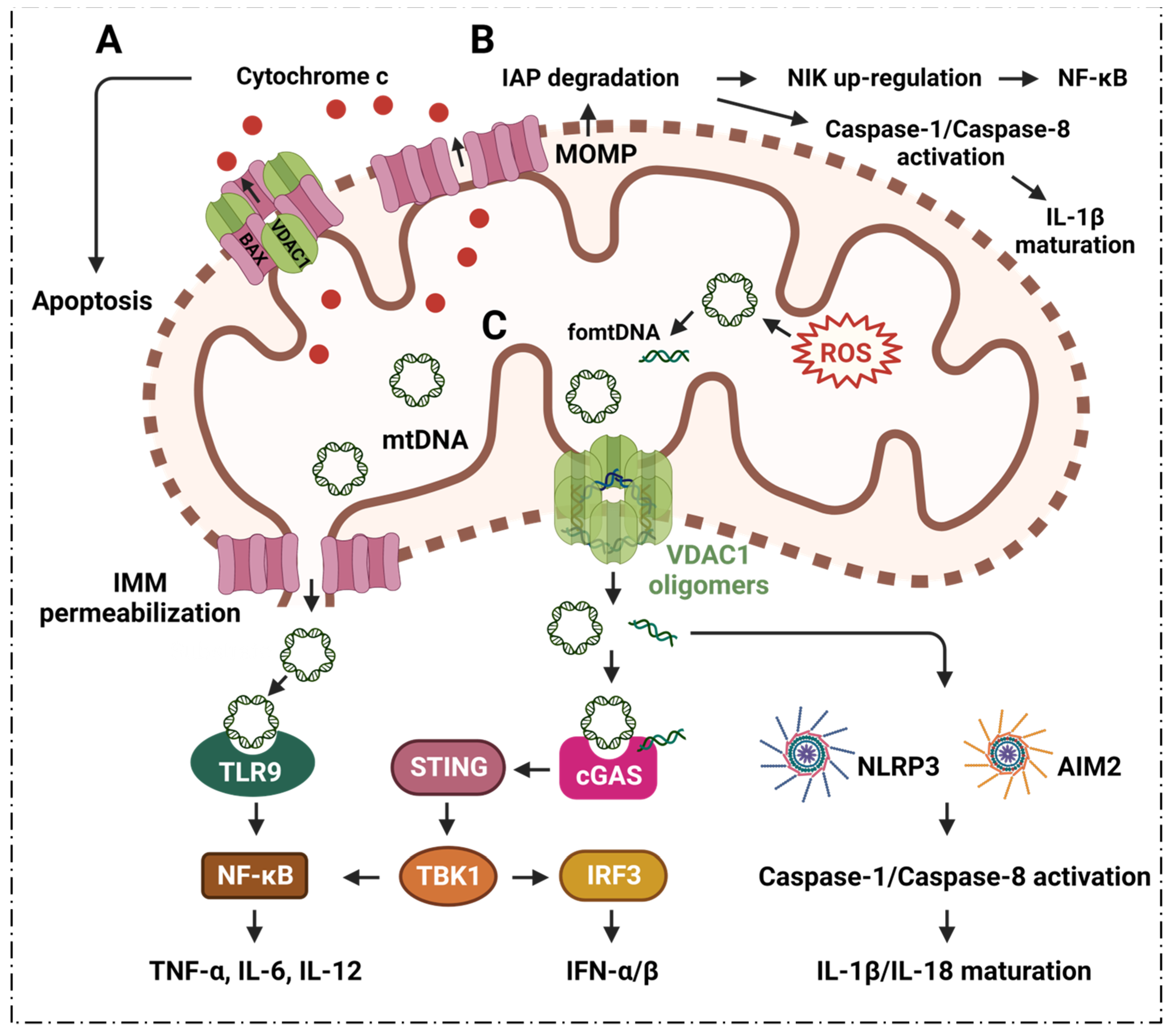
2.2. VDAC1 Mediates Mitochondrial Oxidative Stress in Immune Responses

ROS from mitochondria can be dramatically induced under the stimulation of radiation, cigarette smoke, air pollution, inflammatory factors, tumor necrosis factor, hyperlipidemia, hypoxia, and so on. Notably, the ROS are mainly generated from the respiratory complex that is located in the IMM [50][51]. Malfunctioning mitochondria hyperproduce ROS which negatively affect other components of mitochondria, for example, mtDNA, membrane lipids, oxidative phosphorylation, etc. [52][53]. The mtDNA is mainly localized in the IMM, and mtDNA can easily be oxidized by ROS to generate oxidized mtDNA fragments (fomtDNA) [54]. The released mtDNA acts as ligands for different danger signal sensors, activating the innate immune response (Figure 1). These risk sensors include the cytoplasmic cyclic GMP-AMP synthase (cGAS); Toll-like receptor 9 (TLR9); nucleotide-binding domain and leucine-rich repeat (LRR) containing P3 (NLRP3) inflammasome; and absent in melanoma 2 (AIM2) inflammasome [50]. Through these pathways, mtDNA can induce the secretion of inflammatory cytokines, and induce the recruitment of immune cells at different sites, providing the conditions for inflammation in many diseases [50]. It has been shown that VDAC1 oligomer pores promote MOMP and allow the release of mtDNA into the cytoplasmic matrix in living cells, where mtDNA fragments escape from the mitochondria through direct interactions at the N-terminus of VDAC1 [28]. At the same time, the inhibition of VDAC1 oligomerization eliminates cytoplasmic and circulating mtDNA. Therefore, single-stranded or double-stranded DNA escapes into the cytoplasm through the permeability transition pore that is composed of VDAC1. VDAC1 indirectly participates in mtDNA induction by mediating the translocation of the subsequent mtDNA inflammatory response [28]. VBIT-3 and VBIT-4, as well as VBIT-12, were reported to interact with VDAC1 by disrupting its oligomerization, resulting in altered intracellular Ca2+ concentration and decreased ROS levels, thereby protecting mitochondrial malfunction related to apoptosis and inflammation [55][56]. This response was found to alleviate type 2 diabetes [57], lupus [28], atrial myocardium fibrosis [58], ulcerative colitis [59] and amyotrophic lateral sclerosis [56].

3. Inflammation and VDAC1 Mediates Mitochondrial Ca2+ Transportation

Mitochondrial Ca2+ uptake and release play a key role in cellular physiology by regulating intracellular Ca2+ signaling, energy metabolism and cell death [60]. The transportation of Ca2+ across the inner or outer mitochondrial membranes (IMM, OMM) is mainly mediated by several proteins, including VDAC1, mitochondrial Ca2+ monotransporter (MCU) and Na+-dependent mitochondrial Ca2+ efflux transporter (NCLX) [61][62].
VDAC1 was shown to be highly permeable to Ca2+, and contains a binding site for ruthenium red, thereby inhibiting channel opening [63][64]. VDAC1 may be a key component of the mitochondrial Ca2+ homeostatic mechanism, enhancing the Ca2+ response through different mechanisms. VDAC1 acts as a large conductance channel that allows for the rapid diffusion of Ca2+ across the OMM, thereby allowing the exposure of low-affinity single transporters in the inner membrane to the high Ca2+ microdomains that are generated by the opening of the endoplasmic reticulum (ER)-Ca2+ channel [63][64].
Moreover, VDAC1 has been proposed to be part of a larger complex of members, including the adenine nucleotide transporter, cyclophilin D, peripheral benzodiazepine receptors and Bcl-2 family members [65], which can interact with the ER. The structural components interact with each other, and thus become part of the molecular mechanism of mitochondrial docking with Ca2+. VDAC1 is the major permeation pathway for Ca2+ across the OMM, and VDAC1 mediates Ca2+ transport through the OMM to the IMM space. It can also facilitate Ca2+ transport from the inner mitochondrial membrane space (IMS) into the cytoplasm [61] (Figure 2).
Figure 2. VDAC1 is involved in Ca2+ transportation, lipid metabolism, glycolysis, TCA cycleand in inflammatory responses. (A) VDAC1 regulates Ca2+ transportation, glycolysis, TCA cycle and lipid metabolism. VDAC1 transports Ca2+ between the mitochondria and cytoplasm to maintain calcium homeostasis. In the energy generation system, the VDAC1 pore maintains substrates, metabolites, biomolecules, etc., in a balanced manner to sustain salutogenesis. (B) Ca2+ signaling affects the inflammatory responses of neutrophils, macrophages, dendritic cells and CD4+ T cells. (C) Inflammatory responses of macrophages, dendritic cells and CD4+ T cells are promoted by glycolysis/TCA cycle energy generation pathways. Abbreviations: VDAC1: voltage-dependent anion channel 1; TRPML1: also known as MCOLN1, mucolipin TRP cation channel 1; GRP75: glucose-regulated protein 75; IP3R: inositol 1,4,5-trisphosphate receptor; DJ1: deglycase DJ-1, also known as Parkinson disease protein 7, is encoded by the PARK7 gene in human; RyR2: ryanodine receptor 2; CPT1a: carnitine palmitoyltransferase 1A; ACSL: long-chain acyl-CoA synthase; TCA cycle: tricarboxylic acid cycle; HK: hexokinase; ATP: adenosine triphosphate; ADP: adenosine diphosphate; NADH: nicotinamide adenine dinucleotide hydrogen; PEP: phosphoenolpyruvate; Th: T helper.
Ca2+ is an important regulatory point of barrier function and inflammation. Ca2+ influx is involved in many steps of the inflammatory cascade, including leukocyte rolling, arrest, adhesion, and ultimately, transendothelial migration, etc. [66]. Ca2+ is involved in lymphocytic responses to foreign antigens, and inositol triphosphates (InsP3) are generated as a result of foreign molecules binding to antigen receptors and stimulating Ca2+ release from internal storage [67]. Once these stores are emptied, store-operated Ca2+ channels (SOCs) are activated, allowing lymphocytes to maintain a long-term increase in Ca2+; this usually occurs in the form of a series of regular Ca2+ oscillations that activate nuclear factor of activated T cells (NF-AT) [67]

3.1. Neutrophils

Neutrophils are the most abundant type of white blood cells, and are the first responders to inflammatory stimuli, such as bacterial infection, or tissue damage medium caused by polarization and migration of mediators such as formyl-Met-Leu-Phe (fMLP) and IL-8 [68][69], whose dysfunction often leads to severe infections and inflammatory autoimmune diseases.
In neutrophils, the cytoplasmic free calcium concentration is an important determinant of cell viability, and is a marker of neutrophil activation; it is closely related to a range of neutrophil functions [70]. Rapid cell spreading in neutrophils is induced by Ca2+ signaling [71]; Ca2+ influx activates cytoplasmic calpain, which plays an important role in regulating neutrophil polarization, and in directing their migration toward chemotactic stimuli [68]. The entry of extracellular Ca2+ into neutrophils affects multiple functions, including phagocytosis, ROS production, vesicle secretion and degranulation, β2-integrin activation, and cytoskeletal rearrangement that leads to polarization and migration; these activities play a key role in the occurrence and development of the neutrophil inflammatory response [72][73] (Figure 2B).

3.2. Macrophages

Ultrasound, combined with endogenous protoporphyrin IX derived from 5-aminolevulinic acid (ALA-SDT), induce the apoptosis of macrophages [74]. The inhibition of VDAC1 by 4,4′-diisothiocyanostilbene-2,2′-disulfonic acid (DIDS) was found to prevent ALA-SDT-induced cell apoptosis in THP-1 macrophages [74]. The VDAC1 of the mycobacterium avium phagosome is associated with bacterial survival and lipid export in macrophages [75]. Macrophages are an important part of the innate immune system; their main function is to phagocytose and digest cell debris and pathogens, and they play an important role in inflammatory responses [76][77].
Ca2+ is an essential second messenger in phagocytosis; indeed, elevated cytosolic calcium concentrations are required for efficient phagocytosis and maturation of phagosomes [78]; blocking MCU inhibits macrophage phagocytosis [79]. Studies have shown that Ca2+ influx into macrophages is the trigger for macrophage activation, and that increased Ca2+ concentrations are associated with macrophage differentiation [80]. An influx of extracellular Ca2+ is required to polarize macrophages toward the pro-inflammatory M1 phenotype, while decreasing Ca2+ leads to anti-inflammatory M2 switching [79][81] (Figure 2B).

3.3. Dendritic Cells

Dendritic cells (DCs) are the most powerful antigen-presenting cells in the body. DCs uptake, process and present antigens efficiently that are crucial for initiating T cell responses. They play a central role in initiating, regulating and maintaining immune responses [82].
Ca2+ signaling plays a key role in the function of DCs. Migration of DCs to secondary lymphoid organs is indispensable for subsequent T helper cell-mediated adaptive immunity. It has been shown that chemokine-induced DC migration is Ca2+-dependent [83]. Ca2+ is involved in the regulation of chemokine receptor expression, cell swelling, cytoskeletal changes and amphipod formation activities; DC migration relies tightly on the cytosolic Ca2+ concentration [84]. Activated DCs rapidly up-regulate chemokine receptor 7 (CCR7) expression, and acquire the ability to migrate into afferent lymphatics and drain lymph nodes [85]. CCR7 is a G protein-coupled receptor [86] that regulates DC chemotaxis, survival, migration velocity, cellularity and endocytosis; furthermore, its activation is accomplished by inducing the mobilization of intracellular calcium stores through the inositol 1,4,5-triphosphate (IP3) pathway [87][88]. Ca2+ plays an important role in the inflammatory response because it regulates the function of DCs in various links (Figure 3B).

4. Inflammatory Diseases and VDAC1 in Energy Metabolism

4.1. TCA Cycle

Metabolites in the process of energy metabolism can participate in inflammatory responses through different pathways, affecting the secretion of cytokines, the production of pro-inflammatory mediators, and the activation and differentiation of immune cells. VDAC1 plays an important role in energy metabolism, and participates in the inflammatory response by directly mediating the transport of metabolites during respiration and regulating Ca2+ as well as the activity of respiration-related enzymes (Figure 2A,C). Succinic acid is one of the metabolites that accumulates from the disturbance of the TCA cycle and the breakdown of hyperglutamine. Succinate accumulation leads to macrophage M1 polarization through the direct inhibition of proline hydrolase, prompting HIF-1α and IL-1β secretion [89][90]; it acts as an inflammatory stimulator in an autocrine-dependent manner [89][91]. Lipopolysaccharide (LPS)-induced succinate promotes IL-1β expression via HIF-1α signaling [90][92]. Extracellular succinate induces a pro-inflammatory response in diverse immune cells, increasing the migration and secretion of pro-inflammatory cytokines TNF-α and IL-1β in dendritic cells and macrophages [90].

4.2. Glycolysis

The initial and rate-limiting steps of glycolysis are mainly catalyzed by HK1, most of which is bound to the OMM, mainly through mitochondria formed by VDAC1 and adenine nucleotide translocator (ANT) intermembrane contact sites for transport [93]. It has also been shown that Hexokinase-2 (HK2) binds to VDAC1 on the OMM to facilitate the preferential entry of ATP into HK2 for glycolysis [94]. The binding of HK2 with mitochondrial VDAC1 can be inhibited by chrysin, resulting in decreased glucose uptake and lactate production [95]. VDAC1 is directly involved in the regulation of the glycolytic pathway; it affects the activation, differentiation and migration of various immune cells such as macrophages, DCs, T cells, etc., and affects the production, migration, and release of various cytokines and pro-inflammatory mediators.
VDAC1 can affect mitochondrial respiration, as a result of its important role in controlling the transportation of substances and metabolites. The intermediates in the Krebs cycle have a close relation with the inflammation process [96]. The metabolism of PEP, lactic acid, succinic acid, citric acid, etc., plays an important role in the occurrence and development of inflammation. In conclusion, VDAC1 could become a new therapeutic target for inflammation, and this necessitates further study.

5. Inflammatory Diseases and VDAC1 in Lipid Metabolism

VDAC1 is involved in cholesterol transport, and is generally considered to be part of a complex that mediates fatty acid transport through the OMM [97][98][99]. Meanwhile, VDAC1 also serves as an anchoring site for long-chain acyl-CoA synthase (ACSL), which is associated with the outer surface of the OMM, and for carnitine palmitoyltransferase 1a (CPT1a), which faces the intermembrane space [99] (Figure 2A). ACSL catalyzes the synthesis of fatty acyl-CoA in vivo, which is the first reaction in the human body to utilize fatty acids; meanwhile, CPT1a is involved in the process of transporting long-chain fatty acids into the mitochondria so that fatty acids can be broken down to generate usable energy for cells. It has been reported that CPT1a, ACSL and VDAC1 can form a complex, and that the long-chain fatty acyl-CoA synthesized by ACSL is transferred from the OMM to the intermembrane space through VDAC1; furthermore, CPT1a converts acyl-CoA into long-chain fatty acylcarnitine [99], followed by a series of subsequent oxidation reactions.
It has been found that the phosphorylation state of VDAC1 mediated by glycogen synthase kinase 3 (GSK3) can control the permeability of the OMM [100]. It has been observed that a loss of VDAC1 may cause mitochondria to stop oxidizing fatty acids, and VDAC1 inhibitors can inhibit palmitate oxidation [101][102]. In addition, the VDAC1-based peptide, R-Tf-D-LP4, can stimulate catabolic pathways that are involved in promoting fatty acid transfer to the mitochondria, fatty acid oxidation and increasing the expressions of enzymes and factors that are associated with fatty acid transport to the mitochondria, thereby enhancing β-oxidation and production of energy [101].
VDAC1 is involved in cholesterol transport, and is generally considered to be part of a complex that mediates fatty acid transport through the OMM [97][98][99]. Meanwhile, VDAC1 also serves as an anchoring site for long-chain acyl-CoA synthase (ACSL), which is associated with the outer surface of the OMM, and for carnitine palmitoyltransferase 1a (CPT1a), which faces the intermembrane space [99] (Figure 2A). ACSL catalyzes the synthesis of fatty acyl-CoA in vivo, which is the first reaction in the human body to utilize fatty acids; meanwhile, CPT1a is involved in the process of transporting long-chain fatty acids into the mitochondria so that fatty acids can be broken down to generate usable energy for cells. It has been reported that CPT1a, ACSL and VDAC1 can form a complex, and that the long-chain fatty acyl-CoA synthesized by ACSL is transferred from the OMM to the intermembrane space through VDAC1; furthermore, CPT1a converts acyl-CoA into long-chain fatty acylcarnitine [99], followed by a series of subsequent oxidation reactions.
It has been found that the phosphorylation state of VDAC1 mediated by glycogen synthase kinase 3 (GSK3) can control the permeability of the OMM [100]. It has been observed that a loss of VDAC1 may cause mitochondria to stop oxidizing fatty acids, and VDAC1 inhibitors can inhibit palmitate oxidation [101][102]. In addition, the VDAC1-based peptide, R-Tf-D-LP4, can stimulate catabolic pathways that are involved in promoting fatty acid transfer to the mitochondria, fatty acid oxidation and increasing the expressions of enzymes and factors that are associated with fatty acid transport to the mitochondria, thereby enhancing β-oxidation and production of energy [101]. There are experimental results that show that R-Tf-D-LP4 significantly reduces pathophysiological features, such as hepatocyte ballooning, and inflammation and liver fibrosis in the HFD-32/STAM mouse model that is associated with steatohepatitis and/or NASH; meanwhile, this peptide also reduces the expression of inflammatory macrophages and cytokines (IL-1β and IL-6) in the liver of HFD-32-fed mice [101]. Dysfunction or deletion of VDAC1 will lead to fat deposition and abnormal lipid metabolism, increasing inflammatory macrophages and the expression of cytokines (IL-1β and IL-6). Heightened expression of IL-6 increases autocrine IL-4, which enhances Th2-type immune responses through automated feedback loops, playing an important role in inflammation [101].

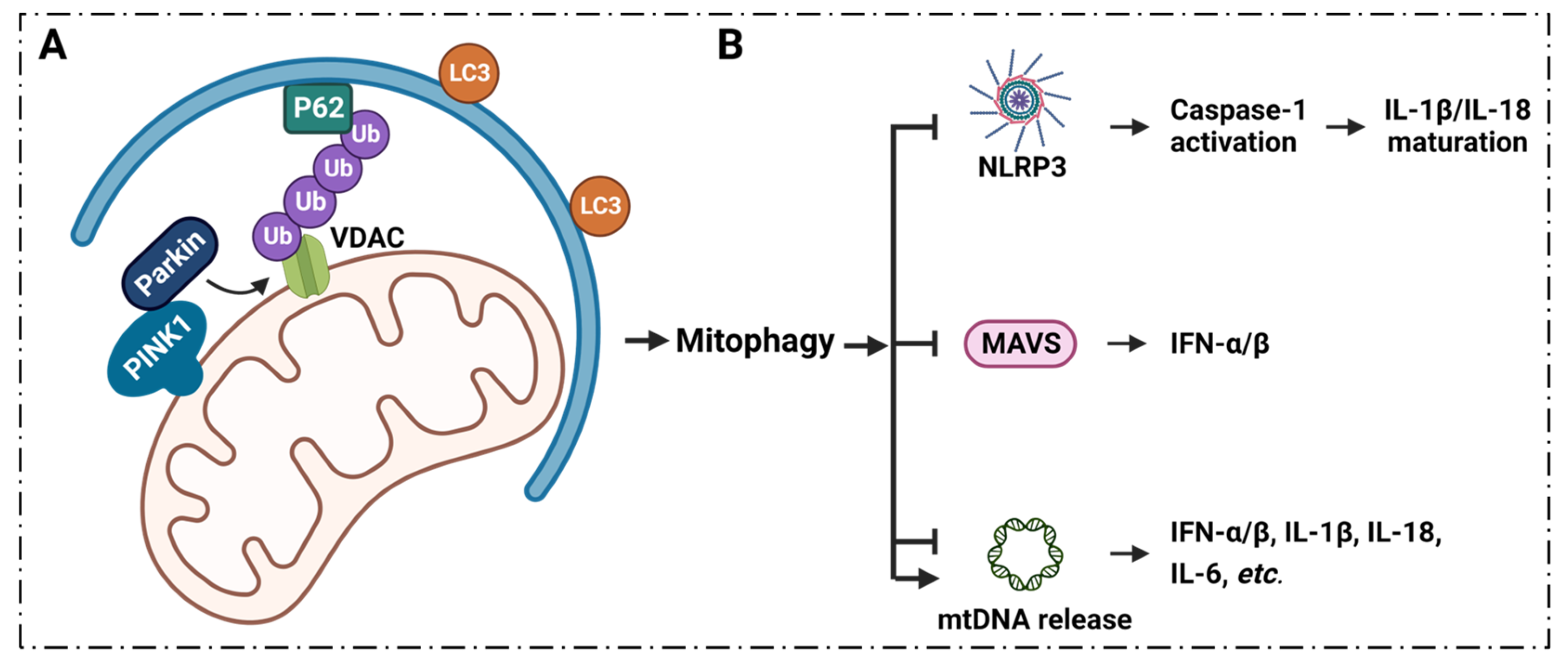
6. Inflammatory Diseases Pathogenesis and VDAC1 in Mitophagy

6.1. Mitophagy Regulates Inflammation via VDAC1

Mitophagy plays a key role in the regulation of inflammatory signaling, and the process of mitophagy limits inflammatory cytokines secretion [32][33][103][104][105] (Figure 3).
Figure 3. VDAC1, PINK1/Parkin signaling and mitophagy. (A) PINK1/Parkin targets damaged mitochondria, ubiquitinates VDAC1, and ultimately degrades damaged mitochondria by promoting mitophagy. (B) Mitophagy modulates NLRP3, MAVS and mtDNA release, affecting the immune response. NLRP3 activates caspase-1 to promote IL-1β/IL-18 maturation. MAVS enhance IFN-α/β expression. Meanwhile, mitophagy suppresses NLRP3, MAVS and mtDNA release, which results in reduced cytokines release. Additionally, mitophagy can also promote mtDNA release, which affects cytokines expression. Abbreviations: IL: interleukin; IFN: interferon; LC3: microtubule-associated proteins 1A/1B light chain 3; MAVS: mitochondrial antiviral signaling protein; NLRP3: nucleotide-binding domain and leucine-rich repeat (LRR) containing P3; PINK1: PTEN-induced putative kinase 1;; Ub: ubiquitin; VDAC1: voltage-dependent anion channel 1.
The PTEN-induced putative kinase 1 (PINK1) and the RING family ubiquitin ligase Parkin were found to be involved in mitophagy [106][107][108][109]. This indicates that induced mitophagy can be accomplished in cells that overexpress Parkin or overexpress PINK1. PINK1/Parkin acts as key regulator of mitophagy, and is vital in controlling infection and the inflammatory response [110]. The interaction of two Parkin domains, RING1 and ubiquitin-like (UBL), affects its activity. UBL binding with RING1 results in the inactive state of Parkin; PINK1 phosphorylates UBL-Ser65, leading to the activation of Parkin to promote substrate ubiquitination, with VDAC1 included [111]

7. Summary and Conclusions, Current Clinical Conditions and Future Perspectives

Mitochondria are fundamental organelles that execute and coordinate various metabolic processes in cells. Mitochondria are key organelles that are associated with cellular functions, and well-functioning mitochondria are critical to maintaining tissue homeostasis [112]. Mitochondrial malfunction is a sign of oxidative stress, inflammation, aging and chronic degenerative diseases [112][113][114]. VDAC1 is an important regulator of mitochondrial function, and acts as a mitochondrial gatekeeper that is responsible for cell fate [115].
Cutting-edge research confirms that VDAC is essential for the apoptotic “Find me signaling” pathway that results from the failure of apoptotic cell clearance, and leads to the pathogenesis of cystic fibrosis, followed by sterile inflammation [116]. Ulcerative colitis (UC) may be promoted by VDAC1 overexpression, and novel interacting targets for the treatment of UC based on VDAC1 are being developed for inflammatory and/or autoimmune diseases [59]. In addition, research suggests that VDAC1 is also related to cardiovascular and cerebrovascular diseases [117]. Furthermore, VDAC1 is widely involved in cancer [24][118], neurodegenerative diseases [119][120], diabetes [57][121], kidney disease [122][123], aging [124] and other areas of study. These all suggest that VDAC1 is a reasonable target to develop the next generation of therapeutic drugs.
 

This entry is adapted from the peer-reviewed paper 10.3390/cells11193174

References

  1. Medzhitov, R. Inflammation 2010: New adventures of an old flame. Cell 2010, 140, 771–776.
  2. Scrivo, R.; Vasile, M.; Bartosiewicz, I.; Valesini, G. Inflammation as “common soil” of the multifactorial diseases. Autoimmun. Rev. 2011, 10, 369–374.
  3. Medzhitov, R. Origin and physiological roles of inflammation. Nature 2008, 454, 428–435.
  4. Hengartner, M.O. The biochemistry of apoptosis. Nature 2000, 407, 770–776.
  5. Nunnari, J.; Suomalainen, A. Mitochondria: In sickness and in health. Cell 2012, 148, 1145–1159.
  6. Pfanner, N.; Warscheid, B.; Wiedemann, N. Mitochondrial proteins: From biogenesis to functional networks. Nat. Rev. Mol. Cell Biol. 2019, 20, 267–284.
  7. Bock, F.J.; Tait, S.W.G. Mitochondria as multifaceted regulators of cell death. Nat. Rev. Mol. Cell Biol. 2020, 21, 85–100.
  8. Tait, S.W.; Green, D.R. Mitochondrial regulation of cell death. Cold Spring Harb. Perspect. Biol. 2013, 5, a008706.
  9. Marchi, S.; Guilbaud, E.; Tait, S.W.G.; Yamazaki, T.; Galluzzi, L. Mitochondrial control of inflammation. Nat. Rev. Immunol. 2022, 1–15.
  10. Schapira, A.H. Mitochondrial diseases. Lancet 2012, 379, 1825–1834.
  11. Gorman, G.S.; Chinnery, P.F.; DiMauro, S.; Hirano, M.; Koga, Y.; McFarland, R.; Suomalainen, A.; Thorburn, D.R.; Zeviani, M.; Turnbull, D.M. Mitochondrial diseases. Nat. Rev. Dis. Prim. 2016, 2, 16080.
  12. Wallace, D.C. Mitochondrial genetic medicine. Nat. Genet. 2018, 50, 1642–1649.
  13. Schein, S.J.; Colombini, M.; Finkelstein, A. Reconstitution in planar lipid bilayers of a voltage-dependent anion-selective channel obtained from paramecium mitochondria. J. Membr. Biol. 1976, 30, 99–120.
  14. Di Rosa, M.C.; Guarino, F.; Conti Nibali, S.; Magrì, A.; De Pinto, V. Voltage-Dependent Anion Selective Channel Isoforms in Yeast: Expression, Structure, and Functions. Front. Physiol. 2021, 12, 675708.
  15. Guardiani, C.; Magrì, A.; Karachitos, A.; Di Rosa, M.C.; Reina, S.; Bodrenko, I.; Messina, A.; Kmita, H.; Ceccarelli, M.; De Pinto, V. yVDAC2, the second mitochondrial porin isoform of Saccharomyces cerevisiae. Biochim. Biophys. Acta Bioenerg. 2018, 1859, 270–279.
  16. De Stefani, D.; Bononi, A.; Romagnoli, A.; Messina, A.; De Pinto, V.; Pinton, P.; Rizzuto, R. VDAC1 selectively transfers apoptotic Ca2+ signals to mitochondria. Cell Death Differ. 2012, 19, 267–273.
  17. Messina, A.; Reina, S.; Guarino, F.; De Pinto, V. VDAC isoforms in mammals. Biochim. Biophys. Acta 2012, 1818, 1466–1476.
  18. Zinghirino, F.; Pappalardo, X.G.; Messina, A.; Guarino, F.; De Pinto, V. Is the secret of VDAC Isoforms in their gene regulation? Characterization of human VDAC genes expression profile, promoter activity, and transcriptional regulators. Int. J. Mol. Sci. 2020, 21, 7388.
  19. Cheng, E.H.; Sheiko, T.V.; Fisher, J.K.; Craigen, W.J.; Korsmeyer, S.J. VDAC2 inhibits BAK activation and mitochondrial apoptosis. Science 2003, 301, 513–517.
  20. Reina, S.; Nibali, S.C.; Tomasello, M.F.; Magrì, A.; Messina, A.; De Pinto, V. Voltage Dependent Anion Channel 3 (VDAC3) protects mitochondria from oxidative stress. Redox Biol. 2022, 51, 102264.
  21. Zinghirino, F.; Pappalardo, X.G.; Messina, A.; Nicosia, G.; De Pinto, V.; Guarino, F. VDAC Genes Expression and Regulation in Mammals. Front. Physiol. 2021, 12, 708695.
  22. Hiller, S.; Garces, R.G.; Malia, T.J.; Orekhov, V.Y.; Colombini, M.; Wagner, G. Solution structure of the integral human membrane protein VDAC-1 in detergent micelles. Science 2008, 321, 1206–1210.
  23. Ham, S.J.; Lee, D.; Yoo, H.; Jun, K.; Shin, H.; Chung, J. Decision between mitophagy and apoptosis by Parkin via VDAC1 ubiquitination. Proc. Natl. Acad. Sci. USA 2020, 117, 4281–4291.
  24. Shoshan-Barmatz, V.; Golan, M. Mitochondrial VDAC1: Function in cell life and death and a target for cancer therapy. Curr. Med. Chem. 2012, 19, 714–735.
  25. Shoshan-Barmatz, V.; De Pinto, V.; Zweckstetter, M.; Raviv, Z.; Keinan, N.; Arbel, N. VDAC, a multi-functional mitochondrial protein regulating cell life and death. Mol. Asp. Med. 2010, 31, 227–285.
  26. Huang, H.; Hu, X.; Eno, C.O.; Zhao, G.; Li, C.; White, C. An interaction between Bcl-xL and the voltage-dependent anion channel (VDAC) promotes mitochondrial Ca2+ uptake. J. Biol. Chem. 2013, 288, 19870–19881.
  27. Baines, C.P.; Kaiser, R.A.; Sheiko, T.; Craigen, W.J.; Molkentin, J.D. Voltage-dependent anion channels are dispensable for mitochondrial-dependent cell death. Nat. Cell Biol. 2007, 9, 550–555.
  28. Kim, J.; Gupta, R.; Blanco, L.P.; Yang, S.; Shteinfer-Kuzmine, A.; Wang, K.; Zhu, J.; Yoon, H.E.; Wang, X.; Kerkhofs, M.; et al. VDAC oligomers form mitochondrial pores to release mtDNA fragments and promote lupus-like disease. Science 2019, 366, 1531–1536.
  29. Xian, H.; Watari, K.; Sanchez-Lopez, E.; Offenberger, J.; Onyuru, J.; Sampath, H.; Ying, W.; Hoffman, H.M.; Shadel, G.S.; Karin, M. Oxidized DNA fragments exit mitochondria via mPTP- and VDAC-dependent channels to activate NLRP3 inflammasome and interferon signaling. Immunity 2022, 55, 1370–1385.e1378.
  30. Wu, J.; Ma, Z.; Raman, A.; Beckerman, P.; Dhillon, P.; Mukhi, D.; Palmer, M.; Chen, H.C.; Cohen, C.R.; Dunn, T.; et al. APOL1 risk variants in individuals of African genetic ancestry drive endothelial cell defects that exacerbate sepsis. Immunity 2021, 54, 2632–2649.e2636.
  31. Onishi, M.; Yamano, K.; Sato, M.; Matsuda, N.; Okamoto, K. Molecular mechanisms and physiological functions of mitophagy. EMBO J. 2021, 40, e104705.
  32. Marek-Iannucci, S.; Ozdemir, A.B.; Moreira, D.; Gomez, A.C.; Lane, M.; Porritt, R.A.; Lee, Y.; Shimada, K.; Abe, M.; Stotland, A.; et al. Autophagy-mitophagy induction attenuates cardiovascular inflammation in a murine model of Kawasaki disease vasculitis. JCI Insight 2021, 6, e151981.
  33. Green, D.R.; Galluzzi, L.; Kroemer, G. Mitochondria and the autophagy-inflammation-cell death axis in organismal aging. Science 2011, 333, 1109–1112.
  34. Sidarala, V.; Pearson, G.L.; Parekh, V.S.; Thompson, B.; Christen, L.; Gingerich, M.A.; Zhu, J.; Stromer, T.; Ren, J.; Reck, E.C.; et al. Mitophagy protects β cells from inflammatory damage in diabetes. JCI Insight 2020, 5, e141138.
  35. Ip, W.K.E.; Hoshi, N.; Shouval, D.S.; Snapper, S.; Medzhitov, R. Anti-inflammatory effect of IL-10 mediated by metabolic reprogramming of macrophages. Science 2017, 356, 513–519.
  36. Camara, A.K.S.; Zhou, Y.; Wen, P.C.; Tajkhorshid, E.; Kwok, W.M. Mitochondrial VDAC1: A Key Gatekeeper as Potential Therapeutic Target. Front. Physiol. 2017, 8, 460.
  37. Suomalainen, A.; Battersby, B.J. Mitochondrial diseases: The contribution of organelle stress responses to pathology. Nat. Rev. Mol. Cell Biol. 2018, 19, 77–92.
  38. Giorgio, V.; Guo, L.; Bassot, C.; Petronilli, V.; Bernardi, P. Calcium and regulation of the mitochondrial permeability transition. Cell Calcium 2018, 70, 56–63.
  39. Urbani, A.; Giorgio, V.; Carrer, A.; Franchin, C.; Arrigoni, G.; Jiko, C.; Abe, K.; Maeda, S.; Shinzawa-Itoh, K.; Bogers, J.F.M.; et al. Purified F-ATP synthase forms a Ca2+-dependent high-conductance channel matching the mitochondrial permeability transition pore. Nat. Commun. 2019, 10, 4341.
  40. Moya, G.E.; Rivera, P.D.; Dittenhafer-Reed, K.E. Evidence for the Role of Mitochondrial DNA Release in the Inflammatory Response in Neurological Disorders. Int. J. Mol. Sci. 2021, 22, 7030.
  41. Gutiérrez-Aguilar, M.; Baines, C.P. Structural mechanisms of cyclophilin D-dependent control of the mitochondrial permeability transition pore. Biochim. Biophys. Acta 2015, 1850, 2041–2047.
  42. Jia, K.; Du, H. Mitochondrial Permeability Transition: A Pore Intertwines Brain Aging and Alzheimer’s Disease. Cells 2021, 10, 649.
  43. Green, D.R.; Reed, J.C. Mitochondria and apoptosis. Science 1998, 281, 1309–1312.
  44. Shimizu, S.; Narita, M.; Tsujimoto, Y. Bcl-2 family proteins regulate the release of apoptogenic cytochrome c by the mitochondrial channel VDAC. Nature 1999, 399, 483–487.
  45. Yang, J.; Liu, X.; Bhalla, K.; Kim, C.N.; Ibrado, A.M.; Cai, J.; Peng, T.I.; Jones, D.P.; Wang, X. Prevention of apoptosis by Bcl-2: Release of cytochrome c from mitochondria blocked. Science 1997, 275, 1129–1132.
  46. Peña-Blanco, A.; García-Sáez, A.J. Bax, Bak and beyond—Mitochondrial performance in apoptosis. FEBS J. 2018, 285, 416–431.
  47. Banerjee, J.; Ghosh, S. Bax increases the pore size of rat brain mitochondrial voltage-dependent anion channel in the presence of tBid. Biochem. Biophys. Res. Commun. 2004, 323, 310–314.
  48. Elmore, S. Apoptosis: A review of programmed cell death. Toxicol. Pathol. 2007, 35, 495–516.
  49. Wang, C.; Youle, R.J. The role of mitochondria in apoptosis. Annu. Rev. Genet. 2009, 43, 95–118.
  50. Pérez-Treviño, P.; Velásquez, M.; García, N. Mechanisms of mitochondrial DNA escape and its relationship with different metabolic diseases. Biochim. Biophys. Acta Mol. Basis Dis. 2020, 1866, 165761.
  51. Alexeyev, M.; Shokolenko, I.; Wilson, G.; LeDoux, S. The maintenance of mitochondrial DNA integrity—critical analysis and update. Cold Spring Harb. Perspect. Biol. 2013, 5, a012641.
  52. Moreira, P.I.; Carvalho, C.; Zhu, X.; Smith, M.A.; Perry, G. Mitochondrial dysfunction is a trigger of Alzheimer’s disease pathophysiology. Biochim. Biophys. Acta 2010, 1802, 2–10.
  53. Schmitt, K.; Grimm, A.; Kazmierczak, A.; Strosznajder, J.B.; Götz, J.; Eckert, A. Insights into mitochondrial dysfunction: Aging, amyloid-β, and tau-A deleterious trio. Antioxid. Redox Signal. 2012, 16, 1456–1466.
  54. Lee, S.R.; Han, J. Mitochondrial Nucleoid: Shield and Switch of the Mitochondrial Genome. Oxidative Med. Cell. Longev. 2017, 2017, 8060949.
  55. Ben-Hail, D.; Begas-Shvartz, R.; Shalev, M.; Shteinfer-Kuzmine, A.; Gruzman, A.; Reina, S.; De Pinto, V.; Shoshan-Barmatz, V. Novel Compounds Targeting the Mitochondrial Protein VDAC1 Inhibit Apoptosis and Protect against Mitochondrial Dysfunction. J. Biol. Chem. 2016, 291, 24986–25003.
  56. Shteinfer-Kuzmine, A.; Argueti-Ostrovsky, S.; Leyton-Jaimes, M.F.; Anand, U.; Abu-Hamad, S.; Zalk, R.; Shoshan-Barmatz, V.; Israelson, A. Targeting the Mitochondrial Protein VDAC1 as a Potential Therapeutic Strategy in ALS. Int. J. Mol. Sci. 2022, 23, 9946.
  57. Zhang, E.; Mohammed Al-Amily, I.; Mohammed, S.; Luan, C.; Asplund, O.; Ahmed, M.; Ye, Y.; Ben-Hail, D.; Soni, A.; Vishnu, N.; et al. Preserving Insulin Secretion in Diabetes by Inhibiting VDAC1 Overexpression and Surface Translocation in β Cells. Cell Metab. 2019, 29, 64–77.e66.
  58. Klapper-Goldstein, H.; Verma, A.; Elyagon, S.; Gillis, R.; Murninkas, M.; Pittala, S.; Paul, A.; Shoshan-Barmatz, V.; Etzion, Y. VDAC1 in the diseased myocardium and the effect of VDAC1-interacting compound on atrial fibrosis induced by hyperaldosteronism. Sci. Rep. 2020, 10, 22101.
  59. Verma, A.; Pittala, S.; Alhozeel, B.; Shteinfer-Kuzmine, A.; Ohana, E.; Gupta, R.; Chung, J.H.; Shoshan-Barmatz, V. The role of the mitochondrial protein VDAC1 in inflammatory bowel disease: A potential therapeutic target. Mol. Ther. J. Am. Soc. Gene Ther. 2022, 30, 726–744.
  60. Bravo-Sagua, R.; Parra, V.; López-Crisosto, C.; Díaz, P.; Quest, A.F.; Lavandero, S. Calcium Transport and Signaling in Mitochondria. Compr. Physiol. 2017, 7, 623–634.
  61. Shoshan-Barmatz, V.; De, S. Mitochondrial VDAC, the Na+/Ca2+ Exchanger, and the Ca2+ Uniporter in Ca2+ Dynamics and Signaling. Adv. Exp. Med. Biol. 2017, 981, 323–347.
  62. Ben-Hail, D.; Palty, R.; Shoshan-Barmatz, V. Measurement of mitochondrial Ca2+ transport mediated by three transport proteins: VDAC1, the Na+/Ca2+ exchanger, and the Ca2+ uniporter. Cold Spring Harb. Protoc. 2014, 2014, 161–166.
  63. Gincel, D.; Zaid, H.; Shoshan-Barmatz, V. Calcium binding and translocation by the voltage-dependent anion channel: A possible regulatory mechanism in mitochondrial function. Biochem. J. 2001, 358, 147–155.
  64. Abu-Hamad, S.; Sivan, S.; Shoshan-Barmatz, V. The expression level of the voltage-dependent anion channel controls life and death of the cell. Proc. Natl. Acad. Sci. USA 2006, 103, 5787–5792.
  65. Griesbeck, O.; Baird, G.S.; Campbell, R.E.; Zacharias, D.A.; Tsien, R.Y. Reducing the environmental sensitivity of yellow fluorescent protein. Mechanism and applications. J. Biol. Chem. 2001, 276, 29188–29194.
  66. Dalal, P.J.; Muller, W.A.; Sullivan, D.P. Endothelial Cell Calcium Signaling during Barrier Function and Inflammation. Am. J. Pathol. 2020, 190, 535–542.
  67. Berridge, M.J.; Bootman, M.D.; Lipp, P. Calcium—A life and death signal. Nature 1998, 395, 645–648.
  68. Lokuta, M.A.; Nuzzi, P.A.; Huttenlocher, A. Calpain regulates neutrophil chemotaxis. Proc. Natl. Acad. Sci. USA 2003, 100, 4006–4011.
  69. Kolaczkowska, E.; Kubes, P. Neutrophil recruitment and function in health and inflammation. Nat. Rev. Immunol. 2013, 13, 159–175.
  70. Immler, R.; Simon, S.I.; Sperandio, M. Calcium signalling and related ion channels in neutrophil recruitment and function. Eur. J. Clin. Investig. 2018, 48 (Suppl. S2), e12964.
  71. Dewitt, S.; Francis, R.J.; Hallett, M.B. Ca²⁺ and calpain control membrane expansion during the rapid cell spreading of neutrophils. J. Cell Sci. 2013, 126 Pt 20, 4627–4635.
  72. Nguyen, G.T.; Green, E.R.; Mecsas, J. Neutrophils to the ROScue: Mechanisms of NADPH Oxidase Activation and Bacterial Resistance. Front. Cell. Infect. Microbiol. 2017, 7, 373.
  73. Bouti, P.; Webbers, S.D.S.; Fagerholm, S.C.; Alon, R.; Moser, M.; Matlung, H.L.; Kuijpers, T.W. β2 Integrin Signaling Cascade in Neutrophils: More Than a Single Function. Front. Immunol. 2021, 11, 619925.
  74. Chen, H.; Gao, W.; Yang, Y.; Guo, S.; Wang, H.; Wang, W.; Zhang, S.; Zhou, Q.; Xu, H.; Yao, J.; et al. Inhibition of VDAC1 prevents Ca²⁺-mediated oxidative stress and apoptosis induced by 5-aminolevulinic acid mediated sonodynamic therapy in THP-1 macrophages. Apoptosis Int. J. Program. Cell Death 2014, 19, 1712–1726.
  75. Danelishvili, L.; Chinison, J.J.J.; Pham, T.; Gupta, R.; Bermudez, L.E. The Voltage-Dependent Anion Channels (VDAC) of Mycobacterium avium phagosome are associated with bacterial survival and lipid export in macrophages. Sci. Rep. 2017, 7, 7007.
  76. Hirayama, D.; Iida, T.; Nakase, H. The Phagocytic Function of Macrophage-Enforcing Innate Immunity and Tissue Homeostasis. Int. J. Mol. Sci. 2017, 19, 92.
  77. Arango Duque, G.; Descoteaux, A. Macrophage cytokines: Involvement in immunity and infectious diseases. Front. Immunol. 2014, 5, 491.
  78. Nunes, P.; Demaurex, N. The role of calcium signaling in phagocytosis. J. Leukoc. Biol. 2010, 88, 57–68.
  79. Tedesco, S.; Scattolini, V.; Albiero, M.; Bortolozzi, M.; Avogaro, A.; Cignarella, A.; Fadini, G.P. Mitochondrial Calcium Uptake Is Instrumental to Alternative Macrophage Polarization and Phagocytic Activity. Int. J. Mol. Sci. 2019, 20, 4966.
  80. Paredes-Gamero, E.J.; Leon, C.M.; Borojevic, R.; Oshiro, M.E.; Ferreira, A.T. Changes in intracellular Ca2+ levels induced by cytokines and P2 agonists differentially modulate proliferation or commitment with macrophage differentiation in murine hematopoietic cells. J. Biol. Chem. 2008, 283, 31909–31919.
  81. Kang, H.; Zhang, K.; Wong, D.S.H.; Han, F.; Li, B.; Bian, L. Near-infrared light-controlled regulation of intracellular calcium to modulate macrophage polarization. Biomaterials 2018, 178, 681–696.
  82. Hilligan, K.L.; Ronchese, F. Antigen presentation by dendritic cells and their instruction of CD4+ T helper cell responses. Cell. Mol. Immunol. 2020, 17, 587–599.
  83. Sumoza-Toledo, A.; Lange, I.; Cortado, H.; Bhagat, H.; Mori, Y.; Fleig, A.; Penner, R.; Partida-Sánchez, S. Dendritic cell maturation and chemotaxis is regulated by TRPM2-mediated lysosomal Ca2+ release. FASEB J. 2011, 25, 3529–3542.
  84. Worbs, T.; Hammerschmidt, S.I.; Förster, R. Dendritic cell migration in health and disease. Nat. Rev. Immunol. 2017, 17, 30–48.
  85. Braun, A.; Worbs, T.; Moschovakis, G.L.; Halle, S.; Hoffmann, K.; Bölter, J.; Münk, A.; Förster, R. Afferent lymph-derived T cells and DCs use different chemokine receptor CCR7-dependent routes for entry into the lymph node and intranodal migration. Nat. Immunol. 2011, 12, 879–887.
  86. Shao, Z.; Makinde, T.O.; Agrawal, D.K. Calcium-activated potassium channel KCa3.1 in lung dendritic cell migration. Am. J. Respir. Cell Mol. Biol. 2011, 45, 962–968.
  87. Rodríguez-Fernández, J.L.; Criado-García, O. The Chemokine Receptor CCR7 Uses Distinct Signaling Modules With Biased Functionality to Regulate Dendritic Cells. Front. Immunol. 2020, 11, 528.
  88. Riol-Blanco, L.; Sánchez-Sánchez, N.; Torres, A.; Tejedor, A.; Narumiya, S.; Corbí, A.L.; Sánchez-Mateos, P.; Rodríguez-Fernández, J.L. The chemokine receptor CCR7 activates in dendritic cells two signaling modules that independently regulate chemotaxis and migratory speed. J. Immunol. 2005, 174, 4070–4080.
  89. Viola, A.; Munari, F.; Sánchez-Rodríguez, R.; Scolaro, T.; Castegna, A. The Metabolic Signature of Macrophage Responses. Front. Immunol. 2019, 10, 1462.
  90. Tannahill, G.M.; Curtis, A.M.; Adamik, J.; Palsson-McDermott, E.M.; McGettrick, A.F.; Goel, G.; Frezza, C.; Bernard, N.J.; Kelly, B.; Foley, N.H.; et al. Succinate is an inflammatory signal that induces IL-1β through HIF-1α. Nature 2013, 496, 238–242.
  91. Littlewood-Evans, A.; Sarret, S.; Apfel, V.; Loesle, P.; Dawson, J.; Zhang, J.; Muller, A.; Tigani, B.; Kneuer, R.; Patel, S.; et al. GPR91 senses extracellular succinate released from inflammatory macrophages and exacerbates rheumatoid arthritis. J. Exp. Med. 2016, 213, 1655–1662.
  92. Mills, E.; O’Neill, L.A. Succinate: A metabolic signal in inflammation. Trends Cell Biol. 2014, 24, 313–320.
  93. Roberts, D.J.; Miyamoto, S. Hexokinase II integrates energy metabolism and cellular protection: Akting on mitochondria and TORCing to autophagy. Cell Death Differ. 2015, 22, 248–257.
  94. Zhang, D.; Yip, Y.M.; Li, L. In silico construction of HK2-VDAC1 complex and investigating the HK2 binding-induced molecular gating mechanism of VDAC1. Mitochondrion 2016, 30, 222–228.
  95. Xu, D.; Jin, J.; Yu, H.; Zhao, Z.; Ma, D.; Zhang, C.; Jiang, H. Chrysin inhibited tumor glycolysis and induced apoptosis in hepatocellular carcinoma by targeting hexokinase-2. J. Exp. Clin. Cancer Res. CR 2017, 36, 44.
  96. Mills, E.L.; Kelly, B.; O’Neill, L.A.J. Mitochondria are the powerhouses of immunity. Nat. Immunol. 2017, 18, 488–498.
  97. Shoshan-Barmatz, V.; Ben-Hail, D.; Admoni, L.; Krelin, Y.; Tripathi, S.S. The mitochondrial voltage-dependent anion channel 1 in tumor cells. Biochim. Biophys. Acta 2015, 1848, 2547–2575.
  98. Shoshan-Barmatz, V.; Pittala, S.; Mizrachi, D. VDAC1 and the TSPO: Expression, Interactions, and Associated Functions in Health and Disease States. Int. J. Mol. Sci. 2019, 20, 3348.
  99. Lee, K.; Kerner, J.; Hoppel, C.L. Mitochondrial carnitine palmitoyltransferase 1a (CPT1a) is part of an outer membrane fatty acid transfer complex. J. Biol. Chem. 2011, 286, 25655–25662.
  100. Martel, C.; Allouche, M.; Esposti, D.D.; Fanelli, E.; Boursier, C.; Henry, C.; Chopineau, J.; Calamita, G.; Kroemer, G.; Lemoine, A.; et al. Glycogen synthase kinase 3-mediated voltage-dependent anion channel phosphorylation controls outer mitochondrial membrane permeability during lipid accumulation. Hepatology 2013, 57, 93–102.
  101. Pittala, S.; Krelin, Y.; Kuperman, Y.; Shoshan-Barmatz, V. A Mitochondrial VDAC1-Based Peptide Greatly Suppresses Steatosis and NASH-Associated Pathologies in a Mouse Model. Mol. Ther. J. Am. Soc. Gene Ther. 2019, 27, 1848–1862.
  102. Turkaly, P.; Kerner, J.; Hoppel, C. A 22 kDa polyanion inhibits carnitine-dependent fatty acid oxidation in rat liver mitochondria. FEBS Lett. 1999, 460, 241–245.
  103. Xu, Y.; Shen, J.; Ran, Z. Emerging views of mitophagy in immunity and autoimmune diseases. Autophagy 2020, 16, 3–17.
  104. Ziegler, P.K.; Bollrath, J.; Pallangyo, C.K.; Matsutani, T.; Canli, Ö.; De Oliveira, T.; Diamanti, M.A.; Müller, N.; Gamrekelashvili, J.; Putoczki, T.; et al. Mitophagy in Intestinal Epithelial Cells Triggers Adaptive Immunity during Tumorigenesis. Cell 2018, 174, 88–101.e116.
  105. Ma, K.; Chen, G.; Li, W.; Kepp, O.; Zhu, Y.; Chen, Q. Mitophagy, Mitochondrial Homeostasis, and Cell Fate. Front. Cell Dev. Biol. 2020, 8, 467.
  106. Matsuda, N.; Sato, S.; Shiba, K.; Okatsu, K.; Saisho, K.; Gautier, C.A.; Sou, Y.S.; Saiki, S.; Kawajiri, S.; Sato, F.; et al. PINK1 stabilized by mitochondrial depolarization recruits Parkin to damaged mitochondria and activates latent Parkin for mitophagy. J. Cell Biol. 2010, 189, 211–221.
  107. Vives-Bauza, C.; Zhou, C.; Huang, Y.; Cui, M.; de Vries, R.L.; Kim, J.; May, J.; Tocilescu, M.A.; Liu, W.; Ko, H.S.; et al. PINK1-dependent recruitment of Parkin to mitochondria in mitophagy. Proc. Natl. Acad. Sci. USA 2010, 107, 378–383.
  108. Shiba-Fukushima, K.; Imai, Y.; Yoshida, S.; Ishihama, Y.; Kanao, T.; Sato, S.; Hattori, N. PINK1-mediated phosphorylation of the Parkin ubiquitin-like domain primes mitochondrial translocation of Parkin and regulates mitophagy. Sci. Rep. 2012, 2, 1002.
  109. Narendra, D.; Tanaka, A.; Suen, D.F.; Youle, R.J. Parkin is recruited selectively to impaired mitochondria and promotes their autophagy. J. Cell Biol. 2008, 183, 795–803.
  110. Cho, D.H.; Kim, J.K.; Jo, E.K. Mitophagy and Innate Immunity in Infection. Mol. Cells 2020, 43, 10–22.
  111. Ham, S.J.; Lee, S.Y.; Song, S.; Chung, J.R.; Choi, S.; Chung, J. Interaction between RING1 (R1) and the Ubiquitin-like (UBL) Domains Is Critical for the Regulation of Parkin Activity. J. Biol. Chem. 2016, 291, 1803–1816.
  112. Picca, A.; Lezza, A.M.S.; Leeuwenburgh, C.; Pesce, V.; Calvani, R.; Landi, F.; Bernabei, R.; Marzetti, E. Fueling Inflamm-Aging through Mitochondrial Dysfunction: Mechanisms and Molecular Targets. Int. J. Mol. Sci. 2017, 18, 933.
  113. Picca, A.; Calvani, R.; Coelho-Junior, H.J.; Marzetti, E. Cell Death and Inflammation: The Role of Mitochondria in Health and Disease. Cells 2021, 10, 537.
  114. Missiroli, S.; Genovese, I.; Perrone, M.; Vezzani, B.; Vitto, V.A.M.; Giorgi, C. The Role of Mitochondria in Inflammation: From Cancer to Neurodegenerative Disorders. J. Clin. Med. 2020, 9, 740.
  115. Shoshan-Barmatz, V.; Shteinfer-Kuzmine, A.; Verma, A. VDAC1 at the Intersection of Cell Metabolism, Apoptosis, and Diseases. Biomolecules 2020, 10, 1485.
  116. Thinnes, F.P. Opening up of plasmalemma type-1 VDAC to form apoptotic “find me signal” pathways is essential in early apoptosis—Evidence from the pathogenesis of cystic fibrosis resulting from failure of apoptotic cell clearance followed by sterile inflammation. Mol. Genet. Metab. 2014, 111, 439–444.
  117. Karachitos, A.; Jordan, J.; Kmita, H. VDAC-Targeted Drugs Affecting Cytoprotection and Mitochondrial Physiology in Cerebrovascular and Cardiovascular Diseases. Curr. Med. Chem. 2017, 24, 4419–4434.
  118. Shoshan-Barmatz, V.; Mizrachi, D. VDAC1: From structure to cancer therapy. Front. Oncol. 2012, 2, 164.
  119. Shoshan-Barmatz, V.; Nahon-Crystal, E.; Shteinfer-Kuzmine, A.; Gupta, R. VDAC1, mitochondrial dysfunction, and Alzheimer’s disease. Pharmacol. Res. 2018, 131, 87–101.
  120. Liu, Y.; Ma, X.; Fujioka, H.; Liu, J.; Chen, S.; Zhu, X. DJ-1 regulates the integrity and function of ER-mitochondria association through interaction with IP3R3-Grp75-VDAC1. Proc. Natl. Acad. Sci. USA 2019, 116, 25322–25328.
  121. Sasaki, K.; Donthamsetty, R.; Heldak, M.; Cho, Y.E.; Scott, B.T.; Makino, A. VDAC: Old protein with new roles in diabetes. Am. J. Physiol. Cell Physiol. 2012, 303, C1055–C1060.
  122. Li, X.; Pan, J.; Li, H.; Li, G.; Liu, B.; Tang, X.; Liu, X.; He, Z.; Peng, Z.; Zhang, H.; et al. DsbA-L interacts with VDAC1 in mitochondrion-mediated tubular cell apoptosis and contributes to the progression of acute kidney disease. EBioMedicine 2022, 76, 103859.
  123. Nowak, G.; Megyesi, J.; Craigen, W.J. Deletion of VDAC1 Hinders Recovery of Mitochondrial and Renal Functions After Acute Kidney Injury. Biomolecules 2020, 10, 585.
  124. Shoshan-Barmatz, V.; Anand, U.; Nahon-Crystal, E.; Di Carlo, M.; Shteinfer-Kuzmine, A. Adverse Effects of Metformin From Diabetes to COVID-19, Cancer, Neurodegenerative Diseases, and Aging: Is VDAC1 a Common Target? Front. Physiol. 2021, 12, 730048.
More
This entry is offline, you can click here to edit this entry!
Video Production Service