Using Nanoparticles in Local Delivery to Cochlea: History
Please note this is an old version of this entry, which may differ significantly from the current revision.

Hearing loss negatively impacts the well-being of millions of people worldwide. Systemic delivery of ototherapeutics has limited efficacy due to severe systemic side effects and the presence of the blood–labyrinth barrier that selectively limits or enables transfer of molecules between plasma and inner ear tissues and fluids. Local drug delivery into the middle and inner ear would be preferable for many newly emerging classes of drugs. Although the cochlea is a challenging target for drug delivery, recent technologies could provide a safe and efficacious delivery of ototherapeutics. Local drug delivery routes include topical delivery via the external auditory meatus, retroauricular, transtympanic, and intracochlear delivery. 

  • drug delivery
  • blood–labyrinth barrier
  • otoprotective therapeutics
  • local delivery
  • cochlea

1. Introduction

The World Health Organization estimates that by 2050, over 700 million people, or one in every ten people, globally will experience disabling hearing loss [1]. Hearing loss negatively impacts personal well-being. Studies have shown that children and adults with hearing loss have a poorer quality of life due to reduced social interactions, isolation, a sense of exclusion, and depression [2], and in older people, this can lead to accelerated cognitive decline [3]. Hearing loss can result from many different types of inner ear disorders, including presbycusis (age-related hearing loss [ARHL]), genetic polymorphisms, trauma, exposure to noise, and ototoxic medications, to name a few. There are several therapeutic strategies to treat inner ear disorders, including systemic or local delivery of therapeutic agents, surgical intervention, and acoustic (e.g., hearing aids) and electric (cochlear implants) amplification [4].
Sensorineural hearing loss results from the dysfunction of the sensory and non-sensory cells, as well as neurons, present in the cochlea. Mammals, including humans, are unable to spontaneously regenerate many of these cell types. An active area of research is focused on preventing hearing loss due to treatment with cisplatin (and its derivatives) in anticancer chemotherapies, or aminoglycoside antibiotics for urgent treatment of severe bacterial infections [5]. Systemic delivery of ototherapeutics presumes that these drugs can reach their targets in their active form and at therapeutic concentrations. However, systemic administration of otoprotective drugs that achieve therapeutic concentrations in the inner ear often has limited efficacy due to unwanted systemic side effects from the high doses needed [6]. Hence, for otoprotective pharmacotherapies to be clinically relevant, it is imperative to develop safe and reliable drugs that can be delivered to the cochlea. 

2. Anatomy

The mammalian ear is a complex sensory organ critical to hearing and maintaining balance (Figure 1). The outer ear includes an external auditory meatus (ear canal) that is slightly curved [7]. The outer and middle ear are separated by the semitransparent tympanic membrane that is thinnest in the center (50–70 µm) and thickest (~100–120 µm) near the peripheral rim [8]. This membrane is concave, with its deepest point projecting into the middle ear cavity. The outer and innermost layers of the tympanic membrane are extensions of the epidermis lining the external auditory meatus and the mucosal epithelial layer lining the middle ear cavity. The middle, extracellular layer of the tympanic membrane (Figure 2A) is formed of radial and circular connective tissue comprised of collagen and elastic fibers and innervated by several cranial nerves [9].
Figure 1. A schematic summary of various drug delivery routes and methodologies to locally deliver therapeutics to the inner ear. Not to scale.
Figure 2. Schematic cross-sections of (A) the tympanic membrane; (B) the oval window membrane; and (C) the round window membrane. Not to scale.
The middle ear is a narrow, air-filled space with an opening in the anterior wall that leads to the nasopharynx—the Eustachian tube (Figure 1). The medial wall separates the middle ear from the inner ear and has two small openings: the oval window and the round window. These are considered the main passage routes for drugs entering the inner ear from the middle ear. There are three middle ear bones (ossicles), the malleus, incus, and stapes, that link the tympanic membrane with the cochlea. This ossicular chain acts as an impedance transformer, converting acoustic air pressure waves into fluid pressure waves at the oval window [7,10].
The cavity of the oval window is closed by the ossified footplate of the stapes, which is attached to the otic capsule by the annular ligament (Figure 2B) [11]. The average dimension of the oval window in humans is 1.26 mm at its narrowest and 2.40 mm at its widest [12]. The oval window has diverse neurovascular structures such as the ptotic facial nerve and the persistent stapedial artery. The oval window is medially bounded by the vestibule [11].
The round window is covered by a thin membrane known as the round window membrane (RWM; Figure 2C). The RWM is made up of a single layer of epithelial cells, with numerous cylindrical-shaped mitochondria, rough endoplasmic reticulum, a Golgi complex, and sparse microvilli in the middle ear cavity. The inner mesothelial cell layer of the RWM faces the perilymphatic space and contains ‘gaps’ between the epithelial cells, allowing direct contact of the intervening connective tissue with perilymph in the cochlear scala tympani [13].
The stapes and oval window, along with the RWM, serve as a physical barrier that protects the inner ear [16]. Locally-administered drugs in the middle ear can pass through the oval or round window membranes to reach the perilymph enclosed within the scala vestibuli or tympani, respectively [17]. The permeability of these membranes is dependent on drug charge, size, concentration, configuration, liposolubility, and thickness [18].

3. Delivery Routes

3.1. Systemic Delivery

Systemic delivery of therapeutics to the inner ear typically involves delivery via the vasculature and crossing the tight junction-coupled blood–labyrinth barrier (BLB). The BLB is fundamentally similar to the blood–brain barrier (BBB) and is well described elsewhere [24]. Paracellular flux across the BLB is not thought to occur under normal physiological conditions but may arise during inflammation, as for the BBB [25]. Drug transport across the BLB could involve similar mechanisms to that across the BBB [24,25,26]. These include (i) diffusion of lipophilic molecules (e.g., solvents) across cellular membranes; (ii) transcellular flux of hydrophilic drugs (e.g., aminoglycosides) via permeation of non-selective cation channels [27,28]; translocation via substrate transporters [29]; or transcytosis through the cell [26].

3.2. Topical Delivery via the External Auditory Meatus

Topical delivery of drugs to the ear canal is used to treat inflammation of the outer and middle ear. Antibacterial and antifungal ointments (or drops) are applied directly to the ear canal up to three times a day. The advantages of this administration route are that it is inexpensive, and it can be performed as an outpatient procedure or at home by the patient. There are a few disadvantages, including apparently limited therapeutic application, as it cannot currently be used for drug delivery to the inner ear as the tympanic and round window membranes are thought to act as barriers, as well as loss of drug from the middle ear via the Eustachian tube [32]. However, ototoxic drugs like gentamicin are not used clinically as ear drops as they can cause serious side effects such as hearing loss and tinnitus when used by patients with a perforated tympanic membrane [33], suggesting that topically-applied therapeutics can reach the inner ear. Moreover, concerns exist regarding the efficacy of topically-administered drugs against bacteria that have infected the middle ear and mastoid cavities [34].

3.3. Transtympanic Delivery

It is a minimally-invasive injection that is typically performed in a clinic setting. The major advantage of transtympanic delivery is that it provides a potential direct route of administration to the inner ear. Typically, transtympanic delivery of drugs is injected onto the round window (and round window niche) and relies on the permeation of compounds through the RWM into the perilymphatic scala tympani in the cochlea. Permeation of the RWM is dependent on various factors, including the drug size, charge, lipophilicity, concentration, and formulation of the substances [18]. Following administration to the middle ear, local absorption takes place via the round window and the oval window into the cochlear perilymph and subsequent diffusion into the vestibular system. Previous studies have demonstrated that prolonging the contact duration of the drug formulation with the RWM can increase drug levels in the inner ear, but the pharmacokinetics for individual drugs remain complex to interpret [35]. Permeation of drugs across the round window membrane depends on passive diffusion and active transport [31]. Manipulations of the RWM have been shown to increase the entry of drugs following transtympanic administration. A variety of agents have been screened that are used to improve drug penetration in other biological systems for their capacity to increase entry into the inner ear. Some of these agents include benzyl alcohol, dimethyl sulfoxide, saponin, caprate, and Poloxamer 407. Benzyl alcohol is commonly used as a preservative in drug formulations and has been found to be the most effective permeabilizing agent [36].

3.4. Retroauricular Delivery

Microcomputed tomography scanning of human temporal bone has revealed multiple large air-filled tuberculae, divided by bony septae. These air-filled spaces known as retroauricular microchannels are located behind the posterior wall of the ear canal and are rich in blood supply [40]. These microchannels have been utilized to deliver drugs to the middle or inner ear via parenteral retroauricular injections [41]. A study in a group of 20 healthy adults given a retroauricular injection of adrenaline found a significant drop in pressure in the middle ear cavity compared to the control group. It is thought that these microchannels enabled the adrenaline to enter the mastoid mucosa to induce vascular constriction and mucosal decongestion, leading to decreased middle ear pressure [42].

3.5. Intracochlear Application

Intracochlear delivery bypasses the tympanic, oval, and round window membranes by infusing drugs directly into perilymph within the cochlea, presumptively providing greater control over drug concentrations within the cochlea [14]. Intracochlear injection through the RWM provides higher and more sustained drug levels compared to transtympanic injection onto the RWM [16]. However, the perforation of the RWM caused by the needle can release inner ear pressure, allowing cerebrospinal fluid (CSF) to enter the scala tympani via the cochlear aqueduct [45].
Other intracochlear drug delivery methods include infusion via an osmotic minipump, or a catheter built into the electrode array of a cochlear implant with an implantable peristaltic pump attached to it [18,46]. The osmotic pump is implanted subcutaneously with a cannula threaded into the middle ear cavity and inserted through the RWM or through the bone directly into the cochlea via a cochleostomy. These pumps can provide continuous infusion of drugs for up to 6 weeks [47]. Osmotic pump delivery of betamethasone to treat a vestibular disorder in guinea pigs showed a shorter duration of recovery than in controls of untreated with no osmotic pump and saline delivered through osmotic pump groups [48]. Osmotic pump delivery of an anti-apoptotic agent, Z-VAD-FMK, infused into the cochlea of guinea pigs for 14 days revealed less noise-induced hearing loss and lower hair cell loss compared to noise-exposed, untreated ears [49]. However, osmotic pumps are limited in that drug delivery cannot be stopped, nor can the flow rate be changed once the pump is started, and the volume of vehicle plus drug infused over time can exceed the volume of perilymph in the inner ear [47]. Recently, a micropump with a better automated control of drugs for intracochlear delivery at safe and slower flow rates without increasing the volume of perilymph in the cochlea has been described [50].
Surgical implants, including cochlear implants, can rehabilitate severe-to-profound sensorineural hearing loss. Recently, intracochlear controlled release drug delivery in combination with cochlear implants has been developed. This can improve the performance of cochlear implants by preventing fibrosis induced by insertion trauma, protecting the neuronal structures and by providing controlled drug release [51]. However, potential limitations include an enhanced risk of infection, particularly meningitis, associated with the surgery [52]; damage to the facial nerve; and loss of residual hearing. An implantable peristaltic pump connected to a cochlear implant electrode array has been developed for long-term delivery and effective dose-control in non-human primates (macaques). The infusion time ranged from 2–24 h to reach maximum peak concentrations, demonstrating feasibility [46].

4. Localized Inner Ear Delivery Methods

Localized drug delivery to the cochlea has the advantages of targeted delivery with higher bioavailability and minimal systemic side effects. The ideal delivery system would be a carrier system that delivers pharmacotherapeutics across the intact tympanic, oval, and/or round window membranes to efficaciously treat inner ear disorders.
Before a carrier system is chosen, advanced knowledge of the preferred drug and its physicochemical properties is required to determine the appropriate formulation and delivery method. In the field of drug discovery, Lipinski’s rule of five is frequently used to predict the absorption and solubility properties of a drug [53,54], and includes molecular weight, lipophilicity, polar surface area, hydrogen bonding, and charge [55]. These properties assist in the design and screening of new candidate drugs by predicting if a chemical compound has the appropriate bioavailability and pharmacokinetics. The rule states that ideal candidate drugs will have a logP ≤ 5 (the partition coefficient of a molecule between aqueous and lipophilic phases [usually water and octanol]), a molecular mass ≤500, ≤10 hydrogen bond acceptors, and ≤5 hydrogen bond donors. Molecules that fail to follow one or more of these rules may have difficulty with bioavailability [56].
Given the uncertainty of how drugs are metabolized in the inner ear, it is paramount to determine the physicochemical properties of candidate drugs for inner ear delivery. These may differ from the well-established Lipinski rule of five for systemic drug development. Thus far, much of our knowledge of these physicochemical properties for inner ear delivery are based on a few empirical studies of selected agents such as local anesthetics, corticosteroids (e.g., dexamethasone), monoclonal antibodies, growth factors, apoptosis inhibitors, and vectors for gene therapy under clinical investigation [57,58]. Many more studies are required to establish the optimal physicochemical properties for efficacious inner ear therapeutics, e.g., the ability to cross the blood–labyrinth barrier, the tympanic, oval, or round window membranes, as well as intracochlear epithelial barriers.

4.1. Developing Different Injectable Solutions like Hydrogels

These hydrogels are fluid-like at room temperature and quickly gelate at body temperature to promote the sustained release of encapsulated drugs, increasing drug contact time with the RWM [61]. Hydrogels have numerous advantages, including increased biocompatibility, adjustable biodegradability, low toxicity, and good swelling behavior [62]. The degree of swelling is a critical parameter, with a higher concentration of polymers leading to greater swelling and slower drug release [63]. In the middle ear, hydrogels are typically injected near the RWM and RWM niche. This enables prolonged diffusion of the released drug across the RWM at appropriate therapeutic concentrations [64]. Hydrogels have been developed in several formulations for inner ear drug delivery, including polymers such as chitosan or PEG-based hydrogel, Poloxamer 407, and hyaluronic acid [65,66,67,68]. Injectable PEG-based hydrogel has been shown to be an effective and safe method for inner ear delivery. In guinea pigs, the dexamethasone concentrations in perilymph were maintained for at least 10 days for the PEG hydrogel as compared to the control sample of free dexamethasone [69]. Hyaluronic acid, when applied to the RWM of guinea pigs prior to delivering an adenovirus, provides an atraumatic and feasible method of delivering transgene into the inner ear [70].

4.2. Poloxamer 407 and Its Mechanism

Poloxamer 407, also known as Pluronic® F-127, is the primary polymer for formulating hydrogels used for inner ear delivery of therapeutics [59]. Poloxamer 407 is an amphiphilic, non-ionic triblock copolymer consisting of a residue of polyoxypropylene (POP) between two units of polyoxyethylene (POE). It is a widely used thermo-sensitive hydrogel due to its non-irritating action on biological membranes and can remain as a gel for several weeks to months [71]. Its thermo-sensitivity is due to the hydrophobic interactions between the copolymer chains of Poloxamer 407. As temperature increases, copolymer chains of Poloxamer 407 aggregate to form a micellar structure due to the dehydration of hydrophobic poly(propylene oxide) units [72] with a micelle diameter in the 20–100 nm range. The hydrophobic core is the drug loading site, creating space for the encapsulation of drugs through chemical interactions. The properties of the inner and outer shell determine the rate of drug release. Different methods are employed for encapsulating drugs in Poloxamer 407, such as direct dissolution, evaporation, and freeze-drying [73]. The gelation of Poloxamer 407 is reversible once gelling conditions, such as temperature, pH, or chemical, are removed [62].

4.3. Nanoparticulate Injection Systems

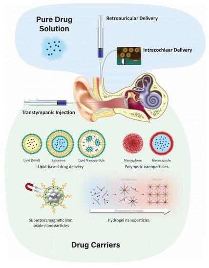
Nanoparticulate drug delivery is one of the most advanced technologies in drug design due to its advantages such as surface modification, improved drug solubility, stability, and bioavailability, as well as sustained controlled drug release at the target site. Injecting nanoparticulates at the targeted site leads to lower systemic toxicity, fewer side effects, improved kinetics of the drug, and extended drug bioavailability [77]. There are two primary nanoparticulate strategies: passive and self-delivery. In passive delivery, drugs are encapsulated in nanocarriers and are slowly released from the carriers. In self-delivery, drugs are conjugated to the carrier for easy delivery, and the drug dissociates from the carrier quickly at the presumptive targeted site, e.g., in the vicinity of tumors [78,79]. A large variety of nanoparticles have emerged, including polymeric, liposomes, and lipid-based structures, among others.

4.3.1. Polymeric Nanoparticles

Biodegradable polymeric microparticles or nanoparticles have been developed for a wide range of therapeutic applications and as inner ear drug delivery systems. They are often based on poly (lactic) co-glycolic acid (PLGA) or chitosan [6] and have advantages over other delivery systems due to their biocompatibility, biodegradability, small size, long shelf life, stability during storage, and highly reproducible formulation methods [59]. Nanoparticles have a diameter of <1 µm and are usually formulated with diameters of 100–300 nm, and for inner ear delivery, ~200 nm or less [4]. Polymeric nanoparticles can also incorporate visualization agents such as fluorescent dyes and MRI contrast agents [100]. Iron oxide nanoparticles have been extensively studied as a contrast agent for MRI. It has a magnetic core and different ligands focus on targeting specific sites or cells. PLGA is one of the more popular polymeric particles that can encapsulate both hydrophobic and hydrophilic drugs for intratympanic delivery and can be transported across the RWM into perilymph via the transcellular pathway [101].

4.3.2. Solid Lipid Nanoparticles

Solid lipid nanoparticles (SLNs) are a novel class of stable nanoparticles that are particularly suitable for the encapsulation of hydrophobic drugs such as curcumin, resveratrol, or quercetin [102]. They can also act as a carrier for hydrophilic drugs when formulated without the use of organic solvents [103]. SLNs are composed of a hydrophobic triglyceride core with an amphiphilic surfactant shell [104]. SLNs are biodegradable, biocompatible, and are non-ototoxic over a wide dose range. Low doses of hydrocortisone encapsulated in SLNs increase their protective effect and prolong the survival of auditory cells treated with cisplatin in vitro [97]. In vivo application of SLNs has shown no interference in hearing threshold or loss of hair cells [105].

4.3.3. Liposomes

Liposomes are composed of two layers of amphipathic molecules with a hydrophilic external layer and an internal surface composed of a phospholipid bilayer [106]. A key advantage of using liposomes for cochlear drug delivery is their ability to control drug release [96]. In an in vivo study, the microinjection of cationic liposome-mediated gene transfer into guinea pig cochleas revealed that transgene expression was steady for up to 14 days in the neurosensory epithelia and surrounding tissues without any toxicity [107]. Others have successfully demonstrated cell–gene delivery of therapeutic agents to the inner ear using a liposome-mediated delivery method [108].

4.3.4. Superparamagnetic Iron Oxide Nanoparticles (SPION)

SPIONs are Fe3O4 particles that can be magnetically controlled to focus on the migration of particles into the inner ear after crossing the RWM [60]. These particles are encapsulated in a polymeric layer of PLGA [109] or chitosan [110] to contain the therapeutic agent. Other polymers such as polyacrylic acid and polyvinylpyrrolidone are used to form a coat around iron oxide nanoparticles to increase stability and improve their magnetic properties [111]. This technique has been demonstrated in vivo in rat and guinea pig models, as well as in vitro in cell lines and the human temporal bone. Biocompatibility and safety have been demonstrated by various methods, including hair cell survival in organotypic cell cultures [112].

4.4. Advantages and Disadvantages of the Nanoparticulate Injection System

Nanoparticles are created from a variety of materials with a diameter range of 10 to several 100 nanometers, and customized to encapsulate various therapeutic agents [113]. Nanoparticles are widely used for non-invasive application, sustained, or controlled release of drugs, drug stabilization, and surface modification for targeting specific organs [114]. Various studies that administered nanoparticles onto the RWM have shown successful delivery of biomaterials into the inner ear [115]. Nanoparticles can enter the perilymph and the endolymph [106] following intratympanic administration and can be targeted to a specific intracochlear site of interest. Nanoparticles, when combined with hydrogel, improve the bioavailability of the drug at the targeted site and prevent rapid drug release [106]. Nanoparticles can also be conjugated to peptides that can penetrate cells, or their surface can be modified to increase their contact with RWM [116]. Challenges for nanoparticle delivery into the inner ear include limited access to the inner ear and poor uptake of drugs by inner ear cells [106].

4.5. Positively-Charged Biomaterials for Local Drug Delivery

The charge of nanoparticles can determine their uptake by inner ear hair cells and their penetration of epithelial membranes. Phospholipid-based nanoparticles with a positive charge of +26 mV were taken up by sensory hair cells at a two-fold higher rate than nanoparticles with a neutral or negative charge. This is likely due to the interaction between positively-charged nanoparticles and negatively-charged lipid plasma membranes [119]. The addition of cationic polyethylene glycol (PEG) to phospholipid-based nanoparticles increases their trafficking across the RWM but has enhanced cytotoxicity [120]. Nanoparticulates containing cationic-PEG to deliver dexamethasone to the RWM in mice provide an anti-inflammatory effect during combined kanamycin and furosemide treatment and higher cellular uptake within the organ of Corti [99]. Positively-charged chitosan nanoparticles enter the inner ear at a faster rate [59]. Positively-charged chitosan nanoparticles containing D-glucosamine and N-acetyl-D-glucosamine facilitate penetration of lipid cell membranes in the inner ear [119]. Other positively-charged nanoparticles, such as 1,2-dioleoyl-3-trimethylammonium-propane (DOTAP), are distributed widely in the inner ear after RWM application [14]. In rodents, positively-charged ferritin readily passes through RWM, while negatively-charged ferritin does not [13].

4.6. Negatively-Charged Biomaterials for Local Drug Delivery

Negatively charged polymers have been frequently used for the preparation of nanoparticles due to their biocompatible properties. PLGA, an anionic polymer, is one of the most successful biodegradable systems for inner ear drug delivery [128]. PLGA with penetration enhancers, such as cell-penetrating peptides, has been used to investigate their impact on cochlear drug delivery in vivo [129]. PLGA nanoparticles coated with Poloxamer-407, with a zeta-potential of −15.90 mV, had a 1.6-fold higher distribution in the cochlea as compared to anionic PLGA nanoparticles coated with chitosan [128]. Negatively charged nanoparticles, with a surface charge of −22.1 mV, were administered intratympanically, diffused across the RWM, and distributed in the basal and middle cochlear turns when visualized by transmission electron microscopy [130]. Negatively-charged gelatin hydrogels composed of highly biocompatible polymers are used for controlled drug release, such as insulin-like growth factor 1 to the inner ear after noise-induced hearing loss in guinea pigs, resulting in increased outer hair cell survival [131]. Zeolitic imidazole nanoparticles have great potential to deliver drugs, proteins, and RNA to the inner ear for the treatment of noise-induced hearing loss. These nanoparticles are anionic carriers with superior cell viability and biocompatibility [132].

5. Conclusions

Local delivery has the advantage of maximizing targeted effects in the inner ear while minimizing systemic toxicity. Intratympanic injections are a relatively minimally invasive procedure. Future research to enhance permeation through the RWM and methods to increase the release duration of therapeutic agents from biomaterials may provide higher and more sustained concentrations of the drug in the cochlea following intratympanic administration. The use of nanoparticles encapsulating therapeutic agents that can target the sensory hair cells in the inner ear is innovative and exciting.
Currently, there are no FDA-approved drugs or licensed therapies on the market for hearing loss due to ototoxicity. As the field of inner ear therapeutics evolves, drug delivery strategies must recognize the relationships between therapeutic agents, formulations, delivery systems, and the disease. Treatment options for hearing loss will undeniably be further refined and optimized in the coming years as new therapeutics become available. Future research is needed to identify new mechanisms of action and delivery that will enable exciting novel treatments for inner ear disorders.
 

This entry is adapted from the peer-reviewed paper 10.3390/ph15091115

This entry is offline, you can click here to edit this entry!
Video Production Service