Your browser does not fully support modern features. Please upgrade for a smoother experience.
Please note this is an old version of this entry, which may differ significantly from the current revision.
Subjects:
Rehabilitation
The aim of this narrative review is to explore the principles of tendon remodeling under resistance-based exercise in both healthy and pathologic tissues. The associated paper also reviews the biomechanical principles of Achilles tendon loading mechanics which may impact an optimized therapeutic exercise prescription for Achilles tendinopathy.
- exercise therapy
- physical therapy modalities
- rehabilitation
- tendons
- tendinopathy
- mechanotransduction
1. Introduction
Resistance-based therapeutic exercise is the cornerstone of non-surgical Achilles tendinopathy (AT) management [1,2]. Understanding how and why such exercises influence the experience of tendon pain and what factors may govern these effects may aid clinicians and researchers in optimizing therapeutic exercise interventions. Additionally, understanding the impact of therapeutic exercises on tendon function and the changes to the morphological, material, and mechanical properties of the tendon is critical for load management. Despite the prevalence of therapeutic exercise AT management, few works have explored tissue loading optimization for individuals with tendinopathy.
Although passive and relatively inelastic structures [3], tendons facilitate joint movement by transferring forces generated by muscles to the skeleton [4]. Specifically, tendons deform under load to store and return strain energy, making them critical during locomotion [5,6]. Human tendons vary considerably throughout the body in terms of structure [7] and mechanical properties [8], largely attributable to the functional demands of different regional loading environments [3,9,10]. The Achilles is the largest, strongest, and thickest tendon in the body [11], often experiencing forces of 5–7 bodyweights per step during running [12,13,14] and up to 7.3 bodyweights during single-leg hopping [15]. With repetitive or intense loading exceeding physiological limits, individuals may develop AT [16,17].
Achilles tendinopathy is defined as consistent pain in the Achilles tendon coupled with a loss of function associated with mechanical loading [18]. The incidence of AT is approximately 0.2–0.3% in adults (i.e., 2–3 per 1000) [19]. The occurrence substantially increases in runners, with incidences of 5.0–10.9% [20,21,22] in recreational runners and up to 52% in male former elite runners [23]. Achilles tendinopathy can either be classified as insertional AT (symptoms localized 0–2 cm from the distal insertion; 20–25% of Achilles tendon injuries) or midportion AT (symptoms localized 2–7 cm proximal to the insertion; 55–65% of Achilles tendon injuries) [1,24]. Diagnosis of insertional AT can often be confounded by additional pathologies, such as Haglund’s deformity, retrocalcaneal bursitis, and retrocalcaneal exostosis [25]. Given structural and functional differences across the Achilles tendon [26], it is important to distinguish between insertional and midportion AT as treatment option efficacy can differ [1,25]. Achilles tendinopathy can result in substantial localized pain and morphological changes to the tendon leading to deficiencies in material properties and mechanical behaviors [27]. If continuously subjected to the same detrimental loading patterns, the tendon structure can deteriorate further increasing the chance of rupture [28].
It is well established that resistance exercise positively remodels the healthy Achilles tendon [29,30,31,32]. Additionally, therapeutic exercise is consistently touted as a standard non-surgical treatment for AT [1,2], largely independent of muscle contraction type (i.e., concentric, eccentric, or isometric) [33,34,35,36]; however, the mechanism of therapeutic action is still a subject of debate and exploration [36,37,38,39]. Resultingly, much of the clinical research for AT has focused on combining resistance exercises with other treatments as opposed to optimizing the exercise program itself [40,41]. Although aspects of loading optimization have been investigated in healthy persons [42,43,44], the translation and applicability of these principles to individuals with AT has not been reported.
2. Anatomy Tailored for Function
This section provides a brief overview of several major anatomical considerations related to the biomechanics of the Achilles tendon and the triceps surae muscle-tendon unit (MTU).
2.1. Achilles Tendon Homeostasis and Structure
As outlined by Thorpe and Screen [3], the Achilles tendon is composed of approximately 20% cellular material and 80% extracellular matrix (ECM). Approximately 55–70% of the ECM is water, with the remaining portion corresponding primarily to highly organized Type I collagen and to a lesser extent Type III, V, and XI collagen, as well as non-collagenous molecules, such as proteoglycans, which promote ECM organization. The ECM is actively regulated by tendon fibroblasts, also known as tenocytes, which present with an elongated morphology and function primarily to control collagen synthesis. Importantly, tenocytes are mechanosensitive and have several force-sensitive provisions, such as integrins and stretch-activated ion channels, which allow them to modulate tendon collagen and non-collagenous content through cell signaling pathways thereby influencing tendon tissue mechanical properties [45]. Tenocytes are distributed both within and between tendon fascicles, which are a distinct unit amongst the tendon structural hierarchy. Amongst the collagen fibers, tenocytes form a three-dimensional network with cellular extensions expanding into the ECM [46], allowing them to sense substrate strain [47] and communicate these signals to adjacent cells via gap junctions [48] thereby promoting load monitoring throughout the tendon.
2.2. Force Transmission within the Achilles Tendon
Components of the tendon microstructure including collagen, elastin, and tenocytes are generally oriented along the longitudinal axis, resulting in anisotropic behavior and high tensile strength [49,50]. Additionally, the fluid within the tendon gives it viscoelastic properties [49,51]. The Achilles tendon is structured to temporarily store and return large amounts of kinetic energy from primarily tensile loads, some exceeding 9 kN [52,53], which is critical for efficient movement [4,49]. The Achilles tendon also optimizes the force generated by the triceps surae muscles by governing the force-length-velocity relationship [54,55]. On the proximal end, the Achilles tendon is the tendinous continuation of the triceps surae which proceeds to medially rotate until inserting distally on the posterior calcaneus [56,57]. As such, the proximal end of the Achilles tendon is cyclically deformed by the triceps surae muscles, while the distal end is fixed to the calcaneus via the enthesis, which serves to mitigate stresses at the hard-soft tissue interface [58]. The primary loading profile of the Achilles tendon underlines that stress and strain vary across the Achilles, but controlling tensile loading along the longitudinal axis is critical [59,60].
Tissue mechanics at all levels of the tendon hierarchy promote the load tolerance of the Achilles tendon [49]. Briefly, the smallest level of the hierarchy is the tropocollagen molecule, which is the structural unit of collagen fibers, and is composed of three polypeptides forming a triple-helix structure stabilized by hydrogen bonds [61]. Tropocollagen molecules are extensible under tensile loading via helix elongation [62] and lateral molecular order increases when tension is applied [63], possibly indicating alignment with the principal loading direction [49]. Staggered tropocollagen molecules self-assemble to form collagen fibrils [49], which take a mature form when covalently cross-linked through the enzymatic action of lysyl oxidase [64,65]. Cross-links are fundamental to the load-bearing capacity of the fibril [66,67], and cross-link density (or lack thereof) directly influences tendon mechanics by governing intra-fibril sliding [68]. At the fiber level, the collagen fibrils are oriented along the longitudinal axis in a distinct pattern known as ‘crimp’, which contributes to load tolerance as the crimp-pattern straightens near the onset of tensile loading [69]. Additionally, collagen fiber sliding appears crucial to tendon elongation [70]. Collagen fascicles are generally considered continuous throughout the tendon [49] and may act primarily as independent load-bearing structures with negligible lateral force transmission at low strain levels [71]. With that said, work investigating mechanical loading at higher load levels (up to the point of rupture) concluded that both the spiral twisting of the fascicles and sliding within the Achilles tendon considerably improve tissue strength by more evenly distributing stresses across the whole tendon [72]. In sum, features throughout the tendon hierarchy are responsible for global tendon elongation, though testing heterogeneity makes it challenging to isolate relative contributions [49].
2.3. Force Transmission within the Triceps Surae Muscle-Tendon Unit
The triceps surae (i.e., medial gastrocnemius [MG], lateral gastrocnemius [LG], and the soleus [SOL]) is responsible for the majority of plantarflexion force generation which enables locomotion [73,74]. While the uniarticular SOL acts as the main plantar flexor muscle [75], the bi-articular gastrocnemius functions to both flex the knee and contributes to ankle plantarflexion [73]. In addition to function, SOL also differs from gastrocnemii in fiber type [76] and architecture [77,78]. The approximate 6:2:1 physiological cross-sectional area (CSA) relationship of the SOL, MG, and LG [79] indicates that the maximal force-production capacity of the SOL is considerably greater than that of the gastrocnemii as physiological CSA is directly linked to muscle force production [80,81]. Each of the triceps surae muscles insert onto the calcaneus by way of three different ‘subtendons’, which originate from each muscle and represent distinct functional portions of the Achilles tendon [56,57] (Figure 1). Both the subtendons and the fascicles comprising them rotate counterclockwise in the right limb and clockwise on the left, though the extent of rotation varies considerably between individuals [56,82,83]. Further, the fascicles tend to fuse distally creating a more uniform tendon structure [72,82]. The difference in muscular force potentials and subtendon transmission pathway through the Achilles may have implications on the incidence of AT through modifications to tendon mechanical properties [84], strain distribution [60,85,86], and shear generated from subtendon sliding [26,86,87].
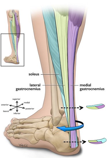
Figure 1. Posterolateral view of the left Achilles tendon and the three subtendons which comprise it. Subtendons rotate in a clockwise fashion traveling distally down the tendon. Cross-sectional views are displayed near the proximal and distal ends of the free tendon, and are based on cadaveric studies [56,82]. The soleus and soleus subtendon are colored teal, the lateral gastrocnemius and associated subtendon chartreuse, and the medial gastroc and its subtendon lavender.
3. Tendon Tissue Remodeling
Despite the complex loading mechanics of the triceps surae MTU, not all loading is detrimental to tendon health. While extrinsic factors contributing to tendon damage appear to be primarily attributable to submaximal cyclic loading, such as those induced by running and other training-related factors [88], targeted tendon loading of adequate magnitude can induce positive changes in tendon morphological, material, and mechanical properties [29,32]. Specifically, mechanotransduction details the body’s ability to translate mechanical loading into structural tissue change via cellular responses [89].
3.1. Healthy Tissue Remodeling
Mechanosensitive cells are responsive to tension, compression, and shear [90]. Loading magnitude [29,32], and perhaps more precisely strain [42,43,44], appears to modulate mechanotransduction in the healthy Achilles tendon. Specifically, strain magnitude, frequency, rate, and duration influence tenocyte biochemical processes [91,92,93] and gene expression [94,95]. For adequately long intervention durations (generally 12 weeks [36]) loads of greater than 70% of maximum voluntary contraction (MVC) [29,32] or strains of 4.5–6.5% [42,43,44] may deliver the appropriate loading-induced tendon stimulus to initiate mechanotransduction pathways; however, the relationship of tendon force and resulting strain can vary substantially between individuals [96,97]. Additionally, strain calculated as the displacement of the gastrocnemius medialis myotendinous junction from its resting length may differ from strain calculated as the change in length of the free tendon, which is more compliant [98,99], and perhaps where the majority of strain occurs. Theoretically, only looking at strain across the free tendon could change the ‘optimal’ adaptation threshold of 4.5–6.5% strain [42,43,44] typically arising from loading programs of greater than 70% of MVC [29,32].
Although the metabolic activity of tendon is low and the structure is typically static, loading-induced stimuli may trigger mechanotransduction and anabolic signaling pathways in the tendon [3]. In particular, the upregulation of insulin-like growth factor (IGF-I), among other growth factors, influences cellular proliferation and matrix remodeling [89,100,101]. Positive matrix remodeling appears to be largely attributable to a net synthesis of type I collagen, thereby making the tendon more load-resistant, though components of the ECM—proteoglycans, glycosaminoglycans, and cross-links—are also influenced by mechanical loading and contribute to macroscopic tendon behaviour through their actions on collagen fibrils [49,100]. Mechanically, longitudinal stiffness (resistance to deformation) increases [29,32,102], and strain for a given tendon force decreases [43,103] in response to increased loading in vivo. Material properties increasing in response to increased loading in vivo include modulus [29,32,102]. Morphologically, tendon CSA increases in response to increased loading in vivo [29,32,102], though limited evidence suggests that transient fluid redistribution may mask this in the short-term [51,104]. Additionally, loading-induced changes may differ along the Achilles tendon as the regional variation in load management [98,105,106] may preferentially activate mechanotransductive pathways leading to region-specific tendon hypertrophy [43,44]. Though still an area of exploration, the opposite could also be the case in that the non-uniform stress distribution within the Achilles tendon could contribute to the location of abnormalities associated with AT [107]. Moreover, while the tendon changes/adaptations described above are primarily related to resistance training, it appears that other types of mechanical loading, such as cyclic loading (e.g., running), can also induce adaptation in the healthy Achilles tendon [108,109]; however, conflicting evidence suggests that some other types of mechanical loading, such as plyometric exercises, may or may not adapt the Achilles tendon in a similar fashion [110,111,112,113,114,115].
3.2. Pathologic Tissue Remodeling
The pathogenesis of tendinopathy appears multifaceted, which has given rise to various pathophysiological theories [36]. Current rhetoric suggests that initial cyclic overloading of the tendon leads to degeneration and disorganization of healthy collagen, which triggers an acute inflammatory response [36,87,101]. If the cyclic overloading is continued without intervention, the tendon pathology worsens through a positive feedback loop of injury to both the original and poor-quality repair tissue, inflammation, and failed repair. Macroscopically, evidence suggests that AT increases tendon CSA [116,117,118] and longitudinal strain [116,117,118], and decreases modulus [116,119], transverse strain [120], longitudinal stiffness [116,118,119], and transverse stiffness [121] in vivo. Taken together, these changes lead to functional deficits across the strength spectrum potentially increasing risk of AT recurrence [122,123,124].
Therapeutic exercise remains one of if not the most effective non-surgical approach for managing AT [1,2]. The suggested mechanism of action is generally considered to be restoration of tendon material, mechanical, and morphological properties similarly to healthy tendon remodeling [36,37,41], thereby improving functional strength [33]. Macroscopically, evidence suggests that targeted mechanical loading decreases tendon thickness [125] and volume [126]; however, there is a paucity of evidence underpinning the restoration of tendinopathic tissue capacity, with most studies focusing on functional and acute analgesic effects [36]. Evidence suggests that abnormal structure (i.e., hypoechoic areas and irregular structure) may normalize in some individuals following a 12-week eccentric exercise protocol [125,127], though the time needed for such changes to occur may vary [38]. Additionally, Cook and colleagues [128] posit that exercise-based adaptation may build capacity in the area of aligned fibrillar structure instead of acting on the area of abnormal structure. Nonetheless, evidence suggests that structural changes do not entirely explain clinical outcomes [129,130]. Building on this idea, O’Neill, Watson, and Barry [37] highlight that tendon structure is not observed to significantly change over the typical intervention period. The authors further suggest that changes in neuromuscular output may explain clinical benefit, and that training should focus on increasing stiffness of the triceps surae MTU, increasing strength, and shifting the length-tension curve of the triceps surae muscles through sarcomerogenesis. Although still an area of exploration, it appears that therapeutic exercise for AT should focus on improving the mechanical and material properties of the entire MTU thereby simultaneously building strength capacity and neuromuscular control [131].
This entry is adapted from the peer-reviewed paper 10.3390/jcm11164722
This entry is offline, you can click here to edit this entry!