Clinical Neurophysiology and Genetics of Dystonia Diagnosis: History
Please note this is an old version of this entry, which may differ significantly from the current revision.
Subjects: Clinical Neurology
Contributor: , , , , ,

Dystonia diagnosis is based on clinical examination performed by a neurologist with expertise in movement disorders. Clues that indicate the diagnosis of a movement disorder such as dystonia are dystonic movements, dystonic postures, and three additional physical signs (mirror dystonia, overflow dystonia, and geste antagonists/sensory tricks). Despite advances in research, there is no diagnostic test with a high level of accuracy for the dystonia diagnosis. Clinical neurophysiology and genetics might support the clinician in the diagnostic process. Neurophysiology played a role in untangling dystonia pathophysiology, demonstrating characteristic reduction in inhibition of central motor circuits and alterations in the somatosensory system. The neurophysiologic measure with the greatest evidence in identifying patients affected by dystonia is the somatosensory temporal discrimination threshold (STDT). Other parameters need further confirmations and more solid evidence to be considered as support for the dystonia diagnosis. Genetic testing should be guided by characteristics such as age at onset, body distribution, associated features, and coexistence of other movement disorders (parkinsonism, myoclonus, and other hyperkinesia). 

  • dystonia
  • clinical diagnosis
  • neurophysiology
  • genetics

. Introduction

Dystonia is a term used to identify hyperkinetic movement disorders in which dystonia is the prominent feature. However, dystonia can also be present in other conditions. According to the etiology, dystonia can be distinguished as acquired, inherited, or idiopathic. The diagnosis of dystonia is based on clinical examination conducted by physicians with expertise in movement disorders through a careful examination of the phenomenology of the condition that allows for a classification of dystonia. For the diagnosis of dystonia syndrome, the examiner should follow the definition of dystonia approved in the last expert consensus [1], articulated in three subdefinitions:
  • Dystonia is a movement disorder characterized by sustained or intermittent muscle contractions causing abnormal, often repetitive, movements, postures, or both.
  • Dystonic movements are typically patterned, twisting, and may be tremulous.
  • Dystonia is often initiated or worsened by voluntary action and associated with overflow muscle activation.
The examiner should focus on the classic five physical signs of dystonia syndromes: two main physical signs (dystonic movements and dystonic posture) and three additional physical signs (mirror dystonia, overflow dystonia and geste antagonists/sensory tricks) [2,3].
The role of laboratory analysis, neuroimaging studies, neurophysiology, and genetic tests is to support the etiology definition of the disease, according to the Axis II of Dystonia classification [1,4].

2. Clinical Neurophysiology

Clinical neurophysiology techniques such as EMG mapping [2,5] allow clinicians to support the diagnosis of dystonia and to explore the activity of individual muscles which is not always easy to achieve with a clinical inspection alone. In addition, clinical neurophysiology with different techniques, such as transcranial magnetic stimulation (TMS) [6,7], transcranial direct current stimulation (tDCS) [8,9], or the newest transcranial focused ultrasound stimulation (tFUS) [10,11,12], allow clinicians to explore in a non-invasive way the brain functions In recent years, these techniques have been widely used as tools to characterize distinctive features and improve diagnostic accuracy for different movement disorders [13], particularly parkinsonian syndromes [14,15,16], tremor syndromes [17,18,19], myoclonus [20], and dystonia [21]. The literature includes several studies that use different neurophysiological tests to assess dystonia [22] (Table 1). Despite the amount of evidence, most of the studies on dystonia neurophysiology have a small sample size and focus on specific forms of dystonia (e.g., DYT-TOR1A); therefore, results are not always generalizable to all forms of dystonia. Neurophysiology assessment is not formally included in the diagnostic process [1]; however, neurophysiological tests can support the diagnosis.
Since the early 1980s, neurophysiology has been used to characterize dystonia pathophysiology. Most studies were performed in focal hand dystonia (FHD) [22]. At first, dystonia was classified as a basal ganglia (BG) disorder; however, in recent years, evidence points to a disorder arising from a complex network system involving the cerebral cortex (motor and sensory area), the basal ganglia, the brainstem, and the cerebellum [43,44], suggesting that is it possible that several structures could be simultaneously involved in the pathogenesis of dystonia subtypes [43,44].
The electromyographic (EMG) pattern observed in dystonia patients records simultaneous activation of agonist and antagonist muscles (co-contraction), prolonged duration of EMG bursts, and involuntary overflow activation of muscles not directly involved in the movement [3,23].
The most relevant neurophysiological feature shared by all dystonia subtypes is the reduced inhibition of central motor circuits [22]. This is demonstrated by characteristics in several structures: (1) at the subcortical level, a reduction of presynaptic inhibition in the spinal cord has been reported in patients with FHD [24]; (2) at the brainstem level, - a reduced inhibition in the blink reflex recovery cycle in blepharospasm patients [25] and an impairment of the trigeminocervical reflex produced by infraorbital nerve stimulation in torticollis patients was noted [26]; and (3) at the motor cortex level, a loss of inhibition was demonstrated with several transcranial magnetic stimulation (TMS) protocols. Several studies reported abnormalities in dystonic patients of paired pulse protocol as short intracortical inhibitions (SICI), that is, an inhibition of motor cortex response produced by a subthreshold conditioning stimulus followed by a supra-threshold stimulus. SICI is reduced in different subtypes of dystonia [27,28,29]. Reduced transcallosal inhibition was also demonstrated in FHD patients with mirror dystonia. In these patients, stimulation of one hemisphere does not suppress motor responses evoked by a stimulus delivered about 10 ms later over the contralateral hemisphere, as observed in normal subjects [30]. Finally, the duration of the cortical silent period (SP), the inhibition of ongoing muscular activity produced by a TMS pulse during muscle contraction, is reduced in dystonic patients [31], and the lack of suppression could be related to some specific tasks [32].
In recent years, the relevance of the cerebellum in dystonia’s pathophysiology has been investigated [45]. The eye blink classic conditioning (EBCC) protocols consist of electric stimulation of the supraorbital nerve. This protocol that involves cerebellar circuits shows impairment in focal dystonia patients [33], while it is normal in inherited dystonia caused by the DYT-TOR1A and DYT-THAP1 gene mutation [34].
Traditionally, dystonia was referred to as a motor disorder; however, several recent studies have provided evidence on the role of the somatosensory system in dystonia pathogenesis. Several studies suggested that abnormalities in the somatosensory system are present in almost all dystonic patients, and several neurophysiology tests investigated these findings. The most relevant discovery is the abnormality in the somatosensory temporal discrimination threshold (STDT) [37]. STDT represents the shorter interval at which two different stimuli are perceived as separate. Cervical dystonia (CD) patients have abnormally increased STDT, and the effect seems higher in CD patients with tremor. In a validation study, 51 CD were compared to essential tremor (ET) patients and Parkinson’s disease (PD) patients. The authors found that compared to ET patients, if STDT is ≤67 ms, it has 100% sensitivity and 100% negative predictive value, while if STDT is ≥120 ms, it has 100% specificity and 100% positive predictive value to differentiate ET from CD. However, no statistically significant differences were found between the PD and CD groups even though evidence suggests that STDT is normal in the early PD phase and becomes abnormal in later stages, while STDT is abnormally increased from the first stages of dystonia disease.
Finally, another possible contribution to dystonic pathophysiology is represented by maladaptive plasticity. Abnormal sensory-motor plasticity was demonstrated using a paradigm termed paired associative stimulation (PAS) In this TMS protocol, cortical stimulation is paired with peripheral nerve stimulation at an interstimulus interval of 25 ms resulting in long-term potentiation-like phenomenon (LTP). This form of LTP is pathologically enhanced in FHD [39]. Maladaptive plasticity could be a key factor in the development of dystonic symptoms and a peculiar feature of dystonic patients as suggested by other studies that did not find the same increased plasticity in DYT-TOR1A carrier subjects [49] and in psychogenic dystonia patients [50]. A pronounced increase of PAS-related plasticity was also reported in Costello syndrome, a genetic syndrome characterized by pronounced dystonia [51,52].
Although all this evidence suggests that dystonia is a complex network disorder involving the brainstem, the basal ganglia, the thalamus, the cortex, and the cerebellum [44], originally dystonia was referred to as basal ganglia disease. Several trials point out that electrical modulation of the basal ganglia network through continuous deep brain stimulation (DBS) in internal globus pallidus (GPi) could improve generalized dystonia symptoms [53]. DBS electrodes were also used to invasively record synchronized neuronal activities, pointing out that in line with other movement disorders, pathological basal ganglia oscillatory activities [54] can be found in dystonic patients [41,42]. This invasive recording of local field potentials (LFP) of basal ganglia revealed that GPi and external globus palidus (GPe) have a decreased discharge rate and irregular firing in dystonic patients [55,56]. In addition, LFP studies demonstrated that pallidus nuclei of dystonic patients show excessive synchronized activities in the 4–10 Hz frequency band [42].

3. Dystonia Genetics

Dystonia genetics is a wide field with continuous updates. After the first description of DYT-TOR1A, several other genes have been proposed as linked with the dystonia phenotype [65]. As in other fields of genetics, after the first years focused on the genetic marker, the focus is moving on to proteomics, searching the causal link between the protein produced by these genes and the phenotype of dystonia. Camargos and Cardoso [66] proposed a model of the “dystonia cell” linking the dystonic genes to the proteins function, based mainly on the classic DYT nomenclature.
The classic DYT nomenclature is based on locus symbols (e.g., DYT 1) and has been used for several years. It is still used in literature and clinical practice [67]. However, the system of locus symbols has been challenged by advances in techniques of genetics research that allow us to define the causative gene, as explained by Marras et al. [68], and the need to renovate the nomenclature system has arisen. The MDS Task Force for the Nomenclature of Genetic Movement Disorders proposed new recommendations, whose use in research and clinical practice is strongly encouraged [69]. This new nomenclature strictly connects the prefix to the predominant phenotype and considers the causative gene rather than the locus symbols (e.g., DYT 1 is now named DYT-TOR1A) [4]. The prefix DYT is used only if dystonia is the prominent disease feature due to a pathogenetic mutation [69]. Otherwise, if another movement disorder is a prominent feature along with dystonia, a double prefix would be assigned (e.g., DYT/PARK-ATP1A3). Indeed, genetic dystonia can be isolated or combined with other movement disorders such as parkinsonism, myoclonus, or other hyperkinesia (Figure 2).
Figure 2. Isolated and combined genetic forms of dystonia [69].
Moreover, in the proposed nomenclature and in the last consensus update on dystonia, the term complex dystonia is used, referring to conditions in which dystonia predominates the clinical phenotype but occurs in the context of a complex disease including symptoms other than movement disorders [1,69]. For example, Wilson disease is named according to the proposed nomenclature with a DYT prefix (DYT-ATP7B), and the same happens for Lesch–Nyhan syndrome and other infantile and childhood onset disease [
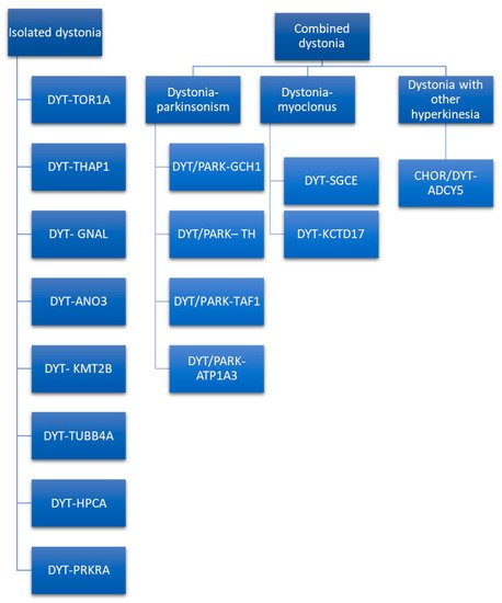
69]. Given that most of isolated hereditary dystonia is recognized as an autosomal dominant inheritance, the mode of transmission cannot be used as the only criterion to make a differential diagnosis. To guide the clinician towards a genetic diagnosis of dystonia, at least clinical phenotype and age of onset should be considered. If dystonia dominates the clinical picture, one of the isolated dystonias may be considered, and the gene mutations involved may be DYT-TOR1A, DYT-THAP1, DYT-GNAL, DYT-ANO3, DYT-KMT2B, DYT-TUBB4A, DYT-HPCA, and DYT-PRKRA [70]. The last-mentioned dystonia is a controversial classification, as it is considered as combined dystonia by some authors [71] and as isolated dystonia by others [70]. Indeed, despite parkinsonism being described in about half the patients, it seemed to be caused not by true parkinsonian features, but by slow movements of dystonic body parts [70]. The isolated form of dystonia could be distinguished according to the age of onset, body distribution, temporal pattern, associated features, responses to drugs, response to DBS, and brain imaging. Regarding age of onset, in infancy, childhood, and adolescence DYT-TOR1A, DYT-THAP1, DYT-KMT2B, DYT-TUBB4A, DYT-PRKRA, and DYT-HPCA are more probable, while DYT-ANO3 and DYT-GNAL begin in early adulthood. In particular, DYT-ANO3 recognizes two peaks of the age of onset: one in infancy/childhood and one in early-late adulthood [70]. Age at onset may by modified by several aspects, e.g., penetrance as is the case of DYT-TOR1A [72]. Hence, age of onset alone cannot be used as the only criteria to orient the diagnosis. According to body distribution, generalized forms of isolated dystonia are mainly due to DYT-TOR1A, DYT-THAP1, DYT-KMT2B, DYT-HPCA, and DYT-PRKRA. Among these, DYT-TOR1A, DYT-HPCA, and DYT-KMT2B usually begin in the lower limbs asymmetrically with secondary generalization. In contrast, DYT-THAP1 may initiate in the upper part of the body, involving cranio–cervical districts, speech difficulties, and the upper limbs, with successive generalizations [73]. If DYT-TOR1A begins in the upper limbs, it tends to be focal. Focal and segmental isolated dystonia are more likely caused by DYT-GNAL and DYT-ANO3. These two forms of dystonia typically begin at the cervical level and may cause head tremor [70]. DYT-GNAL may be suspected if age at onset is in early-late adulthood. In case of early involvement of craniofacial muscles with laryngeal dystonia and speech difficulties, with secondary generalization involving the arms at younger ages, DYT-ANO3 becomes more probable [70]. Another peculiar form of isolated dystonia with focal distribution involving the cervical district and causing spasmodic dysphonia is caused by DYT-TUBB4A. This focal form may successively evolve into a generalized dystonia [74]. Regarding the temporal pattern, except for the last-mentioned dystonia, all the other isolated dystonia follows a persistent temporal pattern. Associated features may guide the clinician in the differential diagnosis. The presence of additional phenotypic characteristic, such as microcephaly, short stature, intellectual disability, abnormal eye movements, myoclonus, dysmorphisms, and psychiatric symptoms, may be suggestive of DYT-KMT2B [70]. Thin face, body habitus, and hobby horse gait are described in the DYT-TUBB4A [75]. None of the isolated forms of dystonia respond to L-Dopa; DYT-TOR1A, DYT-THAP1, DYT-ANO3, DYT-KMT2B, and DYT-HPCA may respond to anticholinergics [70]. Response to alcohol is described in DYT-GNAL and DYT-TUBB4A. It is important to define the genetic etiology of the dystonia because response to DBS varies according to the genetic conditions, and this is an important prognostic factor to be considered when selecting patients for advanced therapy.
Combined dystonia is characterized by the coexistence of another movement disorder in addition to dystonia. The association of dystonia with parkinsonism defines dystonia–parkinsonism. The monogenic forms of dystonia–parkinsonism are DYT/PARK-GCH1, DYT/PARK-TH, DYT/PARK-TAF1, and DYT/PARK-ATP1A3 [71]. Contrary to what has been observed for isolated dystonia, combined dystonia recognizes a different mode of inheritance: autosomal dominant inheritance is characteristic of DYT/PARK-GCH1 and DYT/PARK-ATP1A3, while autosomal recessive inheritance is typical of DYT/PARK-TH. X-linked transmission characterizes DYT/PARK-TAF1 (also known as Lubag syndrome). Among this, it is of paramount importance to diagnose the dopa-responsive dystonia, DYT/PARK-GCH1. Indeed, patients have excellent and sustained response to L-Dopa [80]. Another form of combined dystonia with response to L-Dopa is DYT/PARK-TH. These two forms of dystonia–parkinsonism may be differentiated according to age of onset, as DYT/PARK-GCH1 begins in infancy/childhood, while DYT/PARK-TH may initiate in infancy. Moreover, diurnal fluctuations of parkinsonian symptoms due to circadian variations in dopamine concentration are more pronounced in DYT/PARK-GCH1 than in DYT/PARK-TH [80].
DYT/PARK TAF1 differs from the previous mentioned strains for the age of onset, body distribution of dystonia, and neuroimaging. This form of combined dystonia begins in early to late adulthood and, contrary to DYT/PARK-GCH1 that begins with foot dystonia and then progress cranially, DYT/PARK TAF1 involves mainly the upper body, with characteristic jaw opening dystonia and bulbar involvement. Another difference with respect to the dopa-responsive dystonia is the absence of diurnal fluctuation. Brain imaging shows striatal atrophy and pallidum volume loss, considered an expression of the neurodegenerative nature of the disease. This form recognizes an X-linked transmission, hence is more frequent in males [86,87,88,89].
Combined dystonia also encompasses dystonia associated with myoclonus and other hyperkinetic disorders. To date, two forms of dystonia–myoclonus have received confirmations: DYT-SGCE and DYT-KCTD17. These diseases have several features in common: age of onset is in the first or second decade of life, myoclonic jerks involve the upper body, and in DYT-SGCE also the neck may be involved. In both diseases, dystonia affects the upper part of the body, with involvement of upper limbs and the cranio-cervical region. If in DYT-SGCE myoclonic jerks dominates the clinical picture, in DYT-KCTD17 dystonia seems to be the prominent feature. Interestingly, DYT-SGCE myoclonic symptoms respond to alcohol, while in DYT-KCTD17 this response is absent [71,92].
Dystonia may coexist with other hyperkinetic disorders, such as chorea, as observed in several forms of complex dystonia. Marras et al. [69] also classify CHOR/DYT-ADCY5 as combined dystonia. This disease is characterized by a plethora of hyperkinetic disorders, such as chorea, dystonia, and myoclonus, beginning in early childhood and with a characteristic fluctuating or paroxysmal course. Interestingly, symptoms do not disappear during sleep, resulting in significant disturbances, and may respond to caffeine [93,94]. Response to DBS is lower than in other form of monogenic dystonia [76].

This entry is adapted from the peer-reviewed paper 10.3390/jcm11144184

This entry is offline, you can click here to edit this entry!