Toxoplasma gondii in Foods: History
Please note this is an old version of this entry, which may differ significantly from the current revision.
Contributor: , ,

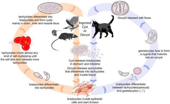
Toxoplasma gondii is an obligate intracellular parasite that causes toxoplasmosis, with approximately one third of the population around the world seropositive. The consumption of contaminated food is the main source of infection. These include meat products with T. gondii tissue cysts, and dairy products with tachyzoites.

  • toxoplasmosis
  • Toxoplasma gondii
  • control
  • food
  • detection

1. Introduction

Toxoplasmosis is a zoonotic disease that is caused by the obligate intracellular parasitic Toxoplasma gondii. This protozoon of the Apicomplexa phyla presents only felines as the definitive host, being the ones where the parasite can complete its life cycle. However, all warm-blooded animals, including mammals and birds, can act as intermediate hosts (Figure 1). In most hosts, T. gondii causes a lifelong latent infection in tissues such as skeletal and heart muscle, and the central nervous system, causing the disease. In humans, infection by T. gondii is particularly important in pregnant women and immunocompromized people. During pregnancy, the risk of fetal infection increases with gestational stage, increasing as gestation progresses [1]. Neonatal manifestations include hydrocephalus, microcephalus, intracranial calcifications, chorioretinitis, cataracts, convulsions, nystagmus, jaundice, petechiae, anemia, enlarged liver and spleen, prematurity, and severe intrauterine growth restriction [2,3]. Ocular manifestations also appear as chorioretinitis and retinal lesions [4]. In immunocompromized people, the neurological symptoms, such as encephalopathy, meningoencephalitis, cerebral mass lesions, headache, confusion, poor coordination, and seizures are usual [5], with toxoplasmic encephalitis being the most frequent manifestation in HIV patients [6], whereas the disseminated toxoplasmosis is more characteristic of transplant patients [7]. However, not only pregnant women and immunocompromized people may suffer the symptoms of Toxoplasma infection. Immunocompetent individuals can develop acute, chronic, and ocular toxoplasmosis. The acute toxoplasmosis is asymptomatic around 80% of individuals [8], and the symptoms in the other 20% includes fever, mononucleosis-like symptoms, with cervical posterior adenopathy, myalgia, and asthenia [9]. Although these symptoms are not relatively serious, the severity of infection depends on genotype of the parasite strain. In fact, infections with a highly virulent strain can produce fatal pneumonitis, myocarditis, meningo-encephalitis, and polymyositis [6]. In chronic toxoplasmosis, tachyzoites form bradyzoite cysts intraneuronal which are controlled but not eliminated by the immune system [10]. The immune response in the brain of patients produces brain inflammation, ventricular dilatation, disrupting neuronal structure and connectivity [11,12]. Although the symptoms of chronic toxoplasmosis have not been unraveled, several studies correlated these manifestations with neuropathies [13,14]. Related to ocular toxoplasmosis, it is the primary cause of infectious uveitis, presenting with retinochoroiditis [15].
Figure 1. Biological cycle of Toxoplasma gondii.
T. gondii has a worldwide geographic distribution and an estimated 30% of the population is seropositive [16]. The genetic diversity of T. gondii around the world has been elevated, so more than 36 genotypes have been found [17]. The transmission of this parasite in humans may result from the ingestion of tissue cysts in raw or undercooked meat of infected animals, ingestion of raw vegetables, water that is contaminated with T. gondii oocysts from cat feces, and by vertical or transplacental transmission [18]. Although, the main route of infection in humans is through ingestion of contaminated food. In fact, it has been described that up to 50% of infections are caused by food transmission using a novel multiplex Polymerase Chain reaction (PCR) assay [19]. A study that was undertaken in school dining rooms of Colombia showed the presence of T. gondii in meat, water, cucumber, and guava juice, both inert and living surfaces [20]. In the last years, the concern about this zoonosis and its transmission has been increasing. In 2018, the EFSA recommended a serological screening of livestock to identify positive farms [21]. In the following year, the EFSA report found that food-borne transmission accounts for 40–60% of T. gondii infections [22]. The last report indicated positive samples of meat, fish, raw mollusks and shellfish, honey, and potable water, and Toxoplasma was included in category III of zoonotic agents to monitor, along with Campylobacter or Yersinia [23].
However, and despite the great health public problem that it poses, there are currently no specific detection criteria for T. gondii in food, and there are no standardized methods or validation procedures for its detection in the food industry. In fact, different direct and indirect detection techniques exist. Cat and mouse bioassays are the reference direct techniques to analyze the viability of the parasite, but these test are not commonly used due to the long time that is taken to obtain results, ethical issues, and great costs [18]. The alternative method are cell cultures which are limited in use because of the variability of the results depending the sample [24]. Other serological methods (indirect detection) have been developed such as immunofluorescent assay (IFAT), enzyme-linked immunosorbent assay (ELISA), latex agglutination tests (LAT), modified agglutination test (MAT), and more recently, a luciferase-linked antibody capture assay (LACA) [23,25]. The latest studies of T. gondii detection in food products have used serological techniques to improve the sensibility of these serological tests using different approaches. For example, Suwan et al. (2022) used a recombinant dense granule antigen 7 protein for the detection of parasites in blood samples [26]. In addition to these serological methods, other molecular techniques have been tested. Some protocols of PCR have been described as nested PCR, real-time PCR, loop-mediated isothermal amplification (LAMP), and others. However, the more sensitive and specific diagnostic tools to detect T. gondii are necessary [27], and the studies about their sensitivity and to unify the detection in different food products are essential to control of parasite infection by food consumption.

2. Methods for T. gondii Detection in Food Products

Although T. gondii is a high priority foodborne zoonotic pathogen around the world, it is not systematically controlled [28]. At present, there are no specific regulations or ISO standards for the detection of T. gondii in any food matrix [21]. Even so, different methods are available to detect tachyzoites, tissue cysts, and oocysts in food products, including immunological and microscopical methods. These methods have an isolate and concentration stage, later applying direct detection methods to the sample. Molecular assays are used to detect the presence of T. gondii DNA in samples, while information on the viability and infectivity can be obtained by in vivo assays (usually in mice) or by in vitro culture techniques. A summary of these methods with sensitivity and type of food product where these methods have been used are shown in Table 1.
Table 1. The table shows different methods for T. gondii detection, sensitivity of method, and type of food product where this method has been used.

Detection Method

Specific Method 1

Type of Food Product

Detection Range

(Sensitivity) 2

References

Animal model bioassay

Cat

Milk

25%

[29,30]


Meat

100%

[31]


Mouse

Milk

100%

[29]


Meat

100% (10 tachyzoites)

[24]


Fresh products

13%

[32]


Bivalve mollusks

2.5%

[33]


Water

100%

[34]

Cell culture


Meat

100% (10,000 tachyzoites)

[24]


Milk

-

[30]

Microscopic method


Meat

-

[31]

Molecular methods

PCR

Meat

47.1%

[35]


Fresh products

95–100%

[36,37]


Water

100%

[36]


Milk

100%

[29,38]


Cheese

100%

[29]


qPCR

Meat

92.3% (limit 0.01 pg)

[39,40]


Fresh products

100% (1 oocyst)

[41,42,43]


Bivalve mollusks

100%

[44]


Water

100%

[44]


LAMP

Lymph nodes

85.7%

[45]


Mussels

5 oocyst/g

[46]


Fresh products

25 oocyst/50 g

[47]


Water

100% (1 fg)

[48,49]

Serological methods

IHA

Meat Juice

100% (10,000 oocysts)

[50]


IFAT

Meat

97%

[51]


Meat Juice

96.9% (10,000 oocysts)

[50]


MAT

Meat

86.6%

[51]


Milk

-

[52]


ELISA

Milk

-

[30]


Meat

91%

[51]


Meat Juice

100% (10,000 oocysts)

[50]


BBMA

Meat

98.5%

[53]

1 PCR: Polymerase chain reaction; qPCR: real-time PCR; LAMP: Loop-mediated isothermal amplification; IHA: indirect hemagglutination antibody; IFAT: indirect fluorescent antibody test; MAT: modified agglutination test; ELISA: Enzyme-Linked Immunosorbent Assay; BBMA: bead-based multiplex assay. 2 The column shows the percentage of samples that were positively detected by the method and the quantity of parasites per quantity of food product that was detected if this data is known. The value (-) means that this data is not known.

3. Prevalence of Toxoplasma gondii in Food Products

Transmission through food is the main system of transmission of T. gondii to humans [18]. Tissues cysts and tachyzoites are responsible for infection thought meat and milk, respectively [56], and sporulated oocysts can contaminate fresh products, shellfish, and water, and infect humans after consumption [88].

3.1. Meat and Meat Products

T. gondii infections have been reported in all meat production animals around the world, although the prevalence depends on the detection method that is used (Table 1). Tissue cysts of parasites in meat are an important source of human infection, due to the fact of that these animals are secondary hosts of the parasite, which can survive long periods of time in these asymptomatic animals, which will later become meat products.
Different techniques are available to detect its presence. The mouse bioassay and PCR are the most widely used direct detection methods, followed by microscopy and the cat bioassay [31]. On the other hand, the MAT, IFAT, and ELISA tests are the most widely used serological methods for the detection of T. gondii infection in cattle and meat products [73]. Table 2 shows the animal and sample that was contaminated, the country of contamination, the method that was used for detection, and prevalence that was found.
Table 2. T. gondii in animals and animal products. The table shows the producer animal, sample contaminated, country of contamination, method that was used for detection, and prevalence that was found.

Animal

Sample Analyzed

Detection Method 1

Number of Samples Tested

Number of Positive Samples (%)

Location

Reference

Sheep

Serum

ELISA

150

26 (17.3%)

Iran

[89]


Serum

ELISA

550

59 (10.8%)

Iran

[90]


Serum

ELISA

1039

179 (17.2%)

Latvia

[91]


Serum

MAT

100

42 (42%)

Lebanon

[92]


Serum

ELISA

64

30 (47%)

Slovakia

[93]


Serum

DAT

252

148 (58.2%)

Ethiopia

[94]


Liver

PCR

150

26 (17.3%)

Iran

[89]


Liver

PCR

90

13 (14.4%)

Iran

[95]


Heart

PCR

150

48 (32%)

Iran

[89]


Brain and heart

MAT

136

10 (7.4%)

India

[96]


Meat juice

ELISA

227

126 (28.6%)

Italy

[97]


Meat juice

MAT

166

11 (6.6%)

China

[98]


Meat

PCR

150

33 (22%)

Iran

[89]


Meat

PCR

438

43 (9.8)

China

[99]


Meat

PCR

150

50 (33.3)

Tunisia

[100]


Meat

ELISA

109

38 (34.9%)

Malaysia

[101]


Meat

PCR

79

34 (43%)

Australia

[102]


Meat

PCR

177

3 (1.7%)

India

[103]

Goat

Serum

ELISA

150

16 (10.7%)

Iran

[89]


Serum

ELISA

185

37 (20%)

Iran

[90]


Serum

ELISA

445

189 (42.5%)

India

[104]


Serum

MAT

80

27 (34%)

Lebanon

[93]


Serum

ELISA

39

8 (21%)

Slovakia

[93]


Serum

LAT

116

64 (55.2%)

Ethiopia

[94]


Liver

PCR

150

24 (16%)

Iran

[89]


Liver

PCR

90

8 (8.8%)

India

[95]


Heart

PCR

150

36 (24%)

Iran

[89]


Brain and heart

MAT

57

4 (7%)

India

[96]


Meat juice

ELISA

51

14 (27.5%)

Italy

[97]


Meat

PCR

150

26 (17.3%)

Iran

[89]


Meat

PCR

254

27 (10.7)

China

[99]


Meat

PCR

120

39 (32.5)

Tunisia

[100]


Meat

ELISA

75

41 (54.7%)

Malaysia

[101]


Meat

PCR

223

3 (1.3%)

India

[104]

Cattle

Serum

ELISA

57

13 (22.8%)

Italy

[105]


Serum

DAT

2411

313 (13%)

Poland

[106]


Serum

ELISA

400

52 (13%)

Iran

[107]


Serum

IFAT

500

2.3 (40.6%)

Brazil

[108]


Meat

PCR

150

29 (19.3)

Tunisia

[100]


Meat

ELISA

392

98 (25%)

Malaysia

[101]


Meat

PCR

48

5 (10.4%)

Brazil

[108]

Pig

Serum

ELISA

653

4 (0.6%)

Finland

[109]


Serum

ELISA

447

73 (16.3%)

Denmark

[110]


Serum

DAT

3111

370 (11.9%)

Poland

[106]


Serum

IFAT

94

44 (46.8%)

Romania

[111]


Serum

ELISA

420

56 (23.3%)

Cuba

[112]


Serum

ELISA

370

14 (3.8%)

Italy

[113]


Serum

ELISA and IFAT

127

56 (44.1%)

Italy

[114]


Serum

MAT

375

8 (2.1%)

Italy

[115]


Serum

ELISA

414

214 (51.7%)

Italy

[116]


Serum

MAT

182

31 (17%)

Serbia

[117]


Serum

MAT and IFAT

356

25 (7%) and 48 (13.5%), respectively

Brazil

[118]


Serum

MAT and IFAT

400

26 (6.5%)

Brazil

[119]


Serum

IFAT

60

44 (77%)

Brazil

[120]


Serum

IHA

784

156 (19.9%)

China

[121]


Tongue

PCR

60

20 (33.3%)

Brazil

[120]


Tongue and muscle

PCR

810

54 (6.7%)

India

[122]


Brain

PCR

339

34 (10%)

China

[123]


Brain

PCR

107

51 (47.7%)

Italy

[116]


Heart

PCR

94

25 (26.6%)

Romania

[111]


Heart

qPCR

103

12 (11.6%)

Italy

[124]


Diaphragm

PCR

45

15 (33.3%)

Serbia

[117]


Diaphragm

PCR

1223

107 (8.7%)

China

[125]


Diaphragm

PCR

60

24 (40%)

Brazil

[120]


Diaphragm

qPCR

103

2 (1.9%)

Italy

[126]


Tissue of seropositive animals

Mouse bioassay

26

18 (69.2%)

Brazil

[119]


Muscle

PCR

60

23 (38.3%)

Brazil

[120]


Meat juice

ELISA

212

33 (15.6%)

Denmark

[110]


Meat

qPCR

118

46 (39%)

Brazil

[126]


Meat

PCR

498

165 (33.1%)

Italy

[64]


Meat

PCR

49

3 (6.1%)

Brazil

[108]


Raw meat products

PCR

3223

175 (5.4%)

Poland

[127]

Chicken

Serum

IFAT

200

72 (36%)

Brazil

[128]


Serum

ELISA

522

34 (6.5%)

India

[129]


Serum

LACA

267

29 (10.9%)

Japan

[85]


Brain

Mouse Bioassay

14

2 (14.3%)

Brazil

[128]


Heart juice

MAT

1185

230 (19.4%)

USA

[130]


Muscle and heart

PCR

522

12 (2.3%)

India

[129]


Meat

PCR

257

21 (8.2%)

China

[131]

Ducks

Meat

PCR

115

9 (7.8%)

China

[131]

Geese

Meat

PCR

42

2 (4.8%)

China

[131]

Rabbit

Brain and heart

PCR

470

13 (2.8%)

China

[132]

Kibbeh

Meat

PCR

44

1 (2.3%)

Brazil

[108]

Water Buffalo

Serum

MAT and ELISA

197

16 (8.1%) and 13 (6.6%), respectively

Romania

[133]

Ostriches (farmed)

Serum

LAT

409

149 (36%)

Czech Republic

[134]

Common quails (farmed)

Serum

MAT

620

59 (9.5%)

China

[135]

Donkey (farmed)

Meat

PCR

618

57 (9.2%)

China

[136]

Tolai hares (farmed)

Serum

PCR

358

29 (8.1%)

China

[137]


Brain

PCR

358

23 (6.4%)

China

[137]

Feral swine

Serum

ELISA

376

34 (9%)

USA

[138]

Wild boar (farmed)

Serum

LAT

882

88 (10%)

China

[139]

Wild boar

Serum

ELISA

331

164 (49%)

Italy

[140]


Serum

ELISA

181

17 (9%)

Finland

[141]


Serum

IFAT

26

20 (76.9%)

Brazil

[142]


Serum

ELISA

306

61 (20%)

Germany

[143]


Tissue

Mouse bioassay

22

1 (4.5%)

Brazil

[142]


Brain

qPCR

141

44 (31.2%)

Italy

[144]


Brain

PCR

263

58 (22%)

Italy

[145]


Heart

qPCR

166

47 (28.3%)

Italy

[144]


Heart

PCR

310

70 (22.6%)

Italy

[145]


Muscle

qPCR

165

40 (24.2%)

Italy

[144]


Muscle

PCR

311

74 (23.8%)

Italy

[145]


Meat juice

ELISA

97

42 (43.3%)

Italy

[146]


Meat

qPCR

306

37 (12%)

Germany

[143]

Venison

Serum

MAT

914

329 (36%)

USA

[147]


Heart

Mouse bioassay

36

11 (30.6%)

USA

[147]

Roe deer

Serum

LAT

356

141 (39.6%)

Spain

[148]


Serum

ELISA

323

130 (40.2%)

Italy

[149]


Serum

ELISA

184

20 (11%)

Germany

[143]


Meat

qPCR

184

11 (6%)

Germany

[143]

Fallow deer

Serum

LAT

372

138 (37.1%)

Spain

[150]


Serum

ELISA

167

17 (10%)

Slovakia

[93]


Meat

qPCR

80

2 (2%)

Germany

[143]

Red deer

Serum

LAT

553

92 (16.6%)

Spain

[148]


Serum

ELISA

96

19 (19.8%)

Italy

[140]


Serum

ELISA

65

4 (6%)

Germany

[143]


Meat

qPCR

65

2 (2%)

Germany

[143]

Southern chamois

Serum

LAT

186

26 (14%)

Spain

[148]

Mouflon

Serum

LAT

209

24 (11.5%)

Spain

[148]


Serum

ELISA

50

12 (24%)

Italy

[140]

Iberian wild goat

Serum

LAT

346

27 (7.8%)

Spain

[148]

Chamois

Serum

ELISA

104

4 (3.8%)

Italy

[140]

Barbary sheep

Serum

LAT

18

1 (5.6%)

Spain

[148]

Moose

Serum

DAT

463

111 (23.9%)

Estonia

[149]

Wild ducks

Brain

qPCR

280

7 (2.5%)

Czech Republic

[150]


Heart

qPCR

280

11 (3.9%)

Czech Republic

[150]


Muscle

qPCR

280

4 (1.4%)

Czech Republic

[150]

Common pheasants

Brain

qPCR

350

8 (2.3%)

Czech Republic

[150]


Heart

qPCR

350

4 (1.1%)

Czech Republic

[150]


Muscle

qPCR

350

3 (0.9%)

Czech Republic

[150]

1 ELISA: Enzyme-Linked Immunosorbent Assay; MAT: modified agglutination test; DAT: direct agglutination test; PCR: Polymerase chain reaction; LAT: latex agglutination test; IFAT: indirect fluorescent antibody test; qPCR: real-time PCR.

3.2. Milk and Dairy Products

Tachyzoites can be shed in the milk of acutely infected animals, so both raw milk and raw dairy products can pose a risk of infection for consumers [165] (Table 3). In fact, one of the factors that is related to infection in the USA is the ingestion of unpasteurized goat’s milk [166]. Different studies show the presence of T. gondii in milk samples from sheep, goat, camels, and donkeys [52,167,168,169,170], where the prevalence can reach up to 43–65% [159]. However, these data differ between production procedures, management, and techniques of detection, increasing with deficient biosecurity levels (related to the application of a health management program, vaccination protocols, correct quarantines, protocols for visitors, etc.) [171], and were higher for serological rather than molecular techniques. In goat, molecular techniques revealed the presence in 20.6% of milk samples, whereas the ELISA showed 63.3% [172]. Other studies showed that relationship between the prevalence of T. gondii antibodies in the goat serum with a prevalence of T. gondii DNA in milk samples [157].
Table 3. T. gondii in milk and dairy products. The table shows the producer animal, sample contaminated, country of contamination, method that was used for detection, and prevalence that was found.

Animal

Sample Analyzed

Detection Method 1

Number of Samples Tested

Number of Positive Samples (%)

Location

Reference

Donkey

Milk

ELISA

418

41 (9.2%)

China

[167]

Goat

Milk

ELISA

30

19 (63.3%)

Italy

[172]


Milk

PCR

60

39 (65%)

Poland

[157]


Milk

ELISA and qPCR

30

27 (90%) and 1 (3.3%), respectively

Egypt

[173]


Bulk tank milk

ELISA

100

59 (59%)

Italy

[172]

Sheep

Milk

PCR

58

1 (1.7%)

Mongolia

[168]


Milk

ELISA and qPCR

30

18 (60%) and 1 (3.3%), respectively

Egypt

[173]

Camel

Milk

PCR

9

8 (88.9%)

Mongolia

[168]


Milk

ELISA and qPCR

30

1 (3.33%) and 0 (0%), respectively

Egypt

[173]

Cattle

Bulk tank milk

ELISA

149

8 (5.4%)

Iran

[174]

1 ELISA: Enzyme-Linked Immunosorbent Assay; PCR: Polymerase chain reaction; qPCR: real-time PCR.

3.3. Fresh Products and Vegetables

Fresh products can become contaminated with T. gondii oocysts from cat feces or contaminated water, and act as a source of infection in humans. Oocyst detection in environmental and food samples is difficult due to complications in separating and concentrating oocysts from complex matrices, such as raw vegetables, so there is a lack of optimized laboratory methods for its detection [41]. However, Dumètre and Dardé (2003) have proposed possible methods for the detection of T. gondii in water, soil, and food samples (mainly, fruit and vegetables), based on methods that are used for other protozoa [176]. Hohweyer et al. (2016) developed an immunomagnetic separation assay (IMS) targeting the cell wall of oocysts, although it is not yet commercially available [67]. In addition to conventional methods such as microscopy, PCR or qPCR, a LAMP test has been developed to detect T. gondii in experimental contaminated baby ready-to-eat lettuces. The detection limit of this method was approximately 25 oocysts per 50 g of lettuce leaves [47]. Recently, special RT-PCR assay has been developed and it was effective to discriminate viable T. gondii, detecting two to nine oocysts per gram of spinach [43].
The first detection of T. gondii DNA in fruits and vegetables was in 2012 [177]. Nowadays, some studies have linked acute outbreaks of human toxoplasmosis with the ingestion of oocysts, where green vegetables have been identified as a possible vehicle of infection which can be contaminated by irrigation water [178,179,180]. In fact, Pinto-Ferreira et al. (2019) undertook a meta-analysis and concluded that vegetables will be the most common possible route of transmission in the future [181]. Contamination by T. gondii has been observed in different vegetables around the word, including lettuce, chicory, rocket, parsley, spinach, pack choi, cabbage, rape, asparagus, endive, Chinese chives, carrots, cucumbers, strawberries, and radish [182] (Table 4).
Table 4. T. gondii in fresh products and vegetables. The table shows the product that was analyzed, country of contamination, method used for detection, and prevalence that was found.

Product Analyzed

Detection Method 1

Number of Samples Tested

Number of Positive Samples (%)

Location

Reference

Mixed-salad packages

qPCR

648 packages

5 (0.8%)

Italy

[183]


PCR

90 packages

8 (8.9%)

Czech Republic

[184]

Leafy greens

qPCR

152

45 (29.6%)

Morocco

[185]

Carrot

qPCR

30

3 (10%)

Morocco

[186]


qPCR

46

9 (19.5%)

Poland

[177]


PCR

93

7 (7.5%)

Czech Republic

[184]

Chicory

PCR

40

2 (5%)

Brazil

[187]

Red cabbage

qPCR

8

1 (1.2%)

China

[42]

Coriander

qPCR

29

8 (27.6%)

Morocco

[186]

Cucumber

PCR

109

13 (11.9%)

Czech Republic

[184]

Lettuce

qPCR

28

3 (10.7%)

Morocco

[186]


qPCR

50

9 (18%)

Poland

[177]


qPCR

71

5 (7%)

China

[42]


PCR

168

5 (3%)

Brazil

[187]

Spinach

qPCR

50

2 (4%)

China

[42]

Parsley

qPCR

29

13 (44.8%)

Morocco

[186]


PCR

5

1 (20%)

Brazil

[187]

Pak Choi

qPCR

34

1 (2.9%)

China

[42]

Radish

qPCR

16

1 (6.3%)

Morocco

[186]


qPCR

60

3 (5%)

Poland

[42]

Rape

qPCR

22

1 (4.5%)

China

[42]

Rocket

PCR

7

1 (14.3%)

Brazil

[187]

1 PCR: Polymerase chain reaction; qPCR: real-time PCR.

3.4. Marine Products

Aquatic environments can be contaminated with wastewater carrying T. gondii oocysts. Mollusks such as clams, mussels, oysters, and scallops, filter-feed and trap phytoplankton in the gills. This filter feeding process can also concentrate waterborne pathogens within their tissues, including oocysts, which can survive for long periods of time in both fresh- and salt-water [55]. For detection in mollusks, samples of whole tissue or organs can be used and the most frequent techniques that are used are those that are based on PCR, generally directed to the B1 gene [189,190]. Various molecular methods have been used for detection in fish, such as PCR, qPCR, and RT-PCR, targeting the same gene, or the 529 bp DNA repeat element. The last method seems more sensitive, with the five oocysts as a low limit of detection. But it is no more specific, requiring direct sequencing for definitive confirmation of T. gondii [191]. In addition, the techniques have been carried out in different matrices, such as the digestive tract, muscle, brain, and even gills, among others [192]. Serological techniques have also been used for the detection in fish, such as ELISA, by detecting IgG and IgM, suggesting the fish are actually infected with T. gondii [193], rather than just serving as paratenic hosts such as shellfish.
The consumption of raw mollusks is considered a risk factor for T. gondii infection. Table 5 shows the prevalence of parasite in different mollusks, bivalves, and fishes.
Table 5. T. gondii in marine products. The table shows the animal, sample contaminated, country of contamination, method that was used for detection, and prevalence that was found.

Animal

Sample Analyzed

Detection Method 1

Number of Samples Tested

Number of Positive Samples (%)

Location

Reference

Bivalve shellfish

Tissue

PCR

2907

82 (2.8%)

China

[194]

Green-lipped mussels

Tissue

PCR

104

13 (16.4%)

New Zealand

[195]

Mediterranean mussel

Gills

qPCR

53 pools at 795 specimens

21 (39.6%)

Turkey

[189]

Clam

Tissue

qPCR

61 pools at 1020 specimens

4 (6.6%)

Tunisia

[190]


Digestive gland

PCR

390

6 (1.5%)

Canada

[196]


Haemolymph

PCR

390

2 (0.6%)

Canada

[196]

Mediterranean scald fish

Gills

PCR

1 pool at 6 specimens

1 (100%)

Italy

[197]

Pacific oyster

Gills

PCR

6 pools at 109 specimens

1 (16.67%)

Italy

[198]

Oyster

Mantle, gills, and rectum

qPCR

1440

447 (31%)

USA

[199]

Bogue

Gills

PCR

26 pools at 260 specimens

4 (15.4%)

Italy

[197]


Intestine

PCR

26 pools at 260 specimens

3 (11.5%)

Italy

[197]


Muscle

PCR

26 pools at 260 fish

6 (23.1%)

Italy

[197]

White seabream

Muscle

PCR

3 pools of 18 specimens

1 (33.3%)

Italy

[197]

European anchovy

Gills

PCR

35 pools at 350 specimens

2 (5.7%)

Italy

[197]


Intestine

PCR

35 pools at 350 specimens

1 (2.9%)

Italy

[197]

European hake

Gills

PCR

15 pools at 90 specimens

1 (6.7%)

Italy

[197]


Muscle

PCR

15 pools at 90 specimens

1 (6.7%)

Italy

[197]

Red mullet

Intestine

PCR

11 pools at 110 specimens

3 (27.3%)

Italy

[197]

American prawn

Muscle

PCR

618

4

China

[197]

Nippon shrimp

Muscle

PCR

813

1

China

[200]

Axillary seabream

Gills

PCR

8 pools at 80 specimens

2 (25%)

Italy

[197]


Intestine

PCR

8 pools at 80 specimens

1 (12.5%)

Italy

[197]


Muscle

PCR

8 pools at 80 specimens

1 (12.5%)

Italy

[197]

Common pandora

Gills

PCR

3 pools at 18 specimens

1 (33.3%)

Italy

[197]


Intestine

PCR

3 pools at 18 specimens

2 (66.7%)

Italy

[197]


Muscle

PCR

3 pools at 18 specimens

1 (33.3%)

Italy

[197]

Thornback ray

Muscle

PCR

1 fish

1 (100%)

Italy

[198]

Red scorpionfish

Intestine

PCR

1 pool at 3 specimens

1 (100%)

Italy

[197]

Blotched picarel

Muscle

PCR

4 pools at 24 specimens

1 (25%)

Italy

[197]

Atlantic horse mackerel

Muscle

PCR

15 pools at 120 specimens

4 (26.7%)

Italy

[197]

1 PCR: Polymerase chain reaction; qPCR: real-time PCR.

4. Control and Food Safety

The control of T. gondii infection must be done at several levels. First, certain risk factors increase the prevalence of the parasite in farm animals. Hygienic management practices and correct management which involves keeping cats away from crops and gardens and animal feed, are essential to control this pathogen in farms [121]. Temperature and humidity control could decrease the survival and distribution of the parasite, as well as a late replacement of the animals, since older animals present higher prevalence than young ones [94,202]. The intensive systems of production present lower prevalence than extensive or semi-intensive ones [171]. In the same way, organic farms present higher prevalence than conventional farms, probably due to due to the high risk of being exposed and infected with environmental oocysts of parasites or from ingested infected rodents [110]. Nevertheless, the most important factor in all production systems seems to be the biosecurity level (control of exposition and infection of animals with environmental parasites and control of domestic animals that are infected near the farms) and early detection [110,112]. Consumption of fresh milk and dairy products are other of factors that cause T. gondii infection in humans. In fact, pasteurization of milk and milk products is also an important control measure. Undoubtedly, stopping consuming these types of products could considerably reduce the prevalence of infection in humans. On the other hand, as occurs in meat products, adequate hygienic and sanitary conditions on farms would lead to this reduction. In fresh products and vegetables, the most common mechanism of contamination is irrigation with water that is contaminated by oocysts, so sanitary control measures in irrigation water would be interesting. Furthermore, washing fresh produce after harvest and before consumption is an important control measure, since the chemical disinfectants are not effective [18].
The control of T. gondii in food production is essential. However, control measures during food inspection are not applied [21]. Currently, different methods of inactivation exist, although in the industry they are not applied directly for the control of this parasite. The most used methods of control are thermal methods, including both high and low temperatures. Heat treatments can destroy oocysts from both sporulated and non-sporulated strains. It is also possible to eliminate bradyzoites and tachyzoites, although the elimination of the first requires higher temperatures and longer times [58,203]. Relationship between raw meat or other animal products have been demonstrated by several studies. In meat products, the main control measure to prevent infection is an adequate cooking and proper prevention of cross-contamination [204]. In fact, T. gondii can be eliminated from meat in 5–6 min at 49 °C, in 44 s at 55 °C, or in 6 s at 61 °C [205]. Different meat products require different temperature conditions for inactivation. For example, beef should be cooked at least 63 °C; whereas pork meat, minced meat, and bushmeat at 71 °C; and poultry at 82 °C. In general, meat should be cooked to at least 67 °C before consumption. In dairy products, the pasteurization of milk, at 63 °C for 30 min is sufficient to eliminate tachyzoites [206]. Rani and Pradhan (2021) published an exhaustive study that was related to the survival of T. gondii during cooking and low temperature storage and concluded that the parasite was not found when the internal temperature reached 64 °C and below −18 °C [207].
However, these elevated temperatures are not applicable to all food matrices. This is the case of vegetables and fresh products [208]. Regarding inactivation by low temperatures, it has been shown that freezing can inactivate tissue cysts of T. gondii. To inactivate isolate tissue cysts, a minimum of three days is required at −20 ºC [209]. In addition to thermal methods, other non-thermal methods can be used for the inactivation, such as high-pressure processing [55,154,210], ionizing radiation [211,212], and curing or salt [34,75]. The inactivation of T. gondii in food for thermal and non-thermal methods has been extensively analyzed in the review that was published by Mirza et al. (2018) [213].
The inactivation of T. gondii in food products has been realized traditionally with high temperatures (thermal methods) and when cured and salted [207,214], whereas the non-thermal methods are presented as emerging technologies for the control of T. gondii in food. High pressure processing (HHP) is a novel method for liquid and solid food products where pressures of 340–550 MPa during 1 min can inactive cysts of the parasite [215]. The second new method is ionizing radiation (IR), which is capable of inactivating or killing T. gondii cysts in meat [58]. However, these methods have not yet been tested in other food matrices or to inactive other parasitic forms.

This entry is adapted from the peer-reviewed paper 10.3390/foods11162542

This entry is offline, you can click here to edit this entry!
Video Production Service