For a single pluripotent cell to expand and differentiate into a specialized and committed cell type in the body, numerous cell-fate decisions and lineage choices must be made during its development. As cells cascade through the landscape of lineage decision steps regulated by epigenetic thresholds, correct genes need to be switched on and genes controlling alternative lineages must be repressed in a timely and dynamic manner.
Polycomb proteins play a crucial role in regulating these processes. There are two main types of Polycomb protein complexes. Both can contribute to repression of transcription of target genes, but they exhibit different histone-modifying activities. Polycomb Repressive Complex 1 (PRC1) contains E3 ligase activity that mediates ubiquitination of lysine 119 residues of histone H2A (H2AK119ub) [
1], whereas Polycomb Repressive Complex 2 (PRC2) catalyzes the mono-, di-, and tri-methylation of lysine 27 on histone H3 tails (H3K27) [
2,
3]. The two complexes are very different in their composition—with the core catalytic component of PRC2 consisting of EZH1/2 (Enhance of Zeste 2 homolog), EED, SUZ12, and RBBP4/7 proteins [
4,
5,
6] and PRC1 composing of at least eight different variants of interacting proteins around the core of RING1A/B (RING1, RNF2) and different PCGF proteins. Both complexes are associated with transcriptional silencing [
7,
8] and they can do so cooperatively, e.g., JARID2 from PRC2 is able to bind to PRC1 deposited H2AK119ub mark [
7,
9,
10,
11,
12].
Seminal work done in mouse and Drosophila has elucidated the roles of PRC1/2 in maintaining pluripotency by repressing the expression of master regulators of differentiation in embryonic stem cells. Intriguingly, they also play a crucial role just a short while after the exit of pluripotency by maintaining the precise repression of Hox genes clusters which are crucial during the body-axis formation and segmentation [
2,
13,
14]. The loss of catalytic subunits, e.g., deletion of EZH1/2 and EED in mouse embryos, leads to gross defects in gastrulation and embryonic lethality [
15,
16,
17]. By contrast, the effect of losing variant subunits, such as MTF2 and JARID2, which interact with the catalytic core at sub-stoichiometric ratios, varies.
Mtf2 null mice display embryonic lethality much later during development, at around E17.5, and
Jarid2 null mice are unable to survive past E14.5 [
18,
19,
20,
21]. In some cases, such as the loss of AEBP2 (PRC2.2), mouse embryos were able to survive but displayed defects in skeletal muscle formation [
22,
23].
2. More Than the Sum of Its Parts—Polycomb Proteins and Their Molecular Functions
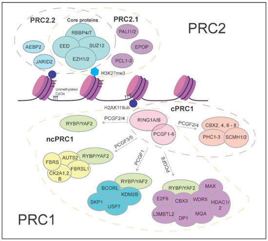
The biochemical properties of Polycomb complexes have important implications for their roles in development. Biochemically distinct variant complexes have been described for both PRC1 and PRC2 (
Figure 1). PRC1 is made up of at least eight different variant forms and they are classified based on different interactors with the E3 Ligase RING1A/B (RING1, RNF2) [
1]. Canonical PRC1 (cPRC1) consists of RING1A/B with SCMH and PHC proteins which are associated with chromatin compaction, PCGF2/4 proteins that ensure the stabilization and integrity of the complex, and CBX proteins which both compact chromatin and aid in targeting of PRC1 to genomic loci [
24,
25,
26,
27,
28]. Non-canonical PRC1, on the other hand, contains either the RING1 and YY1 binding protein (RYBP) or YY1-associated factor 2 (YAF2) protein associating with RING1A/B and different variants of the PCGF proteins [
29,
30,
31]. For example, the PRC1.1 variant complex comprises the ncPRC1 core consisting of RING1A/B and RYBP/YAF2, together with PCGF1 and KDM2B. KDMB2B is crucial in targeting RING1B to unmethylated CpG islands. Functionally, key differences between cPRC1 and ncPRC1 include their ability to interact with H3K27me3 via the chromodomains found in CBX proteins of cPRC1 [
12], and the much lower ubiquitin ligase activity of cPRC1 compared to ncPRC1 complexes. The interaction of CBX proteins of cPRC1 with H3K27me3 underscores the potential for functional interactions with PRC2 activity [
32]. ncPRC1 complexes have other mechanisms of recruitment that are independent of PRC2 and H3K27me3 [
31,
33].
Figure 1. Schematic overview of the different Polycomb group proteins.
PRC2 comprises two major variant complexes, namely PRC2.1 and PRC2.2. Both contain the catalytic core made up of EZH1/2, SUZ12, EED, and RBBP4/7 proteins [
5,
34,
35], but the interaction partners of the core complex are different. PRC2.1 contains one of the Polycomb-like (PCL) proteins—MTF2, PHF1, or PHF19. The PCLs are not directly involved in the catalytic activity of the complex, but MTF2 has been shown to play major roles in the direct recruitment of PRC2 to Polycomb target genes at unmethylated CpG islands [
36,
37]. In addition to a PCL protein, PRC2.1 contains either EPOP (Elongin BC, PRC2 associated protein) or PALI1/2 (PRC2-associated ligand-dependent co-repressor isoform 1/2) [
38,
39]. PRC2.2, on the other hand, contains the AEBP2 and JARID2 proteins, both well studied and established accessory subunits of PRC2 [
22,
40]. AEBP2 has been characterized with nucleosome binding affinity that could stimulate the catalysis of H3K27 methylation [
41]. JARID2, a member of the Jumonji family of proteins that demethylates histone proteins, is known to cooperatively stimulate PRC2 catalytic activity with AEBP2 and facilitate core PRC2 binding to DNA in vitro, particularly towards narrow PRC2 binding domains [
42,
43,
44,
45].
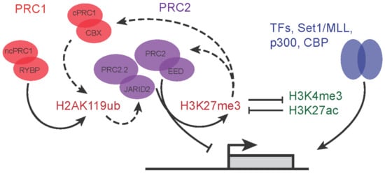
Additionally, JARID2, a part of the PRC2.2 complex, is able to bind to H2AK119ub, which is deposited by PRC1. This increases PRC2 occupancy and deposition of H3K27me3 by PRC2, constituting a positive feedback loop from PRC2 to PRC2 [
7,
9,
11] (
Figure 2). As previously discussed, the recognition and binding of H3K27me3 mark by the CBX proteins of cPRC1 complex outlines an additional layer of communication between PRC1 and PRC2 as cPRC1 can also catalyze H2AK119 ubiquitylation, albeit at lower levels compared to variant PRC1 complexes. These feedback loops (
Figure 2, dotted arrows) highlight the major dependency of individual components on maintaining Polycomb repressive mechanisms, with the reduction of any one component affecting others (PRC1 binding down in PRC2 mutants and vice versa [
45,
46]). One further example of how PRC2 and PRC1 communicate between the complexes is demonstrated by EED, a core component of PRC2, which can bind to H3K27me3 to allosterically reinforce both PRC2.1 (MTF2-directed recruitment) and PRC2.2 (JARID2 binding to H2AK119ub) [
32,
46] activity.
Figure 2. Feedback regulatory loops between activating and repressive complexes. Dashed arrow lines represent the feedback and cooperation between PRC1 and PRC2 variant complexes.
3. Roles of Polycomb Group Proteins in Pluripotency and Early Development
Based on the roles of Polycomb in homeobox gene regulation in
D. melanogaster development [
50,
51,
52,
53], it is no surprise that the Polycomb proteins serve important roles in mammalian development. Early loss-of-function studies in mice have outlined the critical importance of core Polycomb proteins, particularly in early developmental processes such as gastrulation and body-axis formation [
17,
54,
55,
56]. Although the studies were conducted using various loss-of-function methods, the picture emerged that embryos could not survive past gastrulation, without the PRC2 core proteins [
17,
55,
56]. Recent studies identified an important role for PRC2 in restricting trophoblast induction in human naïve pluripotent stem cells in vitro [
57,
58], suggesting a role in the earliest mammalian lineage decision after fertilization. In PRC1, the loss of RING1B protein results in developmental arrest at E6.5, during gastrulation, but RING1A mutants were able to survive and reach birth, although displaying defects in transformation of the axial skeleton and altered levels of Hox gene expression [
59,
60].
On the other hand, the loss of some other non-core subunits, such as the PCLs and JARID2/AEBP2 of PRC2 in mice, revealed a later embryonic lethality [
23,
61]. Though these subunits are essential for normal development, this could suggest that their role in overall PRC2 activity is more subtle, is buffered by other activities at earlier stages, or is more selectively required for specific cell lineages or developmental processes. Their roles may also be contextualized by the expression levels of both PRC2 and the activators they antagonize in specific cell lineages and stages of differentiation.
Table 1 provides an overview of the Polycomb loss-of-function mutations during development, outlining the different cell-types or lineages that the Polycomb proteins were studied in.
Table 1. In vivo and in vitro phenotypes of Polycomb mutants.
| Complex |
Variant Complex |
Non Core Subunit |
In Vivo Developmental Phenotype |
In Vitro Differentiation Phenotype |
Cell Type or Tissue Specificity |
References |
| PRC2 |
PRC2.1 |
MTF2 |
Embryonic lethal due to severe anemia around embryonic day 15.5 |
Mtf2 null cells result in faster differentiation towards all cell types in EBs |
ESC/Gastrula |
Faust et al., 1995: Hojfeldt et al., 2018; Perino et al.,2018; Loh et al., 2021; Rothberg et al., 2018 |
| PHF1 |
ND |
Increased stoichometry relative to EED during NPC differentiation |
Neuronal Precursors |
Kloet et al., 2016 |
| PHF19 |
ND |
Enhanced erythrocyte differentiation |
Mesodermal |
García-Montolio et al., 2021 |
| EPOP |
ND |
ND |
ND |
|
| PALI1/2 |
Lethal between E11.5 and perinatal lethality |
Deregulation and reduction of H3K27me3 on PRC2 target genes |
ND |
Conway, E. et al., 2018 |
| PRC2.2 |
JARID2 |
Lethal between E10.5 - E18.5 |
Stagnated differentiation towards all germ layers |
ND |
Loh et al., 2021 |
| AEBP2 |
Late-embryonic and perinatal lethality; skeletal transformations |
Deregulation and increase of H3K27me3 on PRC2 target genes |
ND |
Grijzenhout, A. et al., 2016;
Kim, H. et al., 2011 |
| cPRC1 |
|
CBX2 |
Male to female sex reversal |
Activator of the testis - determining
gene Sry |
Testes, Sex Organ |
Katoh-Fukui, Y. et al., 1998 |
| CBX4 |
Pre-weaning lethality |
ND |
ND |
|
| CBX6 |
Several transformational defects in late embryonic development |
ND |
Enriched in NPC differentiation |
Dickinson, M. E. et al., 2016 |
| CBX7 |
ND |
Enriched in early ESC stages |
|
|
| CBX8 |
ND |
ND |
Enriched in NPC differentiation |
Morey, L. et al. 2012 |
| PHC1 |
Perinatal lethality |
Destabilized pluripotency by disruption of Nanog long-range
interactions |
ND |
Isono K. et al., 2005 |
| PHC2 |
Posterior transformation; Hox gene deregulation; Viable birth |
ND |
ND |
Isono K. et al., 2005 |
| PHC3 |
Phc3 null animals are born but exhibit cardiac abnormalities |
ND |
ND |
Dickinson, M. E. et al.,
2016 |
| PCGF2 |
Posterior transformation; Hox gene deregulation; Viable birth |
ND |
ND |
Akasaka, T. et al., 1996 |
| PCGF4 |
Perinatal lethality, ataxia, severe haematopoietic defects |
ND |
ND |
van der Lugt, N. M. et al.,
1994 |
| ncPRC1 |
|
CBX3 |
Defects in germ cell development |
Inhibition results in neural differentiation impairment and
diverting towards mesendodermal differentiion |
Neural Precursors |
Abe, K. et al., 2011 |
| PCGF1 |
E12.5 lethality |
Promotes ectoderm and mesoderm differentiation |
Ectoderm and Mesoderm lineages |
Yan Y et al., 2017 |
| PCGF3 |
Double Pcgf3;Pcgf5 deletion results in female-specific embryonic lethality |
ND |
ND |
Almeida, M. et al., 2017 |
| PCGF5 |
|
| PCGF6 |
defects in pre-implantation and periimplantation and in placenta development |
Ablation of Pcgf6 in ESCs leads to robust de-repression of such germ cell-related genes, in turn affecting cell growth and viability. |
pre- and peri-implantation mouse embryo |
Dickinson, M. E. et al.,
2016 |
| KDM2B |
Mid-gestation lethality; posterior transformation of the axial skeleton |
Maintain pluripotency by recruitment of PRC1 to CpGs |
ND |
Boulard, M. et al., 2015 |
| RYBP |
E6.5 Lethality; impaired cell proliferation |
Depletion of Rybp inhibits proliferation and promotes
Neuronal differentiation of embryonic neural progenitor cells
(eNPCs) |
Neural Precursors |
Pirity, M. K. et al., 2015 |
| AUTS2 |
Die before weaning with defects in the nervous, cardiovascular and biliary
systems |
ND |
Neural Ectoderm |
Gao, Z. et al., 2014 |
| L3MBTL2 |
E6.5 lethality; gastrulation defects |
Compromised proliferation and abnormal differentiation of L3mbtl2(-/) embryonic stem (ES) cells |
ESC |
Qin, J. et al., 2012 |
Most of the biochemical and functional studies done in vitro were performed in mouse embryonic stem cells (mESCs). They model the early stages of development well, particularly the inner cell mass of the blastocyst, in which Polycomb proteins play an important role in regulating pluripotency and cellular differentiation. Recent studies have focused more on the mechanisms of how non-core Polycomb proteins work in regulating gene expression in these mESCs.
3.1. PRC2 Variant Complexes and Their Roles in Embryonic Stem Cells
In terms of PRC2, several major landmark papers uncovered the mechanism by which MTF2 (PRC2.1) can directly and selectively bind to a subset of unmethylated CpG-dense DNA regions, with preferential binding to sites with specific DNA shape properties such as an underwound helix as compared to the canonical B-DNA structure [
36,
37]. This finding was supported by crystal structures of the PCL proteins MTF2 and PHF1 bound to DNA, demonstrating a binding of the winged-helix domain to DNA with unmethylated CpGs and reduced helical twist [
37]. The loss of MTF2 in mESCs resulted in globally reduced H3K27me3 levels, and recruitment of EZH2 was also severely reduced at target gene locations [
36,
45,
46,
62]. Unlike MTF2, the loss of other PCL proteins, PHF1 and PHF19 did not elicit a similar level of drastic reduction of PRC2 recruitment in mESCs [
45,
62]. This might be because these proteins are expressed at much lower levels compared to MTF2 in mESCs [
63].
The other PRC2.1 protein that plays an important role in mESCs is EPOP. Deletion of EPOP in mESCs prevents PRC2.1 from interacting with Elongin B/C proteins, which in turn reduces RNA polymerase II elongation on target genes [
64]. Therefore, while the loss of EPOP does not directly translate into loss of H3K27me3 levels, it would set up for a low level of permissive transcription at target genes and affect how PRC2 regulates transcription. The interaction of EPOP with PRC2 is found to be mutually exclusive with PALI1/2 [
38,
40]. This is corroborated by studies demonstrating a dynamic shift of the PRC2.1 interactome between EPOP and PALI1/2 during mESC differentiation [
63]. Unlike EPOP, the ablation of PALI1/2 proteins resulted in some degree of H3K27me3 reduction at target genes [
39]. This indicates the importance of these proteins in maintaining the catalytic function of PRC2, but just like PHF1 and PHF19, their stoichiometry relative to other PRC2.1 proteins is found to be minimal.
4. Roles of Polycomb Complexes in Developmental Cell Lineages
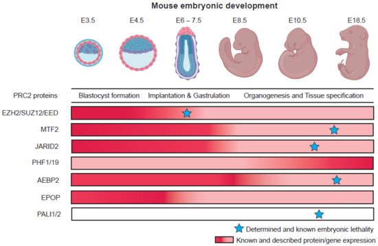
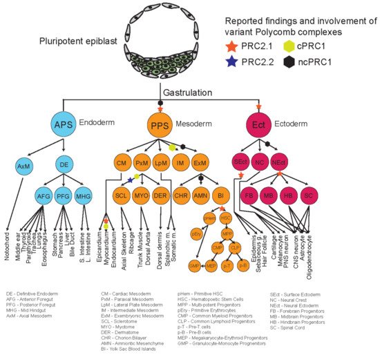
The repertoire of different phenotypes observed for many mutants of different Polycomb proteins, and their relatively delayed phenotypes suggest that some of these variant complex subunits might play more important roles during organogenesis and later developmental stages (Figure 3). Recent studies looking into the roles of different Polycomb proteins during in vitro differentiation shed some light on the potential specific mechanisms and roles of these different proteins during development. Figure 4 provides an overview and developmental roadmap of the known contributions of Polycomb complexes during development. It is apparent that there are still many developmental cell types, particularly from endoderm lineages, that remain to be investigated for contributions of Polycomb complexes.
Figure 3. Differences in pattern of Polycomb group protein expression and their embryonic loss-of-function phenotypes. Embryo pictures reprinted from “Mouse Development”, by BioRender.com (accessed on 15 July 2022). (2022). Retrieved from
https://app.biorender.com/biorender-templates (accessed on 15 July 2022).
Figure 4. Developmental roadmap charting Polycomb function and involvement at different key lineage decision steps.