Type 2 Transglutaminase in Coeliac Disease: History
Please note this is an old version of this entry, which may differ significantly from the current revision.

Coeliac disease (CD) is a multifactiorial enteropathy that affects the small intestine of genetically predisposed individuals. A condition of partial to total atrophy, together with crypt hyperplasia and consistent lymphocytic infiltration, characterises the intestinal mucosa of affected patients. The main environmental trigger is a heterogenic proteic component of some dietary cereals, commonly known as gluten. A strong immune response against gluten, both cellular and humoral, is mounted in CD, accompanied by a humoral autoimmune response against self-proteins, in particular type 2 transglutaminase (TG2).

  • coeliac disease
  • gluten
  • autoimmunity

1. Epidemiology and Clinical Manifestations of CD

Coeliac disease (CD) is widely distributed throughout the world, affecting on average 1% of the population, with differences in incidence due mostly to the dietary habits of certain countries, or to geographical area [6]. This enteropathy can manifest at every stage of life, with a higher frequency in children between 1 and 2 years of age. Classical presentation of CD includes gastrointestinal symptoms, such as malabsorption, chronic diarrhoea, abdominal distension, and pain. In paediatric populations, the disease also presents with failure to thrive and delayed puberty. Other frequent symptoms, mainly due to nutritional deficiencies resulting from the mucosal lesions, are anaemia, osteoporosis, and weight loss [6]. Furthermore, extraintestinal manifestations are also commonly associated with CD, such as liver, skin, endocrine, and neurological disorders [7].

2. The Main Environmental Trigger in CD: Gluten

Common cereal seeds possess abundant storage proteins, 75–80% of which are gluten, a complex mixture of proteins that, due to their high content of Pro and Gln residues in their primary structure, are defined as prolamins. Gluten prolamins of wheat were the first to be identified, and are usually divided into two classes on the basis of their solubility: gliadins (the alcohol-soluble fraction, monomeric) and glutenins (the water-soluble fraction, polymeric) [8]. The equivalents of gliadin proteins in other cereals assume different names, depending on the specific cereal: secalins in rye, hordeins in barley, and avenins in oat. Polypeptide sequences responsible for the immune response in CD individuals are mainly from gliadin and related prolamins [9]. Gliadin comprises three major groups of proteins, namely α/βγ, and ω, according to their electrophoretic mobility (α- and β-gliadins migrate separately but are very similar) [8]. α-gliadins contain the most immunogenic sequences characterised in the context of CD [10]. In particular, the central domain contains the 33-mer segment, comprising six overlapping epitopes relevant for CD pathogenesis. In addition, the high Pro residues content (about 15%) renders gliadin quite resistant to digestive enzymes; as a consequence, numerous large undigested peptides may come into contact with the intestinal barrier, triggering pathways leading to inflammation and immune response [11]. The immune response requires that gliadin peptides are specifically recognised by antigen-presenting cells (APCs) and presented to competent T cells. Immunogenic properties of gliadin sequences are also related to a high content of Gln residues (about 30% of total residues). Some of these Gln residues have a key role in the interaction with the cellular proteins necessary for antigen presentation to the immune system, and may be the target of a post-translational modification (i.e., TG2 deamidation) that renders them more immunogenic [2,5,12].
Even if gluten is the main environmental stimulus in the onset of CD, recent evidence has demonstrated that the alteration in the gut microbiota (a decrease in Bifidobacteria and an increase in Bacteroides has generally been observed) may represent a key environmental factor contributing to the disease [13]. Moreover, the relative timing of gluten introduction into the infant diet and recurrent gastrointestinal viral infections, in particular rotavirus infections, seem to be important in altering the intestinal barrier, thus favouring exposure to gluten [6,12,14].

3. The Genetics of CD

The main genes involved in predisposition to CD are particular haplotypes of the human leukocytes antigen (HLA) system of class II [5]. They codify for integral membrane heterodimers, which have the function of binding gluten peptides on the cell surface and presenting them to CD4+ T lymphocytes. Haplotypes DQ2 and DQ8 are present on APCs in 95% and 5% of CD patients, respectively [15]. However, more than 40% of individuals in the general population present these same haplotypes [16]; thus, it is clear that their presence is a necessary condition but not sufficient on its own to cause CD. Many other non-HLA genes seem to be associated with CD; large scale genomic studies identified several genetic regions harbouring genes controlling immune functions, but altogether they account for only about 15% of the genetic component of the disease [17]. Considering all HLA and non-HLA genes, it is possible to explain about 50% of CD heritability; the remaining part of the heritability could be due to the presence of rare genetic variants with a very low allele frequency, normally excluded from genome-wide association [17]. A recent and promising area of research is the exploration of CD susceptibility beyond genetics: emerging evidences have highlighted a role in CD pathogenesis of specific patterns of DNA methylation and chromatin accessibility, as well as of peculiar transcription patterns of non-coding RNA, specifically micro-RNA and long-noncoding RNA [17].

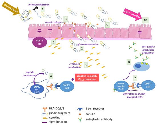
4. The Adaptive and Innate Immune Response in CD

Large partially digested gluten peptides in the gut lumen of CD subjects trigger a cascade of events that ultimately cause mucosal lesions, involving both the adaptive and the innate immune system [12] (Figure 1). One of the first events is gluten-induced zonulin release by intestinal epithelium, which causes an increased permeability, thus favouring gluten translocation to lamina propria [18]. Here, some gliadin peptides are modified by type 2 transglutaminase (TG2) at the level of specific Gln residues, thus becoming more immunogenic. Both unmodified and TG2-modified gliadin peptides are presented by HLA-DQ2/8 APCs, eliciting T helper cell recruitment and activation, with the release of a cascade of pro-inflammatory cytokines, mainly interferon γ (INFγ) and tumour necrosis factor α (TNFα), and activation of B cells secerning anti-gliadin or anti-TG2 antibodies. TH1 and TH2 adaptive responses to gluten are accompanied by an innate immune response with secretion of interleukin (IL)-15, IL8, and keratinocyte growth factor, and with expansion of CD8+ cells; all these events, together with an increased matrix remodelling by metalloproteinases, cause extensive mucosal damage characterised by hyperproliferation, cell death and loss of epithelial barrier architecture [5,6,12]. The innate component of the immune response is strictly related to the presence of specific non-immunogenic peptides, such as the 25-mer, i.e., the sequence 31–55 of α-gliadin and shorter peptide 31–43 (P31–43) and peptide 31–49, which reach lamina propria, where they may induce stress signalling, proliferation, cytoskeleton alterations, and vesicular trafficking modifications [19].
Figure 1. Scheme of the main events of CD pathogenesis. In gut lumen, gluten/gliadin is partially digested (1) and large fragments cross the intestinal barrier (2), triggering an increased intestinal permeability with zonulin release (3). Antigen presentation by HLA-DQ2/8 APCs (4) elicits an adaptive immune response with the release of pro-inflammatory cytokines (5), the recruitment of cytotoxic T cells (6), and the activation of gliadin-specific B cells (7), with the consequent production of anti-gliadin antibodies (8). All together, these events lead to the loss of mucosal integrity (9), which is slowly recovered with a gluten-free diet (10). This scheme does not show the role of TG2 gluten modifications, which are explained in the next figures.

5. Autoimmunity in CD

In the serum and intestinal mucosa of CD patients, antibodies to self-components can be detected; thus, CD displays the characteristics of an autoimmune disease. Some decades ago, Dieterich and colleagues demonstrated that TG2 was the main autoantigen in CD [20]. During CD onset, anti-TG2 antibodies of IgA class appear at a very early stage in the CD intestinal mucosa; in this location, they are detectable before the mucosal changes typical of CD lesions, and before they can be found in serum [21]. Then, they cross the blood vessels and reach various tissues and organs, where they accumulate [22]. Autoantibodies directed against other TG isoforms (TG3 and neuronal TG) have also been found, associated in particular with the occurrence of extra-intestinal manifestations of CD [23,24].

6. The Coeliac Cellular Phenotype

Several recent studies have highlighted that in CD cells, it is possible to observe some constitutive features that are independent from the exposure to gluten, and are also visible far from the intestine, i.e., the main site of inflammation during the response to gluten. The so-called “coeliac cellular phenotype” is an ensemble of characteristics displayed by the CD cells of duodenal biopsies or other cells, such as skin-derived fibroblasts, and can be partially reproduced in cells from control subjects when exposed to α-gliadin P31–43; moreover, this phenotype also includes the fact that some biological effects of P31–43 become evident in CD cells but not in non-CD cells [25,26,27]. It has been demonstrated that CD skin-derived fibroblasts have a higher content of phosphorylated proteins and a higher number of focal adhesions than cells from control subjects [25]. Furthermore, CD enterocytes and dermal fibroblasts show a delay in the progression of vesicular early endosome to late endosome, associated with increased signalling, actin rearrangement, and augmented proliferation [26]. Structural cytoskeleton alterations, associated with an increased Rho A activity, have also been described in the dendritic cells of CD children [28]. Finally, in CD enterocytes, several inflammation markers have been found to become constitutively enhanced [29]. Thus, it is reasonable to assume that CD cells possess constitutive altered pathways and responses that render them more sensible to the effects of gluten peptides and other proinflammatory agents.

7. CD Diagnosis

Currently, a single direct approach to the diagnosis of CD is not available. Genetic tests aimed at finding HLA-DQ2/8 haplotypes have only a negative predictive value, and are employed to support diagnosis. The gold standard for CD diagnosis is obtained by combining the analysis of the duodenal biopsy with the detection of serological markers [30]. Morphological evaluation of biopsy samples highlights the presence of typical features of CD mucosal lesions, i.e., flat and hyperplastic mucosa at various degrees together with an increased concentration of intraepithelial lymphocytes [5]. Currently, two types of non-invasive serological markers can be used to support a correct diagnosis: antibodies raised against the environmental trigger, in particular gliadin, and antibodies against self-components, mainly anti-TG2 antibodies. Moreover, the discovery of CD-associated circulating miRNA associated with CD could open the way for the development of new potential non-invasive biomarkers of CD [31].

Enzymatic and Immunofluorescence Assays

The detection of specific antibodies is routinely achieved by enzyme-linked immunosorbent assays (ELISAs) or by indirect immunofluorescence assays (IFAs). Antibodies related to CD are of both the IgA and IgG classes; however, the highest sensitivity and specificity is reached by tests detecting IgA [32]. Anti-gliadin antibodies were the first serological marker detected and quantified by ELISA, introduced for the screening of at-risk subjects and to support diagnosis. However, ELISA tests used to detect anti-gliadin antibodies were not very specific; thus, other more sensitive and specific serological tests were later developed [6,33]. In particular, a test that showed a higher specificity as an anti-gliadin test, especially in IgA-deficient patients [34], was the test used to search for antibodies against deamidated gliadin peptides (DGP), which are modified by the TG2 enzymatic reaction called deamidation. Currently, tests based on the search for anti-DGP antibodies, together with those used for anti-TG2 antibodies (discussed later) are the most frequently employed serologic tests in clinical CD management. An alternative approach to detecting the autoimmune component is the use of IFAs. In these assays, the presence of autoantibodies in sera causes a specific staining pattern on tissue slides. Searching for anti-reticulin antibodies by IFA was an early method of screening. The term reticulin referred to a reticular component of connective tissue around smooth muscle fibres. A specific reticular pattern obtained by the staining of different rat tissues was considered to be associated with CD. Later, a more sensitive IFA test was employed to screen for the presence of anti-endomysial antibodies (EMA) [35]; endomysium is also a layer of connective tissue around smooth muscle cells, and EMA produce a characteristic staining pattern on slices of monkey oesophagus or of human umbilical cord. With the discovery that TG2 is the main CD autoantigen, it has been clarified that IFA staining patterns were due to the presence of TG2 in tissue slides.

8. CD Therapy

The only currently available therapy for CD is a strict lifelong diet without gluten [6]. Such a diet results in the remission of symptoms in about six months, accompanied by the recovery of intestinal mucosal architecture and the disappearance of the autoimmune antibodies. A gluten-free diet is often difficult to manage, and not only for social reasons. Indeed, gluten-free products and medicines often contain trace amounts of gluten as a consequence of cross-contamination during industrial production. Moreover, gluten-free products are poor in fibre, vitamins, and other micronutrients. Thus, the gluten-free diet tends to be unbalanced and requires additional nutrient supplementation [36]. A minority of adult CD patients are considered to be affected by “refractory CD”, when the disease does not respond to treatment with a gluten-free diet. Symptoms and intestinal atrophy persist even after 12 months of therapy [37]. These patients are at risk of developing serious complications, such as intestinal lymphoma and collagenous sprue. On the bases of increasing insights into CD pathogenesis, novel therapeutic strategies are under investigation, mainly intended as adjuvants to a gluten-free diet, as a rescue treatment after accidental or intentional gluten intake, or to manage refractory CD. One of these new approaches consists in the detoxification of gluten sequences using exogenous endopeptidases. Another possibility is gluten sequestration with polymeric binders, thus preventing immunogenic modification by TG2. Other strategies aim to interfere with pathogenicity patterns, for example by reducing intestinal permeability, by inhibiting the binding of gluten peptides to HLA molecules, by blocking proinflammatory cytokines, etc. Currently, several clinical trials based on above-mentioned strategies are under development, with encouraging results [38].

9.TG2

The TG family includes transferase enzymes (EC 2.3.2.13), which catalyse the formation of an isopeptide bond involving the g-carboxamide group of a Gln residue (the acyl-donor) and the e-amino group of a Lys residue (the nucleophilic acyl-acceptor), generating an intra- or an inter-protein cross-link [[1]]. The biochemical mechanism of the reaction involves a ping-pong kinetics: first, the g-carboxamide group forms a thiol ester with the Cys residue of the enzyme active site with the release of ammonia, then the acyl intermediate is transferred to the nucleophilic aminic substrate. As alternatives to Lys residues as acyl-acceptors in the crosslinking reaction, TGs can use several low molecular-mass primary amines (spermine, spermidine, putrescine, etc.). In the absence of suitable amines and in the presence of a slightly acidic pH, water can act as nucleophile; thus, TGs are able to deamidate Gln to Glu residues [[1]]. In mammals, nine structurally and functionally related genes encoding TGs have been identified [39]. Among them, only one gene encodes for a protein (band 4.2) without catalytic activity. All other eight mammalian TGs are strictly Ca2+-dependent enzymes and share a high sequence identity around the Cys proteinase-active site, comprising the catalytic triad Cys, His, and Asp. Moreover, the structural organization in four sequential domains is highly conserved in TG isoforms: the N-terminal b-sandwich domain is followed by the catalytic core domain (containing the Ca2+ binding sites) and two C-terminal b-barrel domains [2]].

10. TG2 in CD Pathogenesis

Given the wide range of TG2 enzymatic and non-enzymatic functions and its ubiquitous distribution in tissues and cellular compartments, it is not surprising that TG2 has a role in the development of several human pathological conditions, such as cancer, neurodegenerative disorders, inflammatory and cardiovascular diseases, and autoimmune conditions [54–56]. Regarding CD, TG2 represents the trade union between the main environmental trigger (gluten) and the main genetic component (HLA genes) of CD. This enzyme, whose expression is greatly increased in inflamed intestinal sites both intracellularly (in enterocytes) and extracellularly (in the lamina propria) [[3]], recognizes gluten fragments as preferred substrates and catalyses two crucial reactions: deamidation and transamidation of specific Gln residues.

Gluten Modification: Deamidation

Given the high content of Gln residues in gliadin and other gluten proteins, it is evident that TG2 can use them as Gln-donor substrates in transamidating and deamidating reactions. Deamidation is not a common reaction because it requires specific conditions: a slightly acidic pH and a very low concentration of available amines. When and where these conditions may be realised in cells and tissues are still debated. In a recent work, it has been suggested that TG2 released in gut lumen as a consequence of enterocytes shedding could meet luminal gliadin, and in this way could have direct access to adaptive immune B cells at the level of Peyer’s patches [[4]]. TG2 on the cell surfaces of APCs could deamidate gliadin during its turnover by endocytosis at the level of acidic endocytic vesicles. The recent demonstration that TG2 is more abundant on the cell surface and in early endocytic vesicles of CD skin-derived fibroblasts than of control cells supports this hypothesis [27]. In any case, there is no doubt that TG2 recognises specific Gln residues in gliadin sequences and deamidates them, introducing a net negative charge that greatly increases the affinity with which DQ2/DQ8 heterodimers bind gliadin peptides for presentation to T cells [59]. Indeed, the production of high-resolution X-ray crystal structures of representative deamidated gluten peptides in complex with DQ2 and DQ8 has definitively confirmed the importance of the introduction of a negative charge at a specific location in a gluten peptide interacting with the HLA binding groove [60]. As a consequence, a stronger immune response is evoked. An updated list of CD-relevant deamidated gluten epitopes recognised by CD4+ T cells has recently been reported [[5]]. The majority of listed epitopes are DQ2.5-restricted and mainly belong to the a and g gliadins, but some epitopes also belong to the w-gliadins, glutenins, hordeins, secalins, and avenins. Only a few epitopes are DQ2.2-or DQ8-restricted and also belong to the a and g gliadins and glutenins. This discrepancy is likely due to the fact that most gluten epitopes have been discovered in CD patients positive for HLA-DQ2.5 [[6]]. Thus, it is reasonable to expect that further additional epitopes will be defined in the future.

Gluten Modification: Mechanism of Anti-TG2 Antibody Production

As above reported, TG2 is the main autoantigen in CD. To date, no TG2-specific CD4+ helper T cells have been identified. For this reason, it is supposed that gliadin-specific CD4+ helper T cells are essential in the production of anti-TG2 antibodies at the intestinal level, as a consequence of a cross-linking reaction catalysed by TG2 and involving some gliadin peptides [2]. In this case, reactive Gln residues of gliadin peptides are acyl-donor substrates in the transamidating reaction. The aminic second substrates could be represented by Lys residues of TG2 itself or of other intracellular or surface-membrane proteins. Several models supported by experimental data have tried to explain how this activity could be responsible for the autoimmune response. According to the classic hapten-carrier-like model, TG2 forms isopeptide bonds between itself (involving an autoreactive Lys residue) and gliadin peptides, generating complexes that are internalised, processed, and exposed by HLA-DQ2/8 molecules of TG2-specific B cells [[7]]. Then, these cells activate gluten-specific CD4+ T cells, which in turn stimulate B cells to produce anti-TG2 antibodies. In this regard, several isopeptides between TG2 and gliadin fragments have been identified [[8]]. Recently, du Pré et al. developed an elegant transgenic murine model demonstrating that autoreactive anti-TG2 B cells are able to produce autoantibodies when they receive help from T cells [63]. It has also been proposed that complexes between TG2 and gliadin could also be formed involving the Cys residue of the TG2 active site, with the formation of a thioester bond [[9]]. In any case, the hapten-carrier-like model better explains the occurrence of other autoantibodies besides anti-TG2. Indeed, other autoantigens have been identified (actin, desmin, calreticulin, collagen, etc.) and all can be recognised by TG2 as substrates for forming covalent complexes that could be processed by substrate-specific B-cells [65]. Moreover, this model is compatible with the observation that the autoimmune response gradually decreases when a gluten-free diet is adopted. A revised version of the model proposes that TG2 is able to cross-link gliadin peptides directly to B cell receptors on TG2-specific B lymphocytes [[10]]. In a recent work [[11]], it has been demonstrated that the majority of TG2-specific B cells displayed epitope specificity for TG2 N-terminal sequences, and that cell-surface antibodies with such a specificity allowed TG2-cross-linking activity, leading to the formation of covalent complexes with gliadin and consequential deamidation during antigen presentation. Another model on the generation of TG2 antibodies proposes that neoepitopes formed by the transamidating activity of TG2 targeting gluten peptides and itself could recruit T lymphocytes that escape thymic negative selection [[12]]. Finally, some evidence supports the idea that the autoimmune response against TG2 could involve a mechanism of molecular mimicry regarding viral or bacterial antigens [[13]]. 

11. Conclusion

It is doubtless that TG2 represents a key enzyme in the pathogenic mechanisms leading to CD development. At the site of intestinal inflammation, where TG2 is typically overexpressed, its canonical enzymatic activity leads to the formation of isopeptide bonds between itself (or other self-proteins) and gluten peptides, thus provoking a strong autoimmune response. Still uncertain is the role of circulating and intestinal anti-TG2 antibodies in contributing to CD onset; however, antibody detection represents an irreplaceable tool for CD screening programs and diagnosis. In particular circumstances (low pH and virtual absence of primary amines) in the CD mucosa, TG2 can modify specific Gln residues of gluten peptides, transforming a neutral residue into an acidic residue (Glu), which is important in the presentation to the immune system by DQ2/8 molecules and the consequent loss of oral tolerance towards gluten. For these evident reasons, TG2 has become the targetof recent pharmaceutical approaches aimed at reducing deamidation. New generations of increasingly specific and potent TG2 inhibitors are under investigation, some of which have reached the point of clinical experimentation. Finally, given the increasing use of food-grade TGs, particularly TGm, as biotechnological tools in a number of industrial applications, some researchers are exploring the possibility of modifying gluten peptides by transamidating them with nucleophilic amines in an attempt to reduce the immunogenicity of the majority of DQ2/8-restricted epitopes. Finally, it is appropriate to underline that TG2 is a multifunctional enzyme with several catalytic activities and non-catalytic functions. Thus, it cannot be excluded that TG2 may have other unexpected roles in CD pathogenesis, independent of its transamidating and deamidating activity. For example, TG2 is easily targeted on the cell surface by anti-TG2 antibodies, and not only at the intestinal level. Given the biological consequences of such an interaction at the cell surface (for example, increased proliferation and structural and signalling modifications) and potential differences in subcellular TG2 distribution in CD cells, it is reasonable to conclude that TG2 may contribute to CD onset and progression in other peculiar and still unexplored fashions.

This entry is adapted from the peer-reviewed paper 10.3390/ijms23147513

References

  1. Lorand, L.; Graham, R.M. Transglutaminases: Crosslinking enzymes with pleiotropic functions. Nat. Rev. Mol. Cell. Biol. 2003, 4,140–156.
  2. Lorand, L.; Graham, R.M. Transglutaminases: Crosslinking enzymes with pleiotropic functions. Nat. Rev. Mol. Cell. Biol. 2003, 4, 140–156.
  3. Stefania Martucciello; Silvia Sposito; Carla Esposito; Gaetana Paolella; Ivana Caputo; Interplay between Type 2 Transglutaminase (TG2), Gliadin Peptide 31-43 and Anti-TG2 Antibodies in Celiac Disease. International Journal of Molecular Sciences 2020, 21, 3673, 10.3390/ijms21103673.
  4. Rasmus Iversen; Sunniva F. Amundsen; Liv Kleppa; M. Fleur du Pré; Jorunn Stamnæs; Ludvig M. Sollid; Evidence That Pathogenic Transglutaminase 2 in Celiac Disease Derives From Enterocytes. Gastroenterology 2020, 159, 788-790, 10.1053/j.gastro.2020.04.018.
  5. Ludvig M. Sollid; Jason A. Tye-Din; Shuo-Wang Qiao; Robert P. Anderson; Carmen Gianfrani; Frits Koning; Update 2020: nomenclature and listing of celiac disease–relevant gluten epitopes recognized by CD4+ T cells. Immunogenetics 2019, 72, 85-88, 10.1007/s00251-019-01141-w.
  6. Ludvig M. Sollid; Jason A. Tye-Din; Shuo-Wang Qiao; Robert P. Anderson; Carmen Gianfrani; Frits Koning; Update 2020: nomenclature and listing of celiac disease–relevant gluten epitopes recognized by CD4+ T cells. Immunogenetics 2019, 72, 85-88, 10.1007/s00251-019-01141-w.
  7. L M Sollid; Ø Molberg; S McADAM; K E A Lundin; Autoantibodies in coeliac disease: tissue transglutaminase—guilt by association?. Gut 1997, 41, 851-852, 10.1136/gut.41.6.851.
  8. Barbara Lexhaller; Christina Ludwig; Katharina Anne Scherf; Identification of Isopeptides Between Human Tissue Transglutaminase and Wheat, Rye, and Barley Gluten Peptides.. Scientific Reports 2020, 10, 7426-14, 10.1038/s41598-020-64143-9.
  9. Jorunn Stamnæs; Rasmus Iversen; M. Fleur Du Pré; Xi Chen; Ludvig M. Sollid; Enhanced B-Cell Receptor Recognition of the Autoantigen Transglutaminase 2 by Efficient Catalytic Self-Multimerization. PLOS ONE 2015, 10, e0134922, 10.1371/journal.pone.0134922.
  10. Rasmus Iversen; M. Fleur Du Pré; Roberto Di Niro; Ludvig M. Sollid; Igs as Substrates for Transglutaminase 2: Implications for Autoantibody Production in Celiac Disease. The Journal of Immunology 2015, 195, 5159-5168, 10.4049/jimmunol.1501363.
  11. Rasmus Iversen; Bishnudeo Roy; Jorunn Stamnaes; Lene S. Høydahl; Kathrin Hnida; Ralf S. Neumann; Ilma R. Korponay-Szabó; Knut E. A. Lundin; Ludvig M. Sollid; Efficient T cell–B cell collaboration guides autoantibody epitope bias and onset of celiac disease. Proceedings of the National Academy of Sciences 2019, 116, 15134-15139, 10.1073/pnas.1901561116.
  12. Ludvig M Sollid; Bana Jabri; Celiac disease and transglutaminase 2: a model for posttranslational modification of antigens and HLA association in the pathogenesis of autoimmune disorders. Current Opinion in Immunology 2011, 23, 732-738, 10.1016/j.coi.2011.08.006.
  13. Jan Petersen; Laura Ciacchi; Mai T. Tran; Khai Lee Loh; Yvonne Kooy-Winkelaar; Nathan P. Croft; Melinda Y. Hardy; Zhenjun Chen; James McCluskey; Robert P. Anderson; et al. T cell receptor cross-reactivity between gliadin and bacterial peptides in celiac disease. Nature Structural & Molecular Biology 2019, 27, 49-61, 10.1038/s41594-019-0353-4.
More
This entry is offline, you can click here to edit this entry!
Video Production Service