Your browser does not fully support modern features. Please upgrade for a smoother experience.
Please note this is an old version of this entry, which may differ significantly from the current revision.
Subjects:
Biochemical Research Methods
PC-12 cells have been widely used as a neuronal line study model in many biosensing devices, mainly due to the neurogenic characteristics acquired after differentiation, such as high level of secreted neurotransmitter, neuron morphology characterized by neurite outgrowth, and expression of ion and neurotransmitter receptors.
- PC12 cell line
- biosensing
- analytical determination
- neuronal stimulation
1. Introduction
In a normal, healthy, brain the neurons communicate with each other using electrical charges that travel down axons causing the release of chemicals and, through different pathways, perform every function of the brain such as sensations, movements, thoughts, memories, and feelings. The neurons are kept healthy by other cells of the brain, which are closely connected, and miscommunications in one area can disrupt other brain activities, meaning that brain disorders can result in widespread problems. In vitro assays are commonly employed for understanding the pathophysiology processes involved in brain disorders, thus decreasing the complexity of direct in vivo approaches [1].
In particular, primary neurons represent a powerful model to manipulate and observe neurons due to the fact that they maintain the main characteristic features of their tissue of origin, making them a biologically and physiologically relevant tool for the study of neuroscience. However, primary neurons are highly sensitive to isolation conditions and growth environment, do not proliferate, and were limited to short term culture (<5 days) [2]. On the other hand, neuronal cell lines are commonly used for in vitro neurobiology studies because they are easily transfected, possess the ability to proliferate, and can be induced to differentiate into neuron-like cells, expressing neuronal biomarkers and presenting axons and dendrites. For this, commercial immortal neuronal cell lines, which can simulate neurons after differentiation process are available: PC12, SH-SY5Y, bEnd.3, TR-BBB, HBMEC, BV2, etc. Based on the differentiation time (i.e., about 4 days for PC-12 and 18 for days for SH-SY5Y), and the neurogenic characteristics, PC12 is one of the most preferred cell line to be used as model in neurobiology studies [3].
In order to obtain detailed information on the mechanism of intra- or extra-cellular reactions involving cells under different stimuli, mimicking different pathophysiological processes, an appropriate analytical approach is needed. In this regard, the (bio)sensors field represent an excellent tool recognized for fast, low-cost, and innovative methodologies that can be designed for in situ analysis of cell cultures, improving in vitro models, thus allowing the study of molecular mechanisms to be carried out in a simple and reproducible manner [4].
Biosensing represents the detection of target molecules (analytes) based on the chemical principles used by a living system, e.g., molecular recognition. A biosensor is a device that transforms chemical information into an analytically useful signal and contain two connected components: a receptor, consisting in a recognition system (antibody, enzyme, aptamer, cell, etc.,), and a physicochemical transducer. The biological recognition system translates the interaction between the analyte and receptor into a measurable signal transferred by the transducer to a measuring device. Biosensing represents the detection of target molecules (analytes) based on the chemical principles used by a living system, e.g., molecular recognition. A biosensor is a device that transforms chemical information into an analytically useful signal and contains two connected components: a receptor, consisting in a recognition system (antibody, enzyme, aptamer, cell, etc.,), and a physicochemical transducer. The biological recognition system translates the interaction between the analyte and receptor into a measurable signal transferred by the transducer to a measuring device [9].
The immortalized PC-12 cell line demonstrated to be a classical neuronal cell model derived from rat pheochromocytoma with the ability to acquire the sympathetic neurons features in a differentiation process in the presence of nerve growth factor. PC-12 cell line was shown to be the preferred model in neurobiology study using biosensing devices.
2. PC-12 Cell Line
For over half a century, biosensors have been widely investigated and nowadays they are playing an essential role in various sectors such as food and pharmaceutical industry as well as in various medical fields. Integration of cells cultures within biosensors are common practices now and brings great value to the field of medical, pharmaceutical, and biological research.
Neurobiology is a field intensively studied with the help of cell-based biosensors. PC-12 cell line is preferred as model in neurobiology studies because of its neurogenic characteristics (after differentiation) such as neurotransmitters secretion (dopamine, norepinephrine, and other catecholamines), neuron morphology, as well as ion and neurotransmitter receptors.
Being extensively used, PC-12 cells come with the advantage of a great amount of knowledge about culturing conditions and differentiation process. In opposition to SH-SY5Y, PC-12 adh cells have the advantage of taking shorter time to differentiate.
PC-12 is harvested from a pheochromocytoma rat adrenal medulla and is one of the most common neuronal precursor cell lines used in neuroscience research including studying neuronal degenerations, neuronal differentiation, and neural networks. Cultured under normal conditions, PC-12 cells present morphological and physiological characteristics of the adrenal gland cells. When nerve growth factor (NGF) is added to the culture, this type of cells suffers a differentiation process and start to manifest morphological and functional characteristics of sympathetic ganglion neurons, Figure 1.
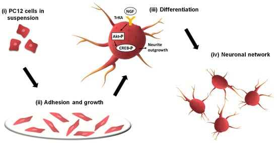
Figure 1. Schematic representation of PC-12 neuronal network formation.
There are two types of PC-12 cell lines, classical PC-12 cells grown in suspension and the adherent line of PC-12 cells originated from selected phenotype of easily adherent cells. To increase the ability of PC-12 in suspension to become adherent it is necessarily to modify with collagen the surface on which there are cultured. Adherent line of PC-12 attaches easily on poly-d-lysine surface as well as on collagen and even on plastic surface [5].
Optimization of nerve growth factor (NGF) concentration added to the culture is of great importance for the differentiation process of PC-12. It has been observed that with the same concentration of NGF, cells in suspension reached full neuronal characteristics within 14 days of incubation and adherent line of PC-12 took 3 to 5 days of incubation to show proper differentiation after which they start to proliferate. This and the fact that PC-12 adherent line attach easily to several types of surfaces makes them a better model to use with cell-based biosensors application [6].
Several cell-based biosensors experiments were employed to investigate neuronal cell morphology, neurodegenerative chemical-induced effects, neuronal response to stimuli (K+, Zn2+, nicotine), intra and extracellular detection of several molecules (ions, neurotransmitters), and expression of neuronal biomarkers [7].
Research regarding synapse formation can be conducted on PC-12 after differentiation because of the capability to form functional synapses with each other. Culturing and differentiating PC-12 cells on microelectrodes set up the chance to study in real time neurological synapses formation and function in a noninvasive way, recording simultaneous extracellular potential of neuronal cells over long periods of time [8].
This entry is adapted from the peer-reviewed paper 10.3390/bios12070500
This entry is offline, you can click here to edit this entry!