Effects of Urolithin A on Pancreatic Diseases: History
Please note this is an old version of this entry, which may differ significantly from the current revision.
Contributor: , , , , , , ,

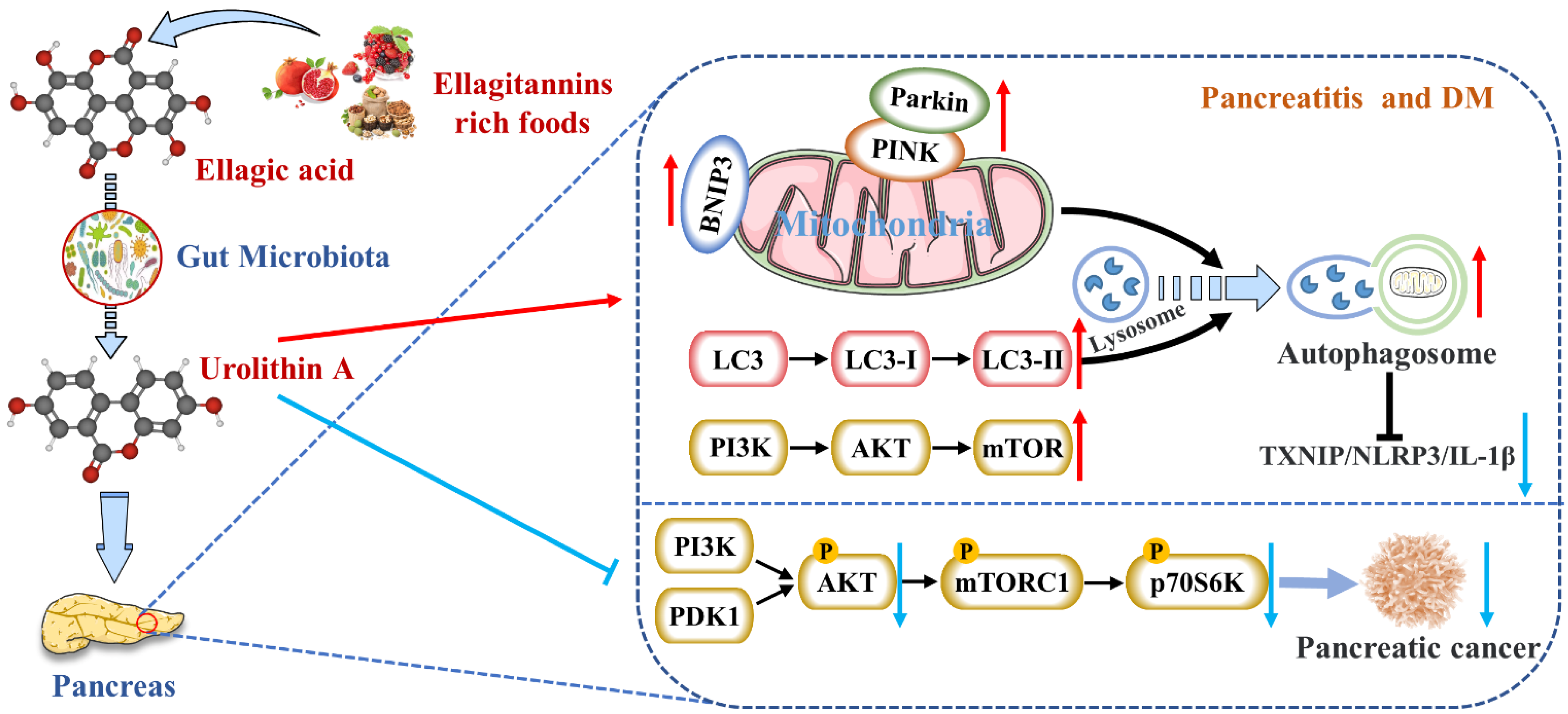
Urolithin A (Uro A) is a dietary metabolite of the intestinal microbiota following the ingestion of plant-based food ingredients ellagitannins and ellagic acid in mammals. Accumulating studies have reported its multiple potential health benefits in a broad range of diseases, including cardiovascular disease, cancer, cognitive impairment, and diabetes.

  • urolithin A
  • pancreas
  • autophagy
  • microbiota
  • diet

1. Reduces the Expression of Pancreatic Inflammatory Factors

The inflammatory microenvironment of the pancreas led to pancreatitis and was the main reason for the decline in endocrine function [1]. Some researchers suggested that if β cells would express high levels of NF-κB signaling marking, cells’ proliferative and regenerative potential were reduced. The NF-κB-expressed β cells also emerged with a premature upregulation of socs2, a gene that inhibits proliferation [2]. It has been widely reported that EA can inhibit pancreatic inflammation (Table 1). In an experimental model of spontaneous chronic pancreatitis, male Wistar Bonn/Kobori rats were fed a diet supplemented with EA (100 mg/kg body weight/day) for ten weeks. They found that EA attenuated pancreatic inflammation and fibrosis by increasing pancreatic weight and decreasing MPO activity (a neutrophil infiltration index), collagen content, transforming growth factor-β1 (TGF-β1) expression, activated pancreatic stellate cells (PSCs), and ED-1-positive cells [3]. Masamune et al. also reported that EA inhibited the production of monocyte chemoattractant protein-1 (MCP-1) and activation of activator protein-1 (AP-1) and MAPK in PSCs, all induced by interleukin (IL)-1β and TNF-α [4]. Meanwhile, EA inhibited PDGF-BB-induced tyrosine phosphorylation of PDGF P-receptors and the downstream ERK and Akt activation in PSCs. In particular, EA inhibited ROS production in PSCs in response to TGF-β1 or platelet-derived growth factor (PDGF) [5].
Table 1. Potential effects and mechanisms of urolithin A and its precursor compounds EA on pancreatic diseases.
Disease Model Treatment Metabolic Response Ref.
Spontaneous CP in male Wistar Bonn/Kobori rats 100 mg/kg BW/day orally administered with EA for 10 weeks
  • Attenuated pancreatic inflammation and fibrosis
  • Increased pancreatic weight
  • Decreased MPO activity, collagen content, TGF-β1 expression; activated PSCs and ED-1-positive cells
[3]
PSCs were isolated from rat pancreas tissue to culture activated, myofibroblast-like phenotype 1–25 μg/mL EA
  • Inhibited PSCs’ proliferation and migration
  • Inhibited MCP-1 production and the expression of smooth muscle actin and collagen genes
  • Inhibited the tyrosine phosphorylation of PDGF P-receptor
  • Inhibited the activation of Akt and MAPKs
[4]
L-arginine induced AP in rats 85 mg/kg orally administered with EA
  • Decreased TOS levels
  • Increased TAC levels
  • Decreased TNF-α, IL-1β, and IL-6 serum levels
  • Healed inflammatory and oxidative stress
[5]
MIN6 β-cell inflammations were induced using 25 mM glucose and 0.5 mM palmitic acid Uro A
  • Inhibited TXNIP/NLRP3/IL-1β inflammation signal
  • Modulated autophagy
  • Down-regulated the PERK and promoted AMPK phosphorylation
[6]
Alcohol-associated CP in C56BL6/J mice Administered during the last 3 weeks of alcohol-associated CP induction
  • Attenuated the severity of ACP
  • Regulated PI3K/AKT/mTOR signaling axis
[7]
DM in male C57BL/6 mice was achieved by a HFD and intraperitoneal STZ injections 50 mg/kg BW/day orally administered with Uro A for 8 weeks
  • Promoted PINK1/Parkin-mediated mitophagy
[8]
Human PDAC cell lines; PDAC mice were achieved by injecting PANC1 cells into the flank of 6-week-old Fox1-nu/nu mice 0–100 μM; 20 mg/kg BW/day (5 days/week) orally administered with Uro A
  • Inhibited the proliferation and migration of PDAC cells
  • Enhanced apoptosis by down-regulating the PI3K/AKT/mTOR pathway
  • Inhibited the PDK1/AKT/mTOR pathway
  • Reduced immunosuppressive TAMs and regulatory T cells
[9]
PKT (Ptf1acre/+; LSL-KrasG12D; Tgfbr2fl/fl) mice, an aggressive genetically engineered PDAC mouse model Orally administered with Uro A for 5 weeks
  • Inhibited AKT, PS6K, and STAT3 signaling
  • Reduced the Ki67-positive tumor cells
  • Increased cleaved caspase-3 expression
[10]
Neonatal STZ-induced non-obese T2DM rats 25–100 mg/kg BW orally administered with EA
  • Stimulated glucose-stimulated insulin secretion from isolated islets
  • Decreased glucose intolerance in diabetic rats
[11]
Although EA had many promising developments, it was poorly absorbed in the human gut, limiting its anti-inflammatory effects. As mentioned above, EA was metabolized by microorganisms into a series of downstream compounds, such as Uro A [12]. A well-known effect of preclinical models exposed to Uro A was the attenuation of harmful inflammatory responses [13]. Uro A showed more potent anti-inflammatory properties than EA or ETs, suggesting that it might be the main compound for treating AP or CP (Table 1). The anti-inflammatory effects were first reported to reduce the mRNA and protein levels of inflammatory marker COX-2 in rats with acute colitis [14]. Zhang et al. had firstly reported that Uro A inhibited the thioredoxin-interacting protein (TXNIP)/Nod-like receptor family pyrin domain containing 3 (NLRP3)/IL-1β inflammation signal in MIN6 β cells by modulating AMPK (Figure 1) [6]. Finally, they testified that Uro A also down-regulated the protein kinase RNA (PKR)-like ER kinase (PERK) and promoted AMPK phosphorylation [15]. The latest research showed that Uro A can attenuate the severity of alcohol-associated chronic pancreatitis (ACP) in C56BL6/J mice by regulating the PI3K/AKT/mTOR signaling axis [7].
Figure 1. Uro A is metabolized by gut microbiota after ingestion of ETs and EA in mammals and has multiple potential health benefits. Uro A can attenuate pancreatic diseases by inhibiting inflammatory signaling pathways, activating autophagy, maintaining the mitochondrial function, and improving the immune microenvironment.

Nevertheless, studies on reducing pancreatic inflammation by Uro A had only been verified in animals and cells without clinical studies. The upstream mediators of Uro A’s anti-inflammatory effects, including the NF-κB and AhR-Nrf2 pathways, were mainly studied in vitro [16]. Nevertheless, the mechanisms of Uro A action in the context of inflammation seemed to vary with tissues and conditions. Hence, the differences in Uro A’s mitigation degree and mechanism on AP and CP need to be further explored.

2. Activates Autophagy and Maintains Mitochondrial Function in the Pancreas

Mitochondrial damage, such as the loss of mitochondrial DNA (mt DNA) integrity, the alteration of mitochondrial morphology, and dysfunction, can lead to cellular senescence and apoptosis [17]. On the one hand, mitochondria acted as both nutrient sensors and signal generators for insulin secretion in β cells. Moreover, nutrients can inhibit the ATP-sensitive K+ (KATP) channel and then enhance insulin secretion either by acting as substrates for mitochondrial ATP synthesis (the triggering pathway) or by regulating Ca2+ channels (the amplifying pathway). On the other hand, mitochondria were the primary source of reactive oxygen species (ROS) at the level of the electron transport chain so that mitochondria might be the main targets of ROS damage [18]. Additionally, many studies have revealed a causal relationship between pancreatic diseases and dysregulation of mitochondrial dynamics (including fusion and fission) [17][19][20]. Thus, mitochondrial damage gave rise to decreased pancreatic function. The most consistent effect of Uro A across species, including cells, worms, mice, and humans, was improved mitochondrial health [13]. This benefit was driven by the clearance and recycling of dysfunctional mitochondria, known as selective autophagy [21]. For example, Uro A increased the expression of mitochondrial autophagy genes lgg-1pink-1, and pdr-1, encoding for LC-3B, and formation of autophagosome vesicles in C. elegans [22].
Interestingly, Pink1 knockdown in microglia eliminated Uro A-mediated reductions in TNF-α and increased IL-10, suggesting that Uro A reduces neuroinflammation by inducing mitochondrial autophagy [23]. Zhang et al. also proved that Uro A inhibited glucolipotoxicity-induced ER stress and the TXNIP/NLRP3/IL-1β inflammation signal in MIN6 β cells by modulating autophagy [6]. Remarkably, the inhibitory effects of Uro A on p62 were stronger than TXNIP-inhibitor verapamil (p < 0.05) [24]. Uro A promoting PINK1/Parkin-mediated mitophagy was also reported in pancreatic cells of diabetic mice [8]. Therefore, Uro A restoring the correct level of mitochondrial autophagy to maintain normal mitochondrial function is highly likely to be the mechanism of Uro A reducing pancreatic diseases (Figure 1).

3. Inhibits Endoplasmic Reticulum Stress in the Pancreas

The misfolding and inhibition of protein folding in the endoplasmic reticulum (ER) lead to the aggregation of unfolded proteins, resulting in ER stress [25]. Li et al. showed that the ER stress and unfolded protein response (UPR) accompanied by the accumulation of protein aggregates emerged as a significant pathway affected by aging, specifically in β cells. Simultaneously, the transcriptomic dysregulation of UPR components was linked to activating transcription factor 6 (ATF6) and inositol-requiring enzyme 1 (IRE1) signaling pathways [26]. ER stress-related apoptosis lead to a reduction in β-cell proliferation and regeneration, ultimately resulting in reduced insulin secretion and increased T2DM morbidity [27]. Therefore, maintaining transcriptional stability and reducing protein homeostasis loss during aging was crucial to recovering pancreatic function. It has been reported that Uro A suppresses glucolipotoxicity-induced ER stress in pancreatic beta cells [6]. However, more studies are needed on Uro A’s upstream and downstream pathways in the pancreas to improve ER stress.

4. Inhibits the Occurrence and Development of Pancreatic Tumors

High intakes of berries rich in ETs, including strawberries, pomegranates, and blueberries, were inversely associated with PDAC incidence [28]. EA, an intestinal metabolite of ellagic tannins, inhibited multiple carcinogenic pathways activated in PDAC, such as COX-2, NF-κB, and Wnt signaling, so that EA successfully arrested cell cycles and reversed epithelial to mesenchymal transition in PDAC [29]. As a downstream compound of EA, Uro A showed more potent antioxidant and anti-inflammatory properties, improving bioavailability and anti-tumor effect [30]. It has been demonstrated that the S473 phosphorylation site of AKT is activated by PI3K [31]. Uro A treatment resulted in a dose-dependent reduction in phospho-AKT (p-AKT) expression in PDAC cell lines, leading to a significant down-regulation of phospho-p70 S6 kinase (p-PS6K) expression regulated by the mTORC1 complex. Therefore, Uro A inhibited the proliferation and migration of PDAC cells and enhanced apoptosis by down-regulating the PI3K/AKT/mTOR pathway [32] (Figure 1). Furthermore, Uro A treatment also down-regulated PDK1 (the upstream target of AKT) and p-GSK3β and p-4E-BP1 (the downstream targets of AKT), suggesting that Uro A effectively inhibited the PDK1/AKT/mTOR [33]. Uro A treatment also reduced immunosuppressive tumor-associated macrophages (TAMs) and regulatory T cells in the engineered PKT mouse model of PDAC. It meant that Uro A treatment attenuated tumor growth and prolonged survival in mice by inducing changes in the immunosuppressive microenvironment of PDAC [9]. Srinivasan et al. also pointed out that Uro A inhibited AKT, PS6K, and STAT3 signaling, thereby reducing the Ki67-positive tumor cells and increasing cleaved caspase-3 expression in the pancreatic tissues of PDAC mice [10]. These results suggest that Uro A is a novel inhibitor/regulator for multi-signal pathways in PDAC and has potential in the prevention and treatment of pancreatic cancer (Table 1).

5. Protects Pancreatic β Cells

There have been many studies on the ameliorative effect of Uro A on DM and its complications. Specifically, Savi et al. first showed that Uro A recovered cardiomyocyte contractility and calcium dynamics in diabetic cardiomyopathy (DCM) rats [34]. Albasher et al. further demonstrated that Uro A prevents streptozotocin (STZ)-induced DCM in rats by activating SIRT1 expression and deacetylase activity [35]. Xiao et al. suggested that Uro A can attenuate DM-related cognitive impairment by ameliorating systemic inflammation and intestinal barrier dysfunction through the N-glycan biosynthesis pathway [36]. This conclusion was also supported by Lee et al. They pointed out that Uro A prevented DM-associated AD by reducing transglutaminase type 2 (TGM2)-dependent mitochondria-associated ER membrane (MAM) formation and maintaining mitochondrial calcium and ROS homeostasis [37]. Xu et al. indicated that Uro A ameliorated diabetic retinopathy by activating the Nrf2/HO-1 pathway to inhibit inflammation and oxidative stress [38]. Zhou et al. found that Phyllanthus emblica L. facilitated vascular function in STZ-induced hyperglycemia rats by regulating Akt/β-catenin signaling, mediated by the ETs metabolites [39].
Insulin resistance is one of the core mechanisms of DM. However, as a complex systemic metabolic disease, insulin resistance alone is not enough to cause DM. Islet dysfunction caused by the decrease in the total amount of islet β cells is also the key to the pathogenesis of DM. Studies have shown that β cells in T2DM can be divided into three main states: susceptibility, adaptation, and failure [40][41][42]. During insulin resistance, β cells compensate for the dysfunction by increasing insulin demand through insulin secretion [8]. When β cells fail to compensate for glucose homeostasis, hyperglycemia occurs. More importantly, EA from Phyllanthus emblica L. increased the size or number of β cells in diabetic rats. EA also directly increased glucose-stimulated insulin secretion from isolated islets, suggesting that EA acted directly on pancreatic β cells to exert anti-diabetic activity, thereby stimulating insulin secretion and reducing glucose intolerance [11]. Histopathological results showed that Uro A had protective effects on β cells, such as improving the pancreatic structure and increasing islet size and number. Ultrastructural damages in DM mice pancreas after Uro A treatment, including ER expansion, mitochondria swelling, cristae fracture, and myelin sheath formation, were also significantly improved [8]. The researchers also discussed earlier that Uro A prevented β-cell apoptosis in T2DM model mice by activating autophagy and regulating the AKT/mTOR signal [6][8][24]. However, the specific mechanisms of Uro A improving β-cell structure and function to mitigate DM risk need to be explored further.
In summary, the metabolism and the roles of Uro A in ameliorating pancreatic diseases have been extensively discussed in this section (summarized in Figure 1). By clarifying Uro A’s metabolism in vivo and Uro A’s mechanisms for protecting the pancreas, it might shed new light on managing pancreatic injuries via plant-based foods rich in ETs and EA.

This entry is adapted from the peer-reviewed paper 10.3390/nu14122549

References

  1. Sandovici, I.; Hammerle, C.M.; Cooper, W.N.; Smith, N.H.; Tarry-Adkins, J.L.; Dunmore, B.J.; Bauer, J.; Andrews, S.R.; Yeo, G.S.; Ozanne, S.E.; et al. Ageing is associated with molecular signatures of inflammation and type 2 diabetes in rat pancreatic islets. Diabetologia 2016, 59, 502–511.
  2. Janjuha, S.; Singh, S.P.; Tsakmaki, A.; Gharavy, S.N.M.; Murawala, P.; Konantz, J.; Birke, S.; Hodson, D.J.; Rutter, G.A.; Bewick, G.A.; et al. Age-related islet inflammation marks the proliferative decline of pancreatic beta-cells in zebrafish. Elife 2018, 7, e32965.
  3. Suzuki, N.; Masamune, A.; Kikuta, K.; Watanabe, T.; Satoh, K.; Shimosegawa, T. Ellagic Acid Inhibits Pancreatic Fibrosis in Male Wistar Bonn/Kobori Rats. Dig. Dis. Sci. 2009, 54, 802–810.
  4. Masamune, A.; Satoh, M.; Kikuta, K.; Suzuki, N.; Satoh, K.; Shimosegawa, T. Ellagic acid blocks activation of pancreatic stellate cells. Biochem. Pharmacol. 2005, 70, 869–878.
  5. Yilmaz, E.E.; Bozdag, Z.; Ibiloglu, I.; Arikanoglu, Z.; Yazgan, U.C.; Kaplan, I.; Gumus, M.; Atamanalp, S.S. Therapeutic effects of ellagic acid on L-arginin induced acute pancreatitis. Acta Cir. Bras. 2016, 31, 396–401.
  6. Zhang, Y.; Aisker, G.; Dong, H.; Halemahebai, G.; Zhang, Y.; Tian, L. Urolithin A suppresses glucolipotoxicity-induced ER stress and TXNIP/NLRP3/IL-1beta inflammation signal in pancreatic beta cells by regulating AMPK and autophagy. Phytomedicine 2021, 93, 153741.
  7. Mehra, S.; Srinivasan, S.; Dai, X.; Dosch, A.; Singh, S.; Bianchi, A.; Silva, I.D.C.; Datta, J.; VanSaun, M.N.; Merchant, N.; et al. Protective Effects of Urolithin A on Alcoholic Chronic Pancreatitis via Inhibiting PI3K/AKT/mTOR Signaling Pathway. Pancreas 2021, 50, 1082.
  8. Tuohetaerbaike, B.; Zhang, Y.; Tian, Y.; Zhang, N.N.; Kang, J.; Mao, X.; Zhang, Y.; Li, X. Pancreas protective effects of Urolithin A on type 2 diabetic mice induced by high fat and streptozotocin via regulating autophagy and AKT/mTOR signaling pathway. J. Ethnopharmacol. 2020, 250, 112479.
  9. Totiger, T.M.; Srinivasan, S.; Jala, V.R.; Lamichhane, P.; Dosch, A.R.; Gaidarski, A.A., III; Joshi, C.; Rangappa, S.; Castellanos, J.; Vemula, P.K.; et al. Urolithin A, a Novel Natural Compound to Target PI3K/AKT/mTOR Pathway in Pancreatic Cancer. Mol. Cancer Ther. 2019, 18, 301–311.
  10. Srinivasan, S.; Jala, V.; Honnenahally, K.; Castellanos, J.; Vermula, P.K.; VanSaun, M.; Merchant, N.; Nagathihalli, N. Urolithin A prevents pancreatic tumor growth and increases survival by inhibiting PI3K/PDK1 and STAT3 signaling. Cancer Res. 2017, 77, 5259.
  11. Fatima, N.; Hafizur, R.M.; Hameed, A.; Ahmed, S.; Nisar, M.; Kabir, N. Ellagic acid in Emblica officinalis exerts anti-diabetic activity through the action on beta-cells of pancreas. Eur. J. Nutr. 2017, 56, 591–601.
  12. Heilman, J.; Andreux, P.; Tran, N.; Rinsch, C.; Blanco-Bose, W. Safety assessment of Urolithin A, a metabolite produced by the human gut microbiota upon dietary intake of plant derived ellagitannins and ellagic acid. Food Chem. Toxicol. 2017, 108, 289–297.
  13. D’Amico, D.; Andreux, P.A.; Valdés, P.; Singh, A.; Rinsch, C.; Auwerx, J. Impact of the Natural Compound Urolithin A on Health, Disease, and Aging. Trends Mol. Med. 2021, 27, 687–699.
  14. Larrosa, M.; González-Sarrías, A.; Yáñez-Gascón, M.J.; Selma, M.V.; Azorín-Ortuño, M.; Toti, S.; Tomas-Barberan, F.; Dolara, P.; Espín, J.C. Anti-inflammatory properties of a pomegranate extract and its metabolite urolithin-A in a colitis rat model and the effect of colon inflammation on phenolic metabolism. J. Nutr. Biochem. 2010, 21, 717–725.
  15. Wu, D.; Yan, Z.B.; Cheng, Y.G.; Zhong, M.W.; Liu, S.Z.; Zhang, G.Y.; Hu, S.Y. Deactivation of the NLRP3 inflammasome in infiltrating macrophages by duodenal-jejunal bypass surgery mediates improvement of beta cell function in type 2 diabetes. Metabolism 2018, 81, 1–12.
  16. Singh, R.; Chandrashekharappa, S.; Bodduluri, H.; Baby, B.V.; Hegde, B.; Kotla, N.G.; Hiwale, A.A.; Saiyed, T.; Patel, P.; Vijay-Kumar, M.; et al. Enhancement of the gut barrier integrity by a microbial metabolite through the Nrf2 pathway. Nat. Commun. 2019, 10, 89.
  17. Vezza, T.; Díaz-Pozo, P.; Canet, F.; de Marañón, A.M.; Abad-Jiménez, Z.; García-Gargallo, C.; Roldan, I.; Solá, E.; Bañuls, C.; López-Domènech, S.; et al. The Role of Mitochondrial Dynamic Dysfunction in Age-Associated Type 2 Diabetes. World J. Men’s Health 2022, 40.
  18. De Tata, V. Age-related impairment of pancreatic Beta-cell function: Pathophysiological and cellular mechanisms. Front. Endocrinol. 2014, 5, 138.
  19. Abuarab, N.; Munsey, T.S.; Jiang, L.-H.; Li, J.; Sivaprasadarao, A. High glucose-induced ROS activates TRPM2 to trigger lysosomal membrane permeabilization and Zn2+-mediated mitochondrial fission. Sci. Signal. 2017, 10, eaal4161.
  20. Kulkarni, S.S.; Joffraud, M.; Boutant, M.; Ratajczak, J.; Gao, A.W.; Maclachlan, C.; Hernandez-Alvarez, M.I.; Raymond, F.; Metairon, S.; Descombes, P.; et al. Mfn1 Deficiency in the Liver Protects Against Diet-Induced Insulin Resistance and Enhances the Hypoglycemic Effect of Metformin. Diabetes 2016, 65, 3552–3560.
  21. Palikaras, K.; Lionaki, E.; Tavernarakis, N. Mechanisms of mitophagy in cellular homeostasis, physiology and pathology. Nat. Cell Biol. 2018, 20, 1013–1022.
  22. Luan, P.; D’Amico, D.; Andreux, P.A.; Laurila, P.-P.; Wohlwend, M.; Li, H.; de Lima, T.I.; Place, N.; Rinsch, C.; Zanou, N.; et al. Urolithin A improves muscle function by inducing mitophagy in muscular dystrophy. Sci. Transl. Med. 2021, 13, eabb0319.
  23. Fang, E.F.; Hou, Y.; Palikaras, K.; Adriaanse, B.A.; Kerr, J.S.; Yang, B.; Lautrup, S.; Hasan-Olive, M.M.; Caponio, D.; Dan, X.; et al. Mitophagy inhibits amyloid-beta and tau pathology and reverses cognitive deficits in models of Alzheimer’s disease. Nat. Neurosci. 2019, 22, 401–412.
  24. Zhang, Y.; Zhang, Y.; Halemahebai, G.; Tian, L.; Dong, H.; Aisker, G. Urolithin A: A pomegranate metabolite, protects pancreatic beta cells from apoptosis by activating autophagy. J. Ethnopharmacol. 2021, 272, 113628.
  25. Marrocco, V.; Tran, T.; Zhu, S.; Choi, S.H.; Gamo, A.M.; Li, S.; Fu, Q.; Cunado, M.D.; Roland, J.; Hull, M.; et al. A small molecule UPR modulator for diabetes identified by high throughput screening. Acta Pharm. Sin. B 2021, 11, 3983–3993.
  26. Li, J.; Zheng, Y.; Yan, P.; Song, M.; Wang, S.; Sun, L.; Liu, Z.; Ma, S.; Izpisua Belmonte, J.C.; Chan, P.; et al. A single-cell transcriptomic atlas of primate pancreatic islet aging. Natl. Sci. Rev. 2021, 8, 127.
  27. Mihailidou, C.; Chatzistamou, I.; Papavassiliou, A.G.; Kiaris, H. Modulation of Pancreatic Islets’ Function and Survival During Aging Involves the Differential Regulation of Endoplasmic Reticulum Stress by p21 and CHOP. Antioxid. Redox Signal. 2017, 27, 185–200.
  28. Veeraraghavan, J.; Natarajan, M.; Lagisetty, P.; Awasthi, V.; Herman, T.S.; Aravindan, N. Impact of Curcumin, Raspberry Extract, and Neem Leaf Extract on Rel Protein-Regulated Cell Death/Radiosensitization in Pancreatic Cancer Cells. Pancreas 2011, 40, 1107–1119.
  29. Cheng, H.; Lu, C.L.; Tang, R.B.; Pan, Y.M.; Bao, S.H.; Qiu, Y.D.; Xie, M. Ellagic acid inhibits the proliferation of human pancreatic carcinoma PANC-1 cells in vitro and in vivo. Oncotarget 2017, 8, 12301–12310.
  30. Tomas-Barberan, F.A.; Gonzalez-Sarrias, A.; Garcia-Villalba, R.; Nunez-Sanchez, M.A.; Selma, M.V.; Garcia-Conesa, M.T.; Espin, J.C. Urolithins, the rescue of “old” metabolites to understand a “new” concept, Metabotypes as a nexus among phenolic metabolism, microbiota dysbiosis, and host health status. Mol. Nutr. Food Res. 2017, 61, 1500901.
  31. Dienstmann, R.; Rodon, J.; Serra, V.; Tabernero, J. Picking the Point of Inhibition: A Comparative Review of PI3K/AKT/mTOR Pathway Inhibitors. Mol. Cancer Ther. 2014, 13, 1021–1031.
  32. Wong, M.H.; Xue, A.; Baxter, R.C.; Pavlakis, N.; Smith, R.C. Upstream and Downstream Co-inhibition of Mitogen-Activated Protein Kinase and PI3K/Akt/mTOR Pathways in Pancreatic Ductal Adenocarcinoma. Neoplasia 2016, 18, 425–435.
  33. Takeuchi, S.; Baghdadi, M.; Tsuchikawa, T.; Wada, H.; Nakamura, T.; Abe, H.; Nakanishi, S.; Usui, Y.; Higuchi, K.; Takahashi, M.; et al. Chemotherapy-Derived Inflammatory Responses Accelerate the Formation of Immunosuppressive Myeloid Cells in the Tissue Microenvironment of Human Pancreatic Cancer. Cancer Res. 2015, 75, 2629–2640.
  34. Savi, M.; Bocchi, L.; Mena, P.; Dall’Asta, M.; Crozier, A.; Brighenti, F.; Stilli, D.; Del Rio, D. In vivo administration of urolithin A and B prevents the occurrence of cardiac dysfunction in streptozotocin-induced diabetic rats. Cardiovasc. Diabetol. 2017, 16, 80.
  35. Albasher, G.; Alkahtani, S.; Al-Harbi, L.N. Urolithin A prevents streptozotocin-induced diabetic cardiomyopathy in rats by activating SIRT1. Saudi J. Biol. Sci. 2022, 29, 1210–1220.
  36. Xiao, Y.; Li, K.; Bian, J.; Liu, H.; Zhai, X.; El-Omar, E.; Han, L.; Gong, L.; Wang, M. Urolithin A Attenuates Diabetes-Associated Cognitive Impairment by Ameliorating Intestinal Barrier Dysfunction via N-glycan Biosynthesis Pathway. Mol. Nutr. Food Res. 2022, 66, 2100863.
  37. Lee, H.J.; Jung, Y.H.; Choi, G.E.; Kim, J.S.; Chae, C.W.; Lim, J.R.; Kim, S.Y.; Yoon, J.H.; Cho, J.H.; Lee, S.-J.; et al. Urolithin A suppresses high glucose-induced neuronal amyloidogenesis by modulating TGM2-dependent ER-mitochondria contacts and calcium homeostasis. Cell Death Differ. 2021, 28, 184–202.
  38. Xu, Z.; Li, S.; Li, K.; Wang, X.; Li, X.; An, M.; Yu, X.; Long, X.; Zhong, R.; Liu, Q.; et al. Urolithin A ameliorates diabetic retinopathy via activation of the Nrf2/HO-1 pathway. Endocr. J. 2022, EJ21-0490.
  39. Zhou, J.; Zhang, C.; Zheng, G.-H.; Qiu, Z. Emblic Leafflower (Phyllanthus emblica L.) Fruits Ameliorate Vascular Smooth Muscle Cell Dysfunction in Hyperglycemia: An Underlying Mechanism Involved in Ellagitannin Metabolite Urolithin A. Evid.-Based Complement. Altern. Med. 2018, 2018, 8478943.
  40. Abdel-Hamid, A.A.M.; El-Firgany, A.E.-D.L. Hydroxychloroquine hindering of diabetic isletopathy carries its signature on the inflammatory cytokines. J. Mol. Histol. 2016, 47, 183–193.
  41. Tuduri, E.; Soriano, S.; Almagro, L.; Garcia-Heredia, A.; Rafacho, A.; Alonso-Magdalena, P.; Nadal, A.; Quesada, I. The Effects of Aging on Male Mouse Pancreatic beta-Cell Function Involve Multiple Events in the Regulation of Secretion, Influence of Insulin Sensitivity. J. Gerontol. A Biol. Sci. Med. Sci. 2022, 77, 405–415.
  42. Pan, F.; He, X.; Feng, J.; Cui, W.; Gao, L.; Li, M.; Yang, H.; Wang, C.; Hu, Y. Peptidome analysis reveals the involvement of endogenous peptides in mouse pancreatic dysfunction with aging. J. Cell. Physiol. 2019, 234, 14090–14099.
More
This entry is offline, you can click here to edit this entry!
ScholarVision Creations