Adipose Tissue Macrophages in Obesity and Insulin Resistance: History
Please note this is an old version of this entry, which may differ significantly from the current revision.
Contributor: , ,

Obesity has become a worldwide epidemic that poses a severe threat to human health. Evidence suggests that many obesity comorbidities, such as type 2 diabetes mellitus, steatohepatitis, and cardiovascular diseases, are related to obesity-induced chronic low-grade inflammation. Macrophages are the primary immune cells involved in obesity-associated inflammation in both mice and humans. Intensive research has yielded tremendous progress in the understanding of the additional roles of adipose tissue macrophages (ATMs) beyond classical M1/M2 polarization in obesity and related comorbidities.

  • adipose tissue
  • macrophage
  • inflammation
  • obesity
  • insulin resistance

1. Introduction

Obesity is a metabolic disease characterized by abnormal and excessive accumulation of body fat. Obesity increases the risk of developing a wide variety of diseases including but not limited to type 2 diabetes mellitus (T2DM) and cardiovascular diseases and has been strongly associated with increased mortality [1][2]. In the past few decades, the prevalence of obesity has increased dramatically in both developing and developed nations around the world [3][4]. More recent statistics indicate that 39% of adults aged 18 years and over were overweight, and 13% were obese worldwide in 2016 [5]. Obesity is a serious public health problem with major health and economic consequences.
Insulin resistance is a key component in the etiology of T2DM, and obesity is clearly the most common cause of insulin resistance in humans [6][7]. With the ongoing worldwide obesity epidemic, there has been a parallel rise in the prevalence of T2DM [8]. It has now been widely recognized that obesity-induced chronic low-grade tissue inflammation, particularly when occurring in adipose tissue, can cause insulin resistance and T2DM [9][10]. Under obese conditions, adipose tissue undergoes a series of dynamic remodeling, including adipocyte hypertrophy, apoptosis, immune cell infiltration, extensive vascularization, and extracellular matrix remodeling [11][12]. Macrophages are the primary immune cells strongly involved in obesity-associated inflammation in both mice and humans [7][13][14][15]. Emerging studies have implicated the crosstalk between adipocytes and adipose tissue macrophages (ATMs) as critical regulators of obesity-associated inflammation and metabolic complications.

2. Adipose Tissue Macrophage (ATM) Subpopulations

White adipose tissue (WAT) is comprised of a versatile group of interacting cells, including adipocytes, immune cells, and other cell types. It is evident that ATMs, the most abundant immune cells in WAT, can even represent up to 40–50% of the cells in obese adipose tissue [13]. ATMs are an extraordinarily heterogeneous population of immune cells with varied and diverse functions (as summarized in Table 1), which have been highlighted as important factors contributing to the pathogenesis of obesity and related comorbidities. Historically, ATMs have been categorized into classically activated (M1-like) and alternatively activated (M2-like) macrophages [16]. However, more and more novel ATM subpopulations have been identified [14][15][17][18][19][20][21][22][23].
Table 1. Adipose tissue macrophage (ATM) subpopulations.
Tumor necrosis factor-α (TNF-α), interleukin-1β (IL-1β), interleukin-6 (IL-6), nitric oxide (NO), interleukin-4 (IL-4), interleukin-10 (IL-10), platelet-derived growth factor (PDGFcc), norepinephrine (NE), solute carrier family 6 member 2 (Slc6a2), monoamine oxidase A (MAOA), sympathetic nervous system (SNS), crown-like structure (CLS).
ATMs were thought to be composed of two main phenotypes: classically activated macrophages and alternatively activated macrophages, which are phenotypically and functionally distinct [16]. The classically activated macrophages represent pro-inflammatory M1-like macrophages, whereas the alternatively activated macrophages are anti-inflammatory M2-like macrophages. M1-like macrophages express F4/80, CD11b, and CD11c and secrete inflammatory factors including tumor necrosis factor-α (TNF-α), interleukin-1β (IL-1β), IL-6, leukotriene B4 (LTB4), and nitric oxide (NO); whereas M2-like macrophages express F4/80, CD11b, CD301, and CD206 and exhibit increased secretion of anti-inflammatory cytokines, such as IL-4 and IL-10 [26][27]. The main roles of ATMs under lean conditions are efferocytosis of dead adipocytes, the production of anti-inflammatory cytokines, the regulation of adipocyte lipolysis, and the restriction of adipocyte progenitor proliferation [28]. Conversely, as obesity progresses, most ATMs are converted from an anti-inflammatory (M2-like) phenotype into a pro-inflammatory (M1-like) phenotype, secreting pro-inflammatory cytokines (such as TNF-α, IL-1β) and causing localized and systemic chronic low-grade inflammation, especially in WAT [29]. The insulin resistance and T2DM would progress under the influence of this inflammatory state [30][31].
ATMs can be further widely divided into adipose tissue-resident macrophages and recruited monocyte-derived macrophages. Tissue-resident macrophages are long-lived and self-renewing cells thought to have originated during embryonic hematopoiesis [32], whereas monocyte-derived macrophages are short-lived cells recruited to adipose tissues during inflammation [33]. Recently, multiple novel populations of adipose tissue-resident macrophages have been discovered in adipose depots [21][22][34]. For example, a new subpopulation of ATMs, TIM4+ adipose tissue-resident macrophages, has recently been revealed to play essential roles in the formation and expansion of adipose tissue during the development and diet-induced obesity [21]. Another novel resident macrophage population (sympathetic neuron-associated macrophages, SAMs) has been discovered in adipose tissue localized around neurons of the sympathetic nervous system (SNS) that mediates noradrenaline clearance and dampens SNS-to-adipocyte communications [22][34].
With the recent advances in scRNA-seq technologies, emerging evidence suggests that ATMs exhibit a wider spectrum of phenotypes and cellular identities than previously described in the context of obesity both in mice and humans. By utilizing scRNA-seq technology, Hill et al. identified three discrete ATM populations (CD11b+ Ly6c+; CD11b+ Ly6c CD9+; CD11b+ Ly6c CD9), two of which (CD11b+ Ly6c+ and CD11b+ Ly6c CD9+) are associated with obesity [17]. CD11b+ Ly6c CD9+ ATMs reside within crown-like structures (CLS) and are lipid-laden and proinflammatory, whereas CD11b+ Ly6c+ ATMs reside outside CLSs and play angiogenic and adipogenic roles [17]. In a more recent study, Jaitin et al. provided a comprehensive single-cell adipose tissue immune atlas in mice and humans and described a novel Trem2+ ATM subpopulation, named lipid-associated macrophages (LAMs), in obese adipose tissue [14]. These LAMs use lipid receptor Trem2 as a sensor of extracellular lipids and play protective functions to counteract adipocyte hypertrophy, inflammation, and metabolic dysfunction [14]. Therefore, obese adipose tissue contains multiple distinct ATM populations with unique origins, tissue distributions, and functions. A comprehensive understanding of ATM heterogeneity in obesity is of great importance for the development of future therapies.

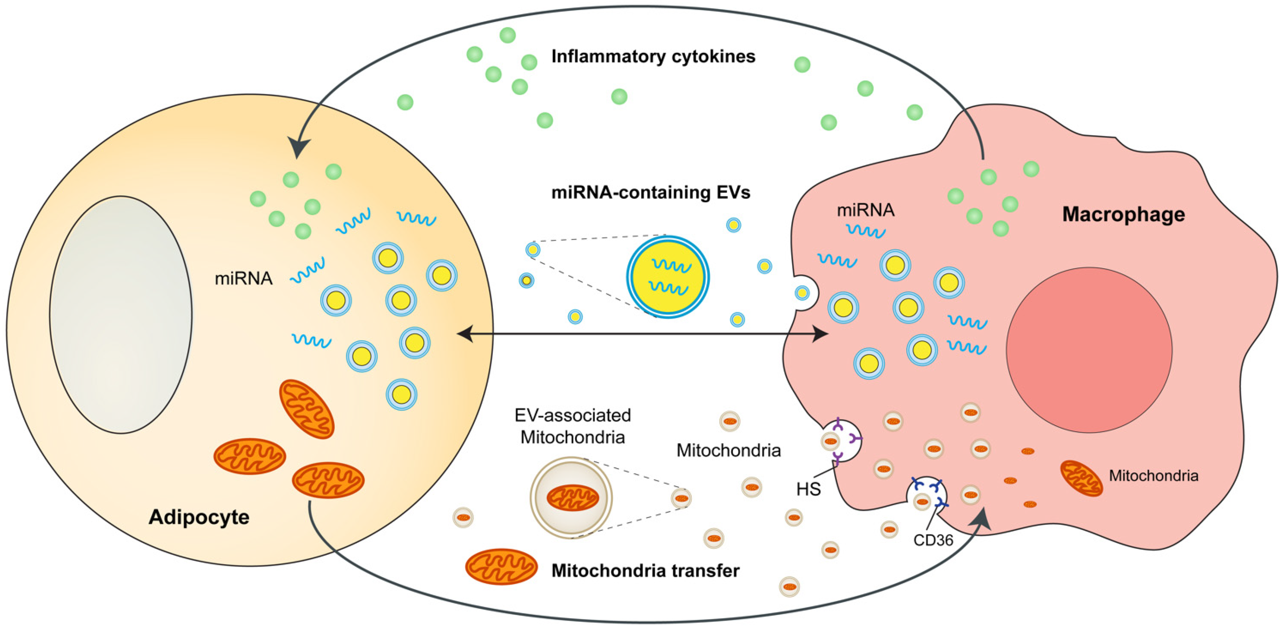
3. Adipocytes and ATMs Crosstalk

The crosstalk between adipocytes and macrophages in adipose tissues is crucial in obesity-induced metabolic complications. Adipocytes and macrophages can interact with each other through a variety of mechanisms, including cytokine and chemokines, microRNA-containing exosomes or microvesicles, and mitochondria transfer. These mechanisms are partly summarized in Figure 1.
Figure 1. Interactions between adipocytes and ATMs in obesity. Adipocytes and macrophages interact with each other through a variety of mechanisms, including cytokine and chemokines, microRNA-containing exosomes or microvesicles, and mitochondrial transfer. ATMs, adipose tissue macrophages; miRNA, microRNA; EVs, extracellular vesicles; HS, heparan sulfates.

4. Adipose Tissue-Resident Macrophages Directly Regulate Adiposity and Energy Storage

In addition to short-lived monocyte-derived macrophages originating from hematopoietic stem cells, there are long-lived tissue-resident macrophages that serve tissue-specific purposes [35]. However, whether adipose tissue-resident macrophages serve their tissue-specific purposes and support the function of energy storage in adipose tissue has not been completely understood.
A new subpopulation of ATMs, CCR2-independent TIM4+ resident macrophages, has recently been reported to modulate adiposity and energy storage in a paracrine manner via the production of platelet-derived growth factor (PDGFcc) in mice WAT [21]. Genetic deletion and pharmacological blockade of PDGFcc reduce adiposity, energy storage, and body weight, and redirect excess lipids mostly toward thermogenesis [21]. This study challenges the outdated M1/M2 macrophage polarization model in which adipose tissue-resident macrophages can serve to sense increased nutritional status and support energy storage, whereas recruited macrophages are responsible for characterizing systemic inflammation of obesity and metabolic complications. Therefore, these data strongly indicate that different developmental subsets of macrophages (including adipose tissue-resident macrophages and recruited monocyte-derived macrophages) exert different functions within adipose tissue and are independent targets of CCR2 and PDGFcc blockade. This study highlighted the additional roles of macrophages beyond classical M1/M2 polarization in obesity development and has the potential to inspire new immunomodulatory therapies that could separately manipulate energy storage and inflammation during obesity.

5. Sympathetic Neuron-Associated Macrophages Indirectly Affect Energy Storage

Adipose tissue is densely innervated by the sympathetic nervous system (SNS), which locally releases noradrenaline into adipose tissue and drives lipolysis and brown or beige adipocyte thermogenesis [24][36][37]. Recent advances in three-dimensional adipose tissue imaging have improved the understanding of how different cell types in adipose tissue are organized and how they interact with one another [38][39]. Moreover, the interwoven relationships between adipocytes, sympathetic nerves, and immune cells in the context of obesity have attracted widespread attention.
In 2017, two landmark studies simultaneously identified noradrenaline-degrading macrophage populations in WAT, which directly modulate the sympathetic innervation of adipocytes [22][23]. Pirzgalska et al. demonstrated that adipose tissue-residing SAMs exhibit specialized morphology for association with SNS neurons in WAT [22]. These SAMs that import and catabolize noradrenaline via noradrenaline transporter (SLC6A2) and degradation enzyme (MAMO) are dramatically increased under obese conditions [22]. Mice with genetic deletion of SLC6A2 from SAMs are resistant to obesity owing to decreased noradrenaline removal, enhanced SNS-to-adipocyte communications, and increased SNS-driven lipolysis [22]. In another study, Camell et al. found that a specialized ATM subpopulation (a SAM-like macrophage population) was activated in aged mice [34]. They further demonstrated these ATMs regulate the age-related reduction in adipocyte lipolysis in adipose tissue by degrading norepinephrine in an inflammasome-dependent manner [34]. In addition, in the more recent study by Wang et al., it is demonstrated that under cold exposure M2-like macrophages secret Slit3, which binds to ROBO1 receptor on sympathetic neurons and stimulates noradrenaline release, leading to enhanced white adipocyte beiging and thermogenesis [40]. These studies identify a sympathetic neuroimmunological role for macrophages in obesity and have opened up a whole new field of neuroimmunometabolism.
It is now well demonstrated that BAT is much more densely innervated by sympathetic nerves than WAT [41]. The triangular relationship between brown adipocytes, macrophages, and SNS has also been recognized. Wolf et al. reported that the nuclear transcription factor MECP2 is an important modulator of BAT function [42]. Mice lacking MECP2 spontaneously develop obesity due to the impairment of BAT function [42]. Further mechanistic investigation indicates that MECP2-deficient macrophage upregulates PlexinA4 expression which prevents the axonal outgrowth of Sema6A+ nerves and diminishes sympathetic innervation of BAT [42].

6. Novel View—Cross-Talk between Perivascular Mesenchymal Cells and ATMs

Although recent research mainly focuses on the role of adipocytes and macrophages in the development of metabolic adipose tissue inflammation, a new study recently highlighted that perivascular mesenchymal cells play a significant role in the regulation of chronic adipose tissue inflammation during obesity.
In the study, Shan et al. utilized scRNA-seq and identified a mouse WAT perivascular cell subpopulation, named fibro-inflammatory progenitors (FIPs) that stimulate pro-inflammatory signaling and modulate accumulation of pro-inflammatory macrophage in the adipose tissue during obesity [43]. These perivascular mesenchymal cells of the adipose tissue are critical “gatekeepers” of macrophage accumulation in obesity. It is also reported that the transcriptional regulator zinc-finger protein 423 (ZFP423) governs the inflammatory response of perivascular mesenchymal cells [43]. Using in vitro studies and in vivo mouse genetic models they determined that ZFP423 modulates NF-κB activity and that expression of ZFP423 in perivascular mesenchymal cells suppresses inflammatory signaling in FIPs and attenuates metabolic inflammation in obesity [43]. These studies highlighted an important role for perivascular mesenchymal cells in the modulation of chronic inflammation in adipose tissue during obesity, and indicate that proinflammatory perivascular mesenchymal cells are potential targets for therapeutic treatment tailored to control obesity and associated co-morbidities.

This entry is adapted from the peer-reviewed paper 10.3390/cells11091424

References

  1. Whitlock, G.; Lewington, S.; Sherliker, P.; Clarke, R.; Emberson, J.; Halsey, J.; Qizilbash, N.; Collins, R.; Peto, R. Body-mass index and cause-specific mortality in 900 000 adults: Collaborative analyses of 57 prospective studies. Lancet 2009, 373, 1083–1096.
  2. Flegal, K.M.; Kit, B.K.; Orpana, H.; Graubard, B.I. Association of all-cause mortality with overweight and obesity using standard body mass index categories: A systematic review and meta-analysis. JAMA 2013, 309, 71–82.
  3. Ng, M.; Fleming, T.; Robinson, M.; Thomson, B.; Graetz, N.; Margono, C.; Mullany, E.C.; Biryukov, S.; Abbafati, C.; Abera, S.F.; et al. Global, regional, and national prevalence of overweight and obesity in children and adults during 1980–2013: A systematic analysis for the Global Burden of Disease Study 2013. Lancet 2014, 384, 766–781.
  4. Afshin, A.; Forouzanfar, M.H.; Reitsma, M.B.; Sur, P.; Estep, K.; Lee, A.; Marczak, L.; Mokdad, A.H.; Moradi-Lakeh, M.; Naghavi, M.; et al. Health Effects of Overweight and Obesity in 195 Countries over 25 Years. N. Engl. J. Med. 2017, 377, 13–27.
  5. World Health Organization. Obesity and Overweight. 2021. Available online: https://www.who.int/news-room/fact-sheets/detail/obesity-and-overweight (accessed on 9 June 2021).
  6. Kahn, S.E.; Hull, R.L.; Utzschneider, K.M. Mechanisms linking obesity to insulin resistance and type 2 diabetes. Nature 2006, 444, 840–846.
  7. Johnson, A.M.; Olefsky, J.M. The origins and drivers of insulin resistance. Cell 2013, 152, 673–684.
  8. World Health Organization. Diabetes. 2021. Available online: https://www.who.int/news-room/fact-sheets/detail/diabetes (accessed on 10 November 2021).
  9. Heilbronn, L.K.; Campbell, L.V. Adipose tissue macrophages, low grade inflammation and insulin resistance in human obesity. Curr. Pharm. Des. 2008, 14, 1225–1230.
  10. Lumeng, C.N.; Saltiel, A.R. Inflammatory links between obesity and metabolic disease. J. Clin. Investig. 2011, 121, 2111–2117.
  11. Marcelin, G.; Silveira, A.L.M.; Martins, L.B.; Ferreira, A.V.; Clement, K. Deciphering the cellular interplays underlying obesity-induced adipose tissue fibrosis. J. Clin. Investig. 2019, 129, 4032–4040.
  12. Choe, S.S.; Huh, J.Y.; Hwang, I.J.; Kim, J.I.; Kim, J.B. Adipose Tissue Remodeling: Its Role in Energy Metabolism and Metabolic Disorders. Front. Endocrinol. 2016, 7, 30.
  13. Weisberg, S.P.; McCann, D.; Desai, M.; Rosenbaum, M.; Leibel, R.L.; Ferrante, A.W., Jr. Obesity is associated with macrophage accumulation in adipose tissue. J. Clin. Investig. 2003, 112, 1796–1808.
  14. Jaitin, D.A.; Adlung, L.; Thaiss, C.A.; Weiner, A.; Li, B.; Descamps, H.; Lundgren, P.; Bleriot, C.; Liu, Z.; Deczkowska, A.; et al. Lipid-Associated Macrophages Control Metabolic Homeostasis in a Trem2-Dependent Manner. Cell 2019, 178, 686–698.e614.
  15. Emont, M.P.; Jacobs, C.; Essene, A.L.; Pant, D.; Tenen, D.; Colleluori, G.; Di Vincenzo, A.; Jorgensen, A.M.; Dashti, H.; Stefek, A.; et al. A single-cell atlas of human and mouse white adipose tissue. Nature 2022, 603, 926–933.
  16. Hill, A.A.; Reid Bolus, W.; Hasty, A.H. A decade of progress in adipose tissue macrophage biology. Immunol. Rev. 2014, 262, 134–152.
  17. Hill, D.A.; Lim, H.W.; Kim, Y.H.; Ho, W.Y.; Foong, Y.H.; Nelson, V.L.; Nguyen, H.C.B.; Chegireddy, K.; Kim, J.; Habertheuer, A.; et al. Distinct macrophage populations direct inflammatory versus physiological changes in adipose tissue. Proc. Natl. Acad. Sci. USA 2018, 115, E5096–E5105.
  18. Burl, R.B.; Ramseyer, V.D.; Rondini, E.A.; Pique-Regi, R.; Lee, Y.H.; Granneman, J.G. Deconstructing Adipogenesis Induced by beta3-Adrenergic Receptor Activation with Single-Cell Expression Profiling. Cell Metab. 2018, 28, 300–309.e4.
  19. Sarvari, A.K.; Van Hauwaert, E.L.; Markussen, L.K.; Gammelmark, E.; Marcher, A.B.; Ebbesen, M.F.; Nielsen, R.; Brewer, J.R.; Madsen, J.G.S.; Mandrup, S. Plasticity of Epididymal Adipose Tissue in Response to Diet-Induced Obesity at Single-Nucleus Resolution. Cell Metab. 2021, 33, 437–453.e435.
  20. Harasymowicz, N.S.; Rashidi, N.; Savadipour, A.; Wu, C.L.; Tang, R.; Bramley, J.; Buchser, W.; Guilak, F. Single-cell RNA sequencing reveals the induction of novel myeloid and myeloid-associated cell populations in visceral fat with long-term obesity. FASEB J. 2021, 35, e21417.
  21. Cox, N.; Crozet, L.; Holtman, I.R.; Loyher, P.L.; Lazarov, T.; White, J.B.; Mass, E.; Stanley, E.R.; Elemento, O.; Glass, C.K.; et al. Diet-regulated production of PDGFcc by macrophages controls energy storage. Science 2021, 373, eabe9383.
  22. Pirzgalska, R.M.; Seixas, E.; Seidman, J.S.; Link, V.M.; Sanchez, N.M.; Mahu, I.; Mendes, R.; Gres, V.; Kubasova, N.; Morris, I.; et al. Sympathetic neuron-associated macrophages contribute to obesity by importing and metabolizing norepinephrine. Nat. Med. 2017, 23, 1309–1318.
  23. Andersson, O.; Korach-Andre, M.; Reissmann, E.; Ibanez, C.F.; Bertolino, P. Growth/differentiation factor 3 signals through ALK7 and regulates accumulation of adipose tissue and diet-induced obesity. Proc. Natl. Acad. Sci. USA 2008, 105, 7252–7256.
  24. Madden, K.S. Sympathetic neural-immune interactions regulate hematopoiesis, thermoregulation and inflammation in mammals. Dev. Comp. Immunol. 2017, 66, 92–97.
  25. Dominguez, H.; Storgaard, H.; Rask-Madsen, C.; Steffen Hermann, T.; Ihlemann, N.; Baunbjerg Nielsen, D.; Spohr, C.; Kober, L.; Vaag, A.; Torp-Pedersen, C. Metabolic and vascular effects of tumor necrosis factor-alpha blockade with etanercept in obese patients with type 2 diabetes. J. Vasc. Res. 2005, 42, 517–525.
  26. Olefsky, J.M.; Glass, C.K. Macrophages, inflammation, and insulin resistance. Annu. Rev. Physiol. 2010, 72, 219–246.
  27. Lumeng, C.N.; Bodzin, J.L.; Saltiel, A.R. Obesity induces a phenotypic switch in adipose tissue macrophage polarization. J. Clin. Investig. 2007, 117, 175–184.
  28. Nawaz, A.; Aminuddin, A.; Kado, T.; Takikawa, A.; Yamamoto, S.; Tsuneyama, K.; Igarashi, Y.; Ikutani, M.; Nishida, Y.; Nagai, Y.; et al. CD206(+) M2-like macrophages regulate systemic glucose metabolism by inhibiting proliferation of adipocyte progenitors. Nat. Commun. 2017, 8, 286.
  29. Hildreth, A.D.; Ma, F.; Wong, Y.Y.; Sun, R.; Pellegrini, M.; O’Sullivan, T.E. Single-cell sequencing of human white adipose tissue identifies new cell states in health and obesity. Nat. Immunol. 2021, 22, 639–653.
  30. Boutens, L.; Stienstra, R. Adipose tissue macrophages: Going off track during obesity. Diabetologia 2016, 59, 879–894.
  31. Russo, L.; Lumeng, C.N. Properties and functions of adipose tissue macrophages in obesity. Immunology 2018, 155, 407–417.
  32. Perdiguero, E.G.; Geissmann, F. The development and maintenance of resident macrophages. Nat. Immunol. 2016, 17, 2–8.
  33. Tsou, C.L.; Peters, W.; Si, Y.; Slaymaker, S.; Aslanian, A.M.; Weisberg, S.P.; Mack, M.; Charo, I.F. Critical roles for CCR2 and MCP-3 in monocyte mobilization from bone marrow and recruitment to inflammatory sites. J. Clin. Investig. 2007, 117, 902–909.
  34. Camell, C.D.; Sander, J.; Spadaro, O.; Lee, A.; Nguyen, K.Y.; Wing, A.; Goldberg, E.L.; Youm, Y.H.; Brown, C.W.; Elsworth, J.; et al. Inflammasome-driven catecholamine catabolism in macrophages blunts lipolysis during ageing. Nature 2017, 550, 119–123.
  35. Ginhoux, F.; Guilliams, M. Tissue-Resident Macrophage Ontogeny and Homeostasis. Immunity 2016, 44, 439–449.
  36. Bachman, E.S.; Dhillon, H.; Zhang, C.Y.; Cinti, S.; Bianco, A.C.; Kobilka, B.K.; Lowell, B.B. betaAR signaling required for diet-induced thermogenesis and obesity resistance. Science 2002, 297, 843–845.
  37. Zeng, W.; Pirzgalska, R.M.; Pereira, M.M.; Kubasova, N.; Barateiro, A.; Seixas, E.; Lu, Y.H.; Kozlova, A.; Voss, H.; Martins, G.G.; et al. Sympathetic neuro-adipose connections mediate leptin-driven lipolysis. Cell 2015, 163, 84–94.
  38. Jiang, H.; Ding, X.; Cao, Y.; Wang, H.; Zeng, W. Dense Intra-adipose Sympathetic Arborizations Are Essential for Cold-Induced Beiging of Mouse White Adipose Tissue. Cell Metab. 2017, 26, 686–692.e3.
  39. Chi, J.; Wu, Z.; Choi, C.H.J.; Nguyen, L.; Tegegne, S.; Ackerman, S.E.; Crane, A.; Marchildon, F.; Tessier-Lavigne, M.; Cohen, P. Three-Dimensional Adipose Tissue Imaging Reveals Regional Variation in Beige Fat Biogenesis and PRDM16-Dependent Sympathetic Neurite Density. Cell Metab. 2018, 27, 226–236.e3.
  40. Wang, Y.N.; Tang, Y.; He, Z.; Ma, H.; Wang, L.; Liu, Y.; Yang, Q.; Pan, D.; Zhu, C.; Qian, S.; et al. Slit3 secreted from M2-like macrophages increases sympathetic activity and thermogenesis in adipose tissue. Nat. Metab. 2021, 3, 1536–1551.
  41. Zeng, X.; Ye, M.; Resch, J.M.; Jedrychowski, M.P.; Hu, B.; Lowell, B.B.; Ginty, D.D.; Spiegelman, B.M. Innervation of thermogenic adipose tissue via a calsyntenin 3beta-S100b axis. Nature 2019, 569, 229–235.
  42. Wolf, Y.; Boura-Halfon, S.; Cortese, N.; Haimon, Z.; Sar Shalom, H.; Kuperman, Y.; Kalchenko, V.; Brandis, A.; David, E.; Segal-Hayoun, Y.; et al. Brown-adipose-tissue macrophages control tissue innervation and homeostatic energy expenditure. Nat. Immunol. 2017, 18, 665–674.
  43. Shan, B.; Shao, M.; Zhang, Q.; Hepler, C.; Paschoal, V.A.; Barnes, S.D.; Vishvanath, L.; An, Y.A.; Jia, L.; Malladi, V.S.; et al. Perivascular mesenchymal cells control adipose-tissue macrophage accrual in obesity. Nat. Metab. 2020, 2, 1332–1349.
More
This entry is offline, you can click here to edit this entry!