1. Introduction
Harmful cyanobacterial blooms (HCBs) caused by cyanobacteria (including
Microcystis,
Anabaena,
Nodularia,
Oscillatoria, and so on) have become a common occurrence in freshwater worldwide [
1,
2]. Among the blooming cyanobacteria,
Microcystis aeruginosa is one of the most common and widespread species [
3]; specifically, it is known to be a representative species due to the dominant production of microcystins [
4,
5]. The rapid and excessive growth of
M. aeruginosa is harmful to drinking water treatments and aquatic ecosystems due to the release of algal organic matters and cyanobacterial toxins [
6,
7]. As a result, the control of HCBs in water sources is a matter of great urgency.
Many approaches have been adopted for
M. aeruginosa removal over the past few decades [
8]. Physical methods including mechanical salvage, physical aeration, and ultrasonic treatment are usually high cost and take a long time; chemical methods such as chemical oxidants are highly efficient and low-cost methods for controlling HCBs within a short time [
9]. However, chemicals may lead to a secondary contamination that may lead to potential threats to the aquatic ecosystem [
10,
11]. Compared with the physical and chemical methods, biological approaches such as plant allelopathy, aquatic animals and anticyanobacterial microorganisms are considered to be an economic and environmentally friendly way for cyanobacteria inhibition/biodegradation [
2,
10,
12]. Among these methods, anticyanobacterial microorganisms are used as efficient biological agents
M. aeruginosa [
13]; furthermore, the microcystins can be biodegraded by certain anticyanobacterial microorganisms at the same time [
6,
14,
15].
2. Anticyanobacterial Modes
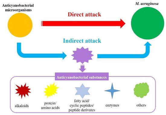
In general, the anticyanobacterial modes by microorganisms are divided into direct attack (bacterial and cyanobacterial cell contact) and indirect attack (the release of anticyanobacterial substances) (
Figure 1) [
10,
32,
72,
118]. To date, although anticyanobacteria can directly kill several different kinds of cyanobacteria, only few has been reported. A wide range of cyanobacteria including
M. aeruginosa,
M. wesenbergii,
M. viridis,
Anabaena flos-aquae,
Oscillatoria tenuis,
Nostoc punctiforme and
Spirulina maxima are lysed by
B. cereus DC22 with the direct attack mode, as well as chlorophyceae (
Chlorella ellipsoidea and
Selenastrum capricornutum) [
89]. In addition to
B. cereus, other anticyanobacteria that destroy
M. aeruginosa with direct attack have also been reported. For example, the anticyanobacterial modes of
Aeromonas bestiarum HYD0802-MK36 [
20],
Chryseobacterium sp. [
40],
Streptomyces globisporus G9 [
83],
Alcaligenes denitrificans [
59], and
Shigella sp. H3 [
60] on
M. aeruginosa are regarded as direct attack, and a number of cyst-like cells are formed in cyanobacteria during the direct attack [
10]. It is speculated that the cyanobacterial cell walls are partially destroyed at the contact point with the anticyanobacteria, and the formation of cyst-like cells is a potential defense system against anticyanobacteria [
2,
10].
Figure 1. Anticyanobacterial modes of microorganisms against M. aeruginosa.
The indirect attack mode has been observed in the numerous metabolites from most of the reported anticyanobacterial microorganisms, and the anticyanobacterial characteristics of these bacteria seem to be unique to
M. aeruginosa. Up to now, the genus
Acinetobacter [
22,
72,
119] and
Exiguobacterium [
44,
45,
96], which firstly attach to
M. aeruginosa and then cause serious damage to the cyanobacterial cell structure and morphology, are recognized as degrading
M. aeruginosa by producing anticyanobacterial substances. Nevertheless, some anticyanobacteria can inhibit or kill green alga and cyanobacteria with an indirect attack simultaneously. For instance,
B. amyloliquefaciens FZB42 can efficiently eliminate
M. aeruginosa,
Anabaena sp.,
A. flos-aquae and
Nostoc sp. by secreting bacilysin [
91]. In line with this genus,
B. amyloliquefaciens T1 produces amino acids to inhibit the growth of four
Microcystis spp., but not of
Anabaena flos-aquae or
Chlorella pyrenoidosa [
49,
94];
S. amritsarensis HG-16 kills
A. flos-aquae,
Phormidium sp. and five
Microcystis spp. by secreting active substances, but has a small inhibitory effect on
C. vulgaris and a promoting effect on
Oscillatoria sp. [
5]. Along with this, the anticyanobacterial modes of
Aquimarina salinaria on green algae and cyanobacterium, which is a direct attack on
C. vulgaris 211-31 and an indirect attack on
M. aeruginosa MTY01, is quite different [
39]. Furthermore, a recent study firstly demonstrated that
Paucibacter aquatile DH15 inhibits
M. aeruginosa by both direct and indirect attacks [
61], which would be interesting and could shed further light on the anticyanobacterial modes by microorganisms.
3. Anticyanobacterial Mechanisms
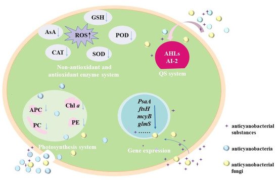
Currently, the anticyanobacterial mechanisms of microorganisms against M. aeruginosa are mainly dependeent on the attack modes, and these mechanisms are revealed with the changes in the photosynthesis system, antioxidant enzymes system, gene expression and QS system (Figure 2).
Figure 2. Anticyanobacterial mechanisms of microorganisms against M. aeruginosa.
3.1. Effects of Anticyanobacterial Microorganisms on Photosynthesis
Photosynthesis, which converts solar energy into chemical energy through the photosynthesis system (PS) II and PS I, is the principal mode of energy metabolism in cyanobacteria [
120]. Anticyanobacterial microorganisms can significantly affect the photosynthesis of
M. aeruginosa cells in several ways, including decreasing the chlorophyll
a (Chl
a) contents and photosynthetic pigments [
56], and the disruption of the electron transport pathway in PS [
23,
93]. Chl
a is one of the important components of cyanobacterial pigments. It is markedly decreased in
M. aeruginosa under the exposure of anticyanobacteria such as
P. aeruginosa [
18,
63],
Streptomyces sp. [
33,
36],
Exiguobacterium sp. [
44,
45], and so on. For the photosynthetic pigments, phycocyanobilin (PC), allophycocyanin (APC) and phycoerythrin (PE) are major indicators of cyanobacterial photosynthetic efficiency and are essential apparatus for light harvesting [
61], and the addition of anticyanobacterium results in a significant decrease in the PC, APC and PE by disrupting the synthesis of an photosynthetic pigments [
56]. In addition, the expressions of
pcA and
apcA genes for PC and APC synthesis in
M. aeruginosa are down-regulated by
Paucibacter aquatile DH15, which shows an inhibition effect on active chlorophyll [
61]. It has been noted that the Chl
a decrease is closely related to the reduction in photosynthetic pigments, and the cyanobacterial membrane is sensitive and easily damaged by anticyanobacterium [
56].
The variations of cyanobacterial energy kinetics have also been evaluated by Chl fluorescence parameters, such as the maximum photochemical quantum yield of PS II (Fv/Fm), the effective quantum yield (Φe), and the maximum electron transport rate (ETRmax) [
41,
95]. With the addition of fermentation filtrate (5%,
v/
v) of
Paenibacillus sp. SJ-73, the Fv/Fm values of
M. aeruginosa PCC7806 and
M. aeruginosa TH1701 dramatically decline from 0.52 and 0.29 to 0 [
95]; similarly, it is only 0.08 (14.3% of the initial value) for
M. aeruginosa FACHB-905 after being treated for 24 h by the fermentation filtrate (5%,
v/
v) of
Raoultella sp. S1 [
23]. Besides, the Φe and ETRmax of
M. aeruginosa 9110 following the treatment of
Chryseobacterium sp. GLY-1106 decrease gradually with time [
41]; the ETRmax values of
M. aeruginosa are also depressed significantly under the stress of
Raoultella sp. S1 [
23] and
Bacillus sp. B50 [
93]. The decreases in Fv/Fm, Φe and ETRmax demonstrate that the photosynthetic system is seriously damaged and the electron transport chain is blocked, resulting in the inhibition of cyanobacterial cell photosynthesis [
55]. In consequence, the possible mechanism underlying the photosynthetic reduction could be due to the reduction in Fv/Fm, Φe and ETRmax in
M. aeruginosa.
3.2. Effects of Anticyanobacterial Microorganisms on Antioxidant Enzymes System
The oxidative damage of the cyanobacterial cells can occur under different environmental stress conditions, and it will results in an increase in reactive oxygen species (ROS), which includes the superoxide anion radical, hydrogen peroxide and hydroxyl radicals [
51,
61]; while excess ROS often leads to oxidative stress, lipid peroxidation, and DNA damage [
56,
121]. The enzymatic antioxidants (such as catalase (CAT), superoxide dismutase (SOD), peroxidase (POD), and so on) and non-enzymatic antioxidants (such as ascorbic acid (AsA) and glutathione (GSH)) are responsible for removing the overproduction of ROS [
2,
31,
41]. For instance,
Streptomyces eurocidicus JXJ-0089 inhibits the growth of cyanobacterial cells in various ways, including promoting ROS production (e.g., O
2•
−), inhibiting the antioxidant synthesis, removing chlorophyll and destroying cell walls [
38].
The ROS of cyanobacteria increases excessively by either the direct attack or indirect attack of anticyanobacterial microorganisms. The O
2•
− content in
M. aeruginosa cells is induced largely by 4 μg mL
−1 3, 4-dihydroxybenzalacetone (DBL) secreted from
Phellinus noxius HN-1 and increased from 0.360 ± 0.001 to 0.400 ± 0.001 μg g
−3 [
104]. The ROS level of
M. aeruginosa NIES 843 treated with
Bacillus sp. AF-1 (cell-free filtrate) was lower than that of the control at the first 48 h but much higher at 72 h, indicating that some evasive mechanisms were taken to prevent the ROS accumulation in cyanobacterial cells at the initial stage [
51]. Similar variations of ROS have been observed in
M. aeruginosa KW after being treated with
Paucibacter aquatile DH15, and the malondialdehyde (MDA) content and SOD activity related to remove ROS also increased at first and then decreased [
61]; The MDA content, CAT and POD activity of
M. aeruginosa FACHB-905 also increased quickly when fermentation liquid (5%,
v/
v) of
P. aeruginosa [
18] and
P. chrysosporium was added quickly [
101]; moreover, the responses of
M. aeruginosa FACHB-905 cells to
Streptomyces sp. KY-34 and
Streptomyces sp. HJC-D1 following a similar pattern with the increases of CAT, SOD and POD, and the MDA further increased during the incubation time [
56,
121]. Although the antioxidants increased immediately to relieve the damage caused by anticyanobacteria, the cyanobacterial cell membrane may have decompose due to the accumulation of MDA [
18,
67,
121].
For the non-enzymatic antioxidants, the variation of GSH is opposite to that of the antioxidase activity. The
Bacillus licheniformis Sp34 induces more GSH production in
M. aeruginosa at first to clear ROS, but the GSH content is much lower at 20 h (compared with the control) [
47]. Such a phenomenon is also obtained in the anticyanobacterial process of
Raoultella sp. S1 [
23]. The prodigiosin from
Hahella sp. KA22 also leads to the variation of GSH content, while the GSH content decreases slightly after exposure for 36 h [
31]. These results demonstrate that the ROS levels and MDA contents decrease under prolonged exposure to anticyanobacteria [
31,
33,
65]; in addition, the non-enzymatic antioxidants also play a critical role in protecting the cyanobacterial cells from oxidative damage under anticyanobacterial stress [
23].
3.3. Effects of Anticyanobacterial Microorganisms on Gene Expression
The relative transcriptional level of some critical genes in cyanobacteria can be dramatically changed by anticyanobacterial microorganisms and substances, including genes related to the synthesis of photosystem reaction center proteins (
PsaA,
psaB,
psbA1 and
psbD1) [
47,
57], peptidoglycan synthesis (
glmS), membrane proteins (
ftsH), antioxidase (
prx) [
100], heat-shock proteins (
grpE) [
100], fatty acids (
fabZ) [
100], cyanotoxin microcystins (
mcyA, m
cyB, m
cyC and m
cyD) [
83,
97], the functions of cell division (
ftsZ) [
93], CO
2 fixation (
rbcL) [
61], and DNA repair (
ftsH and
recA) [
2,
5]. Researchers have reported that the transcription expressions of genes
ftsZ,
psbA1, and
glmS are decreased by DBL that is isolated from
P. noxius HN-1 [
104] and bacilysin that secreted from
B. amyloliquefaciens FZB42 [
91]. The expressions of gene
ftsZ and
psbA are also significantly inhibited by
Bacillus sp. B50 [
93], and the transcriptions of photosynthesis-related genes (
psaB and
psbD1) and CO
2 fixation gene (
rbcL) are inhibited by
B. licheniformis Sp34 [
47], indicating that the metabolisms of
M. aeruginosa are destroyed. Other studies on transcriptomic analysis have demonstrated that the principal subunits of the reaction center (
PsaA and
PsaB) and other subunits (
PsaC,
PsaE,
PsaD,
PsaF and
PsaL) are significantly down-regulated by
B. laterosporus Bl-zj [
57]. It is similar in the case of
S. globisporus G9,
S. amritsarensis and
Raoultella sp. S1, which suppresses the expression of
psbA1, psbD1 or
rbcL [
5,
23,
83]. The reduction in photosynthesis-related gene transcripts might result in an interruption in the electron transport chain and may finally affect the CO
2 fixation process [
61].
Gene such as
mcyB that are involved in microcystins synthesis are also inhibited by
Penicillium spp. [
97], the white-rot fungi
P. chrysosporium [
100,
101] and
P. noxius HN-1 [
104]; moreover, both directly attack the anticyanobacterium (
S. globisporus G9) [
83] and indirectly attack anticyanobacteria (including
S. amritsarensis HG-16 and
Bacillus sp. AF-1) could inhibit microcystins synthesis [
5,
51]. However, the inhibiting ability of
Bacillus sp. AF-1 has not been confirmed with microcystins measurements [
5].
3.4. Regulating the Anticyanobacterial Activity by QS System
QS system is the regulator control system for microorganisms that sense the cell density of their own species and make themselves to coordinate gene expression and physiological accommodation on a community scale [
122,
123]. It is a cell-to-cell communication that relies on the signal molecules [
124], and the accumulated QS signals can bind to the cognate receptors and regulate biological activities and cellular functions [
69,
125]. Previous studies have shown that microbial behaviors such as the secondary metabolites, cell motility and antibiotic resistance are all influenced by QS [
122,
123]; in addition, QS signals that contribute to the interactions between planktonic microalgae and bacteria are summarized as the N-acyl-homoserine lactones (AHLs) [
69], the 2-alkyl-4-quinolones (AQs) [
123], long-chain fatty acids and fatty acid methyl esters (autoinducer-2, AI-2) and dihydroxypentanedione furanone derivates [
12]. It is agreed that most of the anticyanobacterial activities by Gram-negative bacteria (such as
Pseudomonas sp.,
Acinetobacter sp., etc.) are the consequence of bacterial-cyanobacterial QS rather than bacterium-cyanobacteria interactions [
12,
124]. Some species of
Serratia sp. [
109] and
Hahella sp. [
31] can produce prodigiosin to inhibit
M. aeruginosa, and the prodigiosin production is regulated by
LuxI and
LuxR, which are the crucial genes of AHLs [
126]. The QS signal molecule (C4-HSL), which belongs to the classic AHL-based
LuxIR-type QS system of Gram-negative bacteria, is responsible for the synthetic process of the anticyanobacterial compound (3-methylindole) from
Aeromonas sp. GLY-2107 [
69]. During the anticyanobacterial process, the QS systems of Gram-negative bacteria produce AHLs signaling molecules, which are synthesized by the basic regulatory protein of
LuxI [
69,
88,
126].
In contrast, a wide range of the Gram-positive anticyanobacteria (such as
Streptomyces sp.,
Bacillus sp., etc.) generally use AI-2 as the signal molecules in QS systems [
125]. The anticyanobacterium
S. xiamenensis Lzh-2 exhibits QS behavior, and the
LuxS gene is crucial for the AI-2 type QS system; obviously, the anticyanobacterial activity of
S. xiamenensis Lzh-2 is regulated through the
LuxS/AI-2 QS system by inducing the production of anticyanobacterial compounds 2, 3-indolinedione and cyclo(Gly-Pro) [
126]. The AI-2 type QS behavior is present in
Bacillus sp. [
127]. Genomic analysis of
B. subtilis JA has indicated the existence of the
LuxS gene that regulates the pheromone biosynthesis, and the high-molecular-weight anticyanobacterial compounds (>3 kDa) produced by
Bacillus sp. S51107 have been proven to be primarily regulated by the
NprR-
NprX-type (AI-2) QS system [
88]. As a consequence, the AI-2 QS system has been considered as a possible strategy to regulate the behavior of the anticyanobacterial effects of Gram-positive bacteria. Although QS behavior has been reported in recent years, there is still an improved understanding of the interaction between cyanobacteria and anticyanobacterial microorganisms.
This entry is adapted from the peer-reviewed paper 10.3390/microorganisms10061136