Interstitial Brachytherapy for Breast Cancer Treatment: History
Please note this is an old version of this entry, which may differ significantly from the current revision.

Breast cancer represents the second leading cause of cancer-related death in the female population, despite continuing advances in treatment options that have significantly accelerated in recent years. Conservative treatments have radically changed the concept of healing, also focusing on the psychological aspect of oncological treatments. In this scenario, radiotherapy plays a key role. Brachytherapy is an extremely versatile radiation technique that can be used in various settings for breast cancer treatment. Although it is invasive, technically complex, and requires a long learning curve, the dosimetric advantages and sparing of organs at risk are unequivocal. Literature data support muticatheter interstitial brachytherapy as the only method with strong scientific evidence to perform partial breast irradiation and reirradiation after previous conservative surgery and external beam radiotherapy, with longer follow-up than new, emerging radiation techniques, whose effectiveness is proven by over 20 years of experience.

  • brachytherapy
  • muticatheter interstitial brachytherapy
  • APBI
  • accelerated partial breast irradiation
  • breast salvage treatment
  • breast cancer
  • ipsilateral breast recurrence
  • brachytherapy boost
  • breast reirradiation

1. Introduction

Breast cancer (BC) is the most common cancer in the female population, accounting for nearly 25% of all cancer diagnoses worldwide, whose incidence has been continuing to grow by approximately 0.5% per year [1]. Despite innumerable advances in medical and radiation oncology, this kind of tumor still represents the second leading cause of death [1].
Adjuvant radiotherapy (RT) has become increasingly important over the years, as conservative treatments for early-stage breast cancer have been demonstrated to offer the same local control of disease and survival outcomes as radical mastectomy compared to surgery alone [2][3][4][5].
Brachytherapy is a type of radiation technique wherein the radioactive sources are directly implanted into or close to the target tissue. In 1922, Geoffrey Keynes first used ‘interstitial radium needles’ for palliative treatment of breast cancer and achieved a surprising ‘disease control in cancer confined to the breast’ with a 3-year survival rate of 83.5% [6]. Nevertheless, breast multi-catheter interstitial brachytherapy (BCT) was systematically introduced in breast oncology practice in the seventies, acquiring an increasingly important role. Currently, breast BCT is the method with the highest scientific evidence and the longest follow-up. Breast BCT may be considered an extremely precise, versatile, and variable radiation technique. Breast BCT has the advantage of delivering high dose levels in the close proximity of the target volume, thus covering the entire tumor bed, and contemporary guaranteeing a very low dose distribution to the organs at risk (skin, heart, and lung), thus providing excellent local control of disease with low toxicity rates, but also requires a high level of expertise [7]. To date, brachytherapy-based accelerated partial breast irradiation (APBI) is the only one with level 1 evidence to be a valid alternative treatment option to whole breast irradiation (WBI) after breast-conserving surgery (BCS) for low-risk, early-stage breast cancer [7][8][9][10]. Moreover, APBI with multi-catheter brachytherapy has also been proposed for adjuvant re-irradiation of in-breast, ipsilateral tumor recurrences after previous BCS and WBI, with a very low rate of side effects and local recurrence rates comparable to salvage mastectomy [11].

2. Implant Technique and Treatment Delivery

2.1. Catheter Insertion
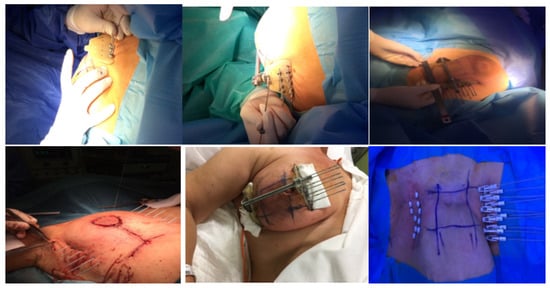
The standard procedure for breast catheter insertion consists of a transcutaneous approach. Metallic needles are manually inserted around the open/close cavity created during a lumpectomy, using a plastic guide template with needle holes to achieve geometric dose distribution. The needles are spaced to form equilateral triangles of 12–20 mm, according to the Paris System [12], then inserted in two to four planes, starting from the inferior plane to ensure an acceptable dose coverage to the deep tumor cavity under direct visualization (intraoperative), or guided by ultrasound images (postoperative). The deepest implant plane should be dorsal to the seroma, while the most ventral one should be placed between the skin surface and the seroma. Special care must be taken so that the needles are positioned at a distance of at least 1 cm from the skin surface to avoid late skin toxicity. At the end of the procedure, in the case of an open cavity (seroma), the needles can be replaced by plastic tubes. The number of applicators and tubes varies according to the size of the tumor cavity and breast anatomy (Figure 1) [13][14]. Once the needle positioning has been completed and the adequacy of the implant has been verified, a computed tomography (CT)-based simulation for target volume delineation and radiotherapy planning will be performed. If no appropriate target volume coverage is detected on the simulation CT scan, a few additional catheters may be inserted freehand without the use of a template.
Figure 1. Implant technique: manual insertion of metallic needles.

2.2. Target Definition and Delineation
Recently, guidelines for patients’ selection and brachytherapy target volume delineation after breast-conserving surgery with both a closed and an open cavity, as well as dose recommendations according to risk factors, were provided by the GEC-ESTRO Breast Cancer Working Group [15][16][17].
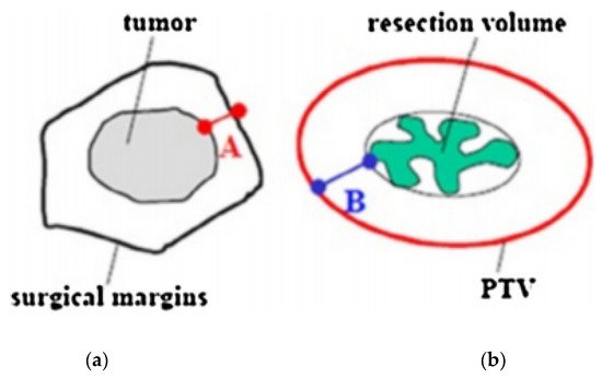
A CT scan with a 2–3 mm slice thickness is required to locate the surgical clips, which are needed to properly outline the target volume. Treatment planning begins with the delineation of an estimated target volume, taking into account preoperative imaging (mammography, breast ultrasound, and breast magnetic resonance if available), the surgical scar, the position of the surgical clips, and surgical margins. The clinical target volume (CTV) is defined with the addition of an isotropic, a total safety margin of 20 mm to the estimated target volume, and subtraction of the surgical margin. The thoracic wall and the skin must not be a part of the CTV. No additional margin to obtain the planning target volume (PTV) is necessary if the tumor bed and surgical clips are clearly visible. In the case of uncertainties ranging from 5 to 10 mm, additional margins can be delineated (Figure 2) [18][19].
Figure 2. Definition of safety margins. (a) Minimal resection margin. (b) Safety margin, > 20 mm minus A. PTV: planning target volume.

2.3. Dosimetry

The total dose to the target volume is nowadays delivered in the following two different ways: low-intensity pulses repeated every hour for up to a few days (pulse-dose-rate (PDR) brachytherapy); or a few, consecutive, high-dose fractions (HDR), the most used. Various radioisotopes with specific properties in terms of half-life and energy can be used. The most commonly applied in modern brachytherapy are iridium-192, cobalt-60, iodine-125, and palladium-103.
In order to select an appropriate isodose, the dose distribution has to be uniquely normalized. The dwell times are calculated on the basis of volumetric dose constraints. In the case of HDR and PDR BCT, geometric optimization for volume implants should keep the dose non-uniformity ratio (V100/V150) below 0.35 (0.30 ideally) [13]. The volume of PTV receiving 100% of the prescribed dose must be greater than 90% (coverage index ≥ 0.9), with a volume of PTV receiving 150% of the prescribed dose (V150%) less than 30%, and a volume receiving 200% of the prescribed dose (V200%) less than 15%, dose non-homogeneity ratio (V150/V100) < 0.35 (ideally 0.30). The maximum acceptable dose to the skin surface should be less than 70% of the prescribed dose. Table 1 summarizes the GEC-ESTRO normal tissue dose constraints [20] (Table 1).
Table 1. Recommended dose–volume limits for OAR-s.
Organs Constraints
Ipsilateral no target breast tissue V90 < 10%
V50 < 40%
Skin D1 cm3 < 90%
D0.2 cm3 < 100%
Ribs D0.1 cm3 < 90%
D1 cm3 < 80%
Heart MHD < 8%
D0.1 cm3 < 50%
Ipsilateral lung MLD < 8%
D0.1 cm3 < 60%
Abbreviations: MHD: mean heart dose, MLD: mean lung dose. Skin volume is defined as a 5 mm shell below the body contour.

3. Brachytherapy Doses

In 2018, the ESTRO-ACROP expert panel published the following recommendations for breast brachytherapy doses [20].
Recommended radiation schedules for HDR-BCT-based lumpectomy boost are as follows: a biologically equivalent total dose (BED2 for alpha/beta ratio = 4–5 Gy) in the range of 10–20 Gy from 1 to 4 fractions should be selected.
The panel of experts preferably recommends 2 × 4–6 Gy, or 3 × 3–5 Gy scheduled 2 times per day, with an interval between fractions of at least 6 h, and a total treatment time of 1–2 days, or a single fraction of 7–10 Gy, depending on the desired total EQD2.
Recommended schedules for APBI/accelerated partial breast reirradiation (APBrI) with HDR are as follows: 10 fr 3.4 Gy, or 8 fr 4 Gy, or 7 fr 4.3 Gy. With PDR-Brachytherapy: pulsed-dose 0.5–0.8 Gy/pulse, total dose 50 Gy, scheduled every hour, 24 h per day, total treatment time of 4–5 days.
Recommended schedules for lumpectomy boost with PDR-BCT: pulsed-dose 0.5–0.8 Gy/pulse, total dose 10–20 Gy, scheduled every hour, 24 h per day, total treatment time 1–2 days.

4. Advantages and Disadvantages of the Technique

The effectiveness of brachytherapy is based on the very high radiation dose directly delivered to the target volume by placing radiation sources in close proximity to or inside the tumor mass/tumor bed. A unique characteristic of this technique is the rapid dose fall-off outside the sources at the end of the implant, thus limiting dose exposure to the surrounding normal tissues. Brachytherapy offers dosimetric advantages with very sharp radiation dose gradients compared to conventional external beam radiation (EBRT) techniques.
As the source moves at the same time as the target, an additional margin is not necessary to cover the set-up uncertainties due to the organ motion, with a subsequent reduction of the planning treatment volume (PTV) and a smaller amount of healthy tissue receiving high doses, hence a reduction in side effects [20]. As a result, brachytherapy combines optimal tumor-to-normal tissue gradients while minimizing the integral dose to the remaining patient’s body tissue [21][22][23][24]. Brachytherapy is preferred in women with large breast sizes and deep tumor masses because the integral dose delivered with electron beams or EBRT is high, with a high risk of unacceptable lung and/or heart dose. Several studies have shown that, from a dosimetric point of view, brachytherapy boost better protects organs at risk (OARs) from medium to high radiation doses in deeply seated lumpectomy beds, compared to EBRT and high-energy electron beams [24]. Actually, brachytherapy is also the radiation technique with the highest level of scientific evidence regarding APBI and APBrI for ipsilateral breast recurrence after curative treatment [25].
Nevertheless, brachytherapy is also burdened with side effects, which may be minor to intense, depending on the delivered dose, the breast tumor site, and the size of the treated volume. Acute reactions (inflammation and irritation at the treatment site) are frequently in view of the very high doses delivered [25][26]. However, the significant decrease in the irradiated volume compared to other radiation techniques contributes to the good long-term functional outcome reported in the literature, with the potential for lower rates of normal tissue fibrosis (which is one of the mechanisms underlying organ dysfunction) [25][26]. Moreover, as it is an invasive treatment, there is a not negligible risk of infection and perioperative pain. The high specialization of the technique, requiring a long learning period to acquire the skills to guarantee the correct positioning of the catheters, may be considered the main limitation of brachytherapy. The Breast BCT procedure also requires specialized equipment able to perform the procedure under aseptic conditions, a dedicated operating room to properly handle the implant, and together with dedicated facilities that meet the radiobiological protection criteria.

This entry is adapted from the peer-reviewed paper 10.3390/cancers14102564

References

  1. Siegel, R.L.; Miller, K.D.; Fuchs, H.E.; Jemal, A. Cancer Statistics, 2021. CA Cancer J. Clin. 2021, 71, 7–33.
  2. Fisher, B.; Bauer, M.; Margolese, R.; Poisson, R.; Pylch, Y.; Redmond, C.; Fisher, E.; Wolmark, N.; Deutsch, M.; Montague, E.; et al. Five-year results of a randomized clinical trial comparing total mastectomy and segmental mastectomy with or without radiation in the treatment of breast cancer. N. Engl. J. Med. 1985, 312, 665–673.
  3. Fisher, B.; Anderson, S.; Bryant, J.; Margolese, R.; Deutch, M.; Fisher, E.R.; Jeong, J.H.; Wolmark, N. Twenty-year follow-up of a randomized trial comparing total mastectomy, lumpectomy, and lumpectomy plus irradiation for the treatment of invasive breast cancer. N. Engl. J. Med. 2002, 347, 1233–1241.
  4. Veronesi, U.; Saccozzi, R.; Del Vecchio, M.; Banfi, A.; Clemente, C.; De Lena, M.; Gallus, G.; Greco, M.; Luini, A.; Marubini, E.; et al. Comparing radical mastectomy with quadrantectomy, axillary dissection, and radiotherapy in patients with small cancers of the breast. N. Engl. J. Med. 1981, 305, 6–11.
  5. Veronesi, U.; Cascinelli, N.; Mariani, L.; Greco, M.; Saccozzi, R.; Luini, A.; Aguilar, M.; Marubuni, E. Twenty year follow-up of a randomized study comparing breast-conserving surgery with radical mastectomy for early breast cancer. N. Engl. J. Med. 2002, 347, 1227–1232.
  6. Keynes, G. The place of radium in the treatment of cancer of the breast. Ann. Surg. 1937, 106, 619–630.
  7. Polgar, C.; Fodor, J.; Major, T.; Sulyok, Z.; Kásler, M. Breast-conserving therapy with partial or whole breast irradiation: Ten-year results of the Budapest randomized trial. Radiother. Oncol. 2013, 108, 197–202.
  8. Ott, O.J.; Hildebrandt, G.; Potter, R.; Hammer, J.; Lotter, M.; Resch, A.; Sauer, R.; Strnad, V. Accelerated partial breast irradiation with multi-catheter brachytherapy: Local control, side effects and cosmetic outcome for 274 patients. Results of the GermanAustrian multi-centre trial. Radiother. Oncol. 2007, 82, 281–286.
  9. Strnad, V.; Hildebrandt, G.; Pötter, R.; Hammer, J.; Hindemith, M.; Resch, A.; Spiegl, K.; Lotter, M.; Uter, W.; Bani, M.; et al. Accelerated partial breast irradiation: 5-year results of the German-ustrian multicenter phase II trial using interstitial multicatheter brachytherapy alone after breast-conserving surgery. Int. J. Radiat. Oncol. Biol. Phys. 2011, 80, 17–24.
  10. Polgar, C.; Major, T.; Fodor, J.; Sulyok, Z.; Somogyi, A.; Lovey, K.; Nemeth, G.; Klaser, M. Accelerated partial-breast irradiation using high-dose-rate interstitial brachytherapy: 12-year update of a prospective clinical study. Radiother. Oncol. 2010, 94, 274–279.
  11. Hannoun-Levi, J.M.; Resch, A.; Gal, J.; Kauer-Dorner, D.; Strnad, V.; Niehoff, P.; Loessl, K.; Kovács, G.; Van Lim-bergen, E.; Polgár, C.; et al. Accelerated partial breast irradiation with interstitial brachytherapy as second conservative treatment for ipsilateral breast tumour recurrence: Multicentric study of the GEC-ESTRO Breast Cancer Working Group. Radiother. Oncol. 2013, 108, 226–231.
  12. Pierquin, B.; Wilson, J.-F.; Chassagne, D. The Paris System. In Modern Brachytherapy; Pierquin, B., Wilson, J.F., Chassagne, D., Eds.; Masson: Paris, France, 1987.
  13. Major, T.; Fröhlich, G.; Lövey, K.; Fodor, J.; Polgár, C. Dosimetric experience with accelerated partial breastirradiation using image-guided interstitial brachytherapy. Radiother. Oncol. 2009, 90, 48–55.
  14. Polgar, C.; Strnad, V.; Major, T. Brachytherapy for partial breast irradiation: The European experience. Semin. Radiat. Oncol. 2005, 15, 116–122.
  15. Polgar, C.; Van Limbergen, E.; Potter, R.; Kovacs, G.; Polo, A.; Lyczek, J.; Hildebrandt, G.; Niehoff, P.; Guinot, J.L.; Guedea, F.; et al. Patient selection for accelerated partial-breast irradiation (APBI) after breastconserving surgery: Recommendations of the Group Europeen de Curietherapie-European Society for Therapeutic Radiology and Oncology (GEC-ESTRO) breast cancer working group based on clinical evidence (2009). Radiother. Oncol. 2010, 94, 264–273.
  16. Strnad, V.; Hannoun-Lévi, J.-M.; Guinot, J.-L.; Lössl, K.; Kauer-Dorner, D.; Resch, A.; Kovacs, G.; Major, T.; Van Limnergen, E. Recommendations from GEC ESTRO Breast Cancer Working Group (I): Target definition and target delineation for accelerated or boost partial breast irradiation using multicatheter interstitial brachytherapy after breast conserving closed cavity surgery. Radiother. Oncol. 2015, 115, 342–348.
  17. Major, T.; Gutiérrez, C.; Guix, B.; van Limbergen, E.; Strnad, V.; Polgar, C. Recommendations from GEC ESTRO Breast Cancer Working Group (II): Target definition and target delineation for accelerated or boost partial breast irradiation using multicatheter interstitial brachytherapy after breast conserving open cavity surgery. Radiother. Oncol. 2016, 118, 199–204.
  18. Strnad, V.; Ott, O.J.; Hildebrandt, G.; Kauer-Dorner, D.; Knauerhase, H.; Major, T.; Lyczek, J.; Guinot, J.L.; Dunst, J.; Gutierrez miguelez, C.; et al. 5-year results of accelerated partial breast irradiation using sole interstitial multicatheter brachytherapy versus whole-breast irradiation with boost after breast-conserving surgery for low-risk invasive and in-situ carcinoma of the female breast: A randomised, phase 3, non-inferiority trial. Lancet 2016, 387, 229–238.
  19. Sato, K.; Shimo, T.; Fuchikami, H.; Takeda, N.; Kato, M.; Okawa, T. Catheter-based delineation of lumpectomy cavity for accurate target definition in partial-breast irradiation with multicatheter interstitial brachytherapy. J. Contemp. Brachyther. 2019, 11, 108–115.
  20. Strnad, V.; Major, T.; Polgar, C.; Lotter, M.; Guinot, J.L. ESTRO-ACROP guideline: Interstitial multi-catheter breast brachytherapy as Accelerated Partial Breast Irradiation alone or as boost—GEC-ESTRO Breast Cancer Working Group practical recommendations. Radiother. Oncol. 2018, 128, 411–420.
  21. Chargari, C.; Van Limbergen, E.; Mahantshetty, U.; Deutsch, E.; Haie-Meder, C. Radiobiology of brachytherapy: The historical view based on linear quadratic model and perspectives for optimization. Cancer Radiother. 2018, 22, 312–318.
  22. Georg, D.; Kirisits, C.; Hillbrand, M.; Dimopoulos, J.; Potter, R. Image-guided radiotherapy for cervix cancer: High-tech external beam therapy versus high-tech brachytherapy. Int. J. Radiat. Oncol. Biol. Phys. 2008, 71, 1272–1278.
  23. Yanez, L.; Ciudad, A.M.; Mehta, M.P.; Marisglia, H. What is the evidence for the clinical value of SBRT in cancer of the cervix? Rep. Pract. Oncol. Radiother. 2018, 23, 574–579.
  24. Otahal, B.; Dolezel, M.; Cvek, J.; Simetka, O.; Klat, J.; Knibel, L.; MOlenda, L.; Skacelikova, E.; Hlavka, A.; Felti, D. Dosimetric comparison of MRI-based HDR brachytherapy and stereotactic radiotherapy in patients with advanced cervical cancer: A virtual brachytherapy study. Rep. Pract. Oncol. Radiother. 2014, 19, 399–404.
  25. Chargari, C.; Deutsch, E.; Blanchard, P.; Gouy, S.; Martelli, H.; Guerin, F.; Dumas, I.; Bossi, A.; MOrice, P.; Viswanathan, A.; et al. Brachytherapy: An overview for clinicians. CA Cancer J. Clin. 2019, 69, 386–401.
  26. Sato, K.; Shimo, T.; Fuchikami, H.; Takeda, N.; Kato, M.; Okawa, T. Predicting adherence of dose-volume constraints for personalized partial-breast irradiation technique. Brachytherapy 2021, 20, 163–170.
More
This entry is offline, you can click here to edit this entry!
Video Production Service