Field-Effect Transistor-Based Biosensors for Environmental Monitoring: History
Please note this is an old version of this entry, which may differ significantly from the current revision.

The precise monitoring of environmental contaminants and agricultural plant stress factors, respectively responsible for damages to our ecosystems and crop losses, has become nowadays a topic of uttermost importance. This is also highlighted by the recent introduction of the so-called "Sustainable Development Goals" of the United Nations, which aim at reducing pollutants while implementing more sustainable food production practices leading to a reduced impact on all ecosystems. In this context, the standard methods currently used in these fields represent a sub-optimal solution, being expensive, laboratory-based techniques, and typically requiring trained personnel with high expertise. Recent advances in both biotechnology and material science, have led to the emergence of new sensing (and biosensing) technologies, enabling low-cost, precise, and real-time detection. An especially interesting category of biosensors is represented by field-effect transistor-based biosensors (bio-FETs), which enable the possibility of performing in-situ, continuous, selective, and sensitive measurements of a wide palette of different parameters of interest. Furthermore, bio-FETs offer the possibility of being fabricated using innovative and sustainable materials, employing various device configurations, each customized for a specific application. In the specific field of environmental and agricultural monitoring, the exploitation of these devices is particularly attractive as it paves the way to early detection and intervention strategies useful to limit, or even to completely avoid negative outcomes (such as diseases to animals or ecosystems losses). 

  • bio-FETs
  • environmental pollutants
  • plant stresses
  • transistors

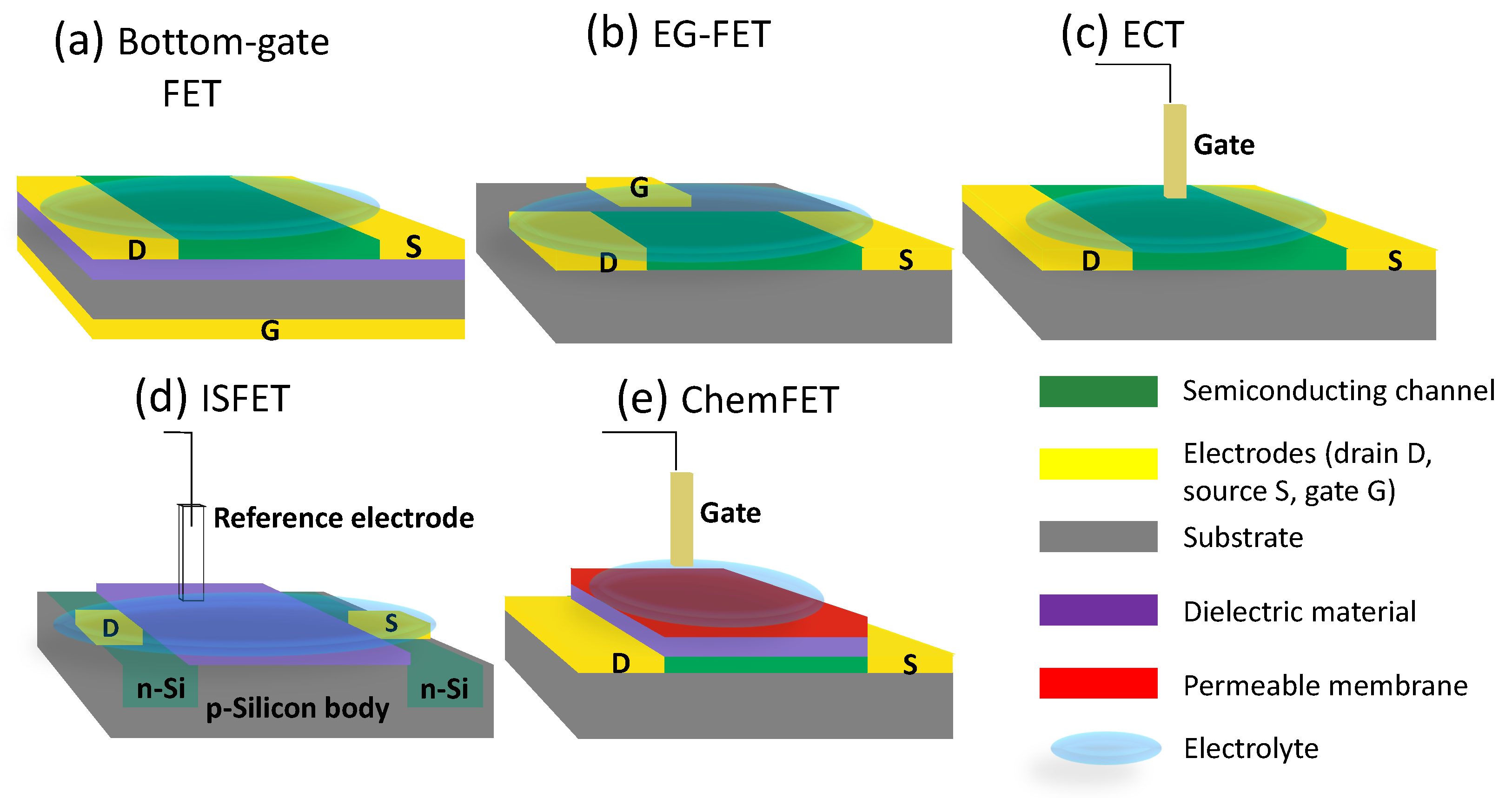
1. Bio-FET Operation and Configurations

A field-effect transistor (FET) is an active device that is composed of three electrodes (i.e., source, drain, and gate), a thin insulating layer (made of a dielectric material), and a semiconducting channel, which is the active layer of the device [1][2]. The gate electrode (which in some bio-FET configurations, as explained later, can also be an external reference electrode) is insulated from the semiconducting channel and the other two electrodes by the insulating layer, while the drain and source are connected through the semiconducting material. The so-called metal-oxide-semiconductor FETs (MOSFETs), where the gate electrode is insulated from the silicon (Si) semiconducting channel by a silicon dioxide (SiO2) layer, are the most commonly used types of FET devices [3][4]. In MOSFETs, Si is the fundamental part as it acts not only as the semiconductor but also as the substrate (body) while also offering two of its regions to realize the source and drain electronics doping processes (i.e., the process in which impurities are added to a semiconductor to alter its electrical properties) [5]. The MOSFET semiconducting channel is in fact formed between these two regions when the right voltage is applied to the gate [6]. MOSFETs form the basis of modern electronics, as demonstrated by the outstanding technological developments in the field [3][4]. Beside the major Si mainstream, alternative semiconductor materials, which are also suitable for flexible, stretchable, and/or biocompatible substrates (i.e., the materials on which the device is “built on”), are nowadays available and used in the realization of FETs [7][8]. These alternative semiconducting and substrate materials are essential building blocks of the so-called thin-film transistors (TFTs), a special type of FETs. TFTs typically employ inorganic (e.g., amorphous metal oxides) or organic materials as semiconductors [6]. The main difference between regular MOSFETs and TFTs relies exactly on these alternative non-Si semiconductors, which are not forming the substrate (body) themselves. In fact, in TFTs, thin films of the semiconductive layer are deposited on the substrate (which can be made of a variety of materials) [6][9]. When compared to TFTs, MOSFETs have drastically higher carrier mobility (i.e., the performance metrics measuring how fast electrons or holes can move in the semiconducting material under applied electric field [10]) because of the different materials that are used. TFTs are vastly used in other applications (and not just in the biosensor field), such as optical display systems, power transmission systems, and data transmission systems [6]
Despite the possibilities of having different configurations, the basic working principles of FETs are basically the same and are described in the following paragraphs. As mentioned before, there can be two types of semiconducting materials, n-type or p-type; in n-type materials, there is a surplus of negative charge carriers (i.e., electrons) in the semiconductor; on the contrary, in p-type materials, there is a lack of electrons, which can be regarded as a surplus of positive charge carriers (i.e., holes) [11][12]. When a voltage is applied to the gate electrode (in a specific direction dictated by the type of semiconductor), a flow of charge carriers (i.e., an electrical current) is allowed from the drain to the source through the semiconducting channel [11]. In more detail, the modulation of current between the source and drain is achieved through the semiconducting channel because of the field-effect mechanisms, which is the capacitive injection of carriers close to the dielectric–semiconductor interface [1][6]. In TFTs, this mechanisms is achieved by an accumulation layer and not an inversion region, like in the case of MOSFETs.
Transistor device behavior is commonly represented using its transfer and output characteristics. Transfer curves are obtained by measuring the current (drain-source, IDS) versus the gate-source voltage (VGS) while keeping a fixed drain-source voltage (VDS); output curves are instead obtained by measuring IDS and plotting it versus VDS, usually for several fixed values of VGS. Examples of transfer and output curves of a device with a p-type semiconductor material are shown. Changes to the gate-source voltage result in current variations over many orders of magnitude [13]; while the voltage control is essential in electronics to realize circuits, other alternative mechanisms allow different applications. In fact, besides voltage control, current variation (and amplification) can also be caused by surface effects, local electric fields, redox (reduction-oxidation) reactions, and other chemical reactions in the bulk solution [1].
As mentioned already, there are different possible configurations/architectures for FET devices, especially in the case of TFTs. Here, the most common ones that can be used for bio-FETs are highlighted and briefly compared (depicted in Figure 1). Besides the most traditional bottom-gate FETs, the others are electrolyte-gated FETs (EG-FETs) [1], electrochemical transistors (ECTs) [14], ion-sensitive FETs (ISFETs) [15], and chemically sensitive FETs (ChemFETs) [16]. As it can be seen from Figure 1, in all these configurations (except for bottom-gate), the gate electrode is separated from the rest of the structure by an electrolyte, which could be a solid polymer, an ion-gel, or, more commonly, a liquid solution [1][17][18]. An electrolyte is an electrical conducting medium that contains ions (i.e., an atom or molecule with a net electric charge) [18]. This property is important because in the majority of the cases, the analyte of interest is found in a liquid solution. Additionally, this is also convenient to interface physiological solutions.
Figure 1. Some of the most common structures of FET devices that are used as biosensors, i.e., as bio-FETs. All structures have source and drain electrodes (in yellow), a semiconducting channel (in green), and a substrate (in gray). (a) Bottom-gate FET; the substrate is Si, while the dielectric material is SiO2. (b) EG-FET (planar configuration); gate is in-plane with source/drain and is insulated through the electrolyte solution. (c) ECT; the gate is an external electrode and is dipped in the electrolyte solution. (d) ISFET; the gate electrode is replaced by a reference electrode (very often an Ag/AgCl electrode). (e) ChemFET; the gate electrode is separated from the source and drain by an electrolytic solution, and a semi-permeable membrane is present at the gate interface.
In the studies, when a bottom-gate FET was the employed structure, the devices were fabricated on a Si/SiO2 wafer; in most of these cases, the wafer (especially the Si body) acts as the gate electrode, while the dielectric material is SiO2. It is important to notice that in most cases a semiconducting material was deposited on the substrate to form the channel. In Figure 1a, this structure is depicted.
In EG-FETs, when a voltage is applied to the gate electrode, the ions contained in the electrolytic solution rearrange themselves, leading to the formation of two electric double layers (EDLs) at the two liquid–solid interfaces (gate electrode–electrolyte and semiconducting material–electrolyte) [1][19][20]. In this situation, the bulk electrolyte solution behaves like an insulator [21][22]. An example is presented in Figure 1b; in this case, the gate electrode is in the same plane as the source and drain, thus, this configuration is called in-plane EG-FET [1][23]. This is not always the case, as the gate electrode could also be external, such as a reference electrode, a needle, or a microwire [1][24].
In ECTs, the active material is represented mostly by a charge permeable layer, while the gate electrode (very often external to the structure) is submerged in the electrolyte solution (Figure 1c). The conductivity and the doping of the active film are changed by the injection of ions from the electrolyte into the active material by controlling VGS and VDS [5][14]. As opposed to EG-FETs, the doping changes in ECTs occurs on the whole volume of the channel rather than at the thin inter-facial area [14]. Thus, low voltages applied to the gate can lead to big variations in the drain current, which make this structure a strong amplifier in comparison to other FET ones.
ISFET was the first silicon-based sensor introduced by Bergveld [15]. It could be defined as a MOSFET, where the metal gate is substituted by a reference electrode and the dielectric layer is sensitive to the analyte changes, which is the main difference from the just-mentioned structures [15][25][26]. ISFETs commonly use a sensitive membrane as a dielectric (e.g., Si3N4) to detect ions in liquid solutions and are usually applied for pH sensing. In ISFETs, a surplus of charge regions are created using an electric field to increase or decrease the local conductivity. A representation of ISFETs is found in Figure 1d.
ChemFETs are considered an ISFET covered with a membrane (over the dieletric layer) that is selective to a specific ion analyte (or sometimes also gases); the permeable membrane is present at the gate interface and can be modulated by specific chemical stimulus [16][25][27]. ChemFETs are used to sense chemical concentrations in a solution [28]. A representation of a ChemFET is found in Figure 1e.
The choice of the FET structure depends mainly on: (A) the final application, such as the need to use flexible, stretchable or biocompatible materials, and (B) the analyte of interest, as some configurations are more convenient to measure pH than others, for example (e.g., ISFET).

2. Bio-FETs in Agricultural Plants Applications

Modern monitoring platforms (i.e., Agriculture 4.0) [29] are composed of different core technology layers, which are the following: (a) the physical layer composed of sensors to acquire data and actuators to automate some practices using robots with specific automation mechanisms [30], sometimes also including unmanned aerial vehicles (UAVs) [31]; (b) the connectivity layer using the Internet of Things (IoT) to continuously connect the data acquired from sensors based on standard wireless communication protocols with different frequency bands, transmission ranges, data rates, and energy consumption rates [32]; (c) the cloud computing layer, which offers a platform (hardware, software, data storage, and security) service for the big data produced by the IoT [33]; (d) the data layer, which includes big data analytics, machine learning (ML) algorithms, and artificial intelligence (AI), typically playing an important role in transforming data into knowledge by aggregating, processing, and visualizing the large data matrices and maximizing the prediction output that may improve decision making [34]; (e) the decision support system layer, represented as the final layer by an application that is friendly to the end-user for decision making [29].
Sensors are considered an indispensable basic layer for the development of these platforms. In agricultural monitoring, sensors are used to monitor the soil (temperature, pH levels, pollutants, nutrients/fertilizers, moisture, conductivity, and salinity), the environmental weather (temperature, humidity, atmospheric pressure, wind speed, and wind direction), the plants (biotic and abiotic stresses, metabolites, pH, ions) but also livestock and other parameters of relevance, such as agricultural machinery. The scope of this section is to give an overview of the most interesting bio-FETs developed for plant monitoring because of the important role plants play in providing food, oxygen, shelter, chemicals, fiber, medicine, energy, and other applications related to reducing soil erosion and water draining.
Plants respond to stress in different ways, generally suffering from two types of stresses: abiotic and biotic. The plant response happens in different physiological stages. At the beginning of stress, plants start to behave differently from their normal metabolism/ behaviour, with a deviation inducing an alarming reaction represented by complex biological, chemical, and electrical signals that lead to an increase in catabolism (e.g., breaking down of metabolite compounds) over anabolism (e.g., sugar synthesis). Afterwards, plants enter in a new phase where they resist the stress by adopting repair processes. Later on, when the stress reaches its maximum, it leads to either a plant disease, low performance, or death. The final phase is represented by removing the stress, thus leading to a recovery stage [35]. All the aforementioned phases are regulated through biological, chemical, and electrical signals, which can be sensed in different plant organs using appropriate instrumentation, different techniques, or a selective biomarker. It is crucial when developing a sensor for plants to consider the position in which the sensor will be applied because each organ needs a specific approach. Plants have different organs (e.g., leaf, stem, and fruit) to be monitored. Leaves are in charge of photosynthesis, organizing the transpiration rate, making up sugars, and releasing several VOCs [36]. Some fruits are important for reproducing the plants thanks to their seeds, while several kinds of fruits represent an edible part of the plant. As leaves and some fruits emit some VOCs and are in contact with the outside environment, they could be a good place to monitor gaseous VOC biomarkers or could be sensitive part to residual pesticides that can remain on their surface; thus, it is attractive to place the sensors on them. On the other hand, the stem provides the basic plant structure and is considered a network point to connect different parts of the plant using phloem and xylem tubes. These vascular tissues are filled with sap—a plant fluid that is composed of different compounds (e.g., water, nutrients and metabolites). In xylem, the sap moves in the direction from the root to the shoot, while in phloem, it moves from leaves (that are responsible for photosynthesis) to different parts of the plant. Thus, monitoring sap is very attractive thanks to the specific changes that happen in its composition while plants are under different stresses.

2.1. Abiotic Stresses

Abiotic stresses are caused by non-living factors such as drought, temperature changes, nutrient deficiency (e.g., potassium), hypoxia (e.g., oxygen scarcity), light, wounds, anoxia (e.g., total oxygen absence), and salinity. These stresses sometimes are harmful and could lead to huge yield losses. Thus, monitoring it is extremely important in order to detect it early, which can lead to an early intervention [37][38]. Relevant studies to detect abiotic stresses are highlighted here.
Coppedè et al. developed an OECT made on a cotton thread substrate, called ”bioristor” (i.e., transistor used for plant physiology and biology), with the organic polymer PEDOT:PSS as the active material to monitor plant sap electrolyte content over a period of 22 days [39]. With the bio-FET, they showed a periodic change in IDS, which indeed corresponded to ion changes that happen during the circadian cycle of the plants. In fact, a reduction in the IDS was obtained when the VGS was increased, which could be attributed to the de-doping of the PEDOT:PSS by plant sap. Because of the substrate biological origin, cotton was convenient and more accepted by the plant, even if an immune response of the plant was observed around the inserted zone but without affecting the plant growth or morphology. The bioristor was used later for different applications in several studies to monitor the changes in the ionic composition of the plants’ xylem sap in real-time and then to correlate the changes to different plant stresses [40][41].
For example, Amato et al. implanted the bioristor in the trunk of olive trees (Olea europaea) to monitor the water flux density (WFD) and vapor pressure deficit (VPD) [40]. This device and how it was applied to the trunk are depicted in Figure 2c, with measurements performed over 10 days [40]. They showed that the response (the change in measured IDS compared to IDS0, which was IDS at VGS of 0 V) was inversely proportional to the water flux density and correlated with the vapor pressure deficit. The change in this case was also attributed to the change in sap composition, which directly affects the de-doping of PEDOT:PSS.
Figure 2. Examples of bio-FETs developed for plant monitoring. (a) OECT developed to monitor plant xylem metabolites. Top: device setup; the sensor was inserted into the xylem of the tree stem. Bottom: Instantaneous monitoring of sucrose (orange), glucose (cyan), and control (black) for 2 days. Bright areas correspond to daytime, while dark areas are related to nighttime. Re-adapted with permission from [42]. Copyright 2022, Elsevier. (b) EG-FET for the detection of plant viruses. Top: Cross-view of final device. Bottom: Transfer characteristics of the sensor after being exposed to various plum pox virus (PPV) concentrations (from 0 to 50 μg/mL); a decrease in current is shown with increasing concentrations of PPV. Re-adapted with permission from [24]. Copyright 2022, Elsevier. (c) OECT for the detection of ions in olive trees. Top: a representation of the sensor setup inserted into the stem of an olive tree. Bottom: diurnal fluctuations of the sensor response R (o) and plant transpiration (E) (cyan line) sensed for 48 h. Re-adapted with permission from [40]. (d) Bottom-gate FET for monitoring microalgae membrane depolarization. Top: representation of the sensor functionalized with algae cells and infected with viruses introduced with syringe. Bottom: the current changes upon virus infection. Reprinted with permission from [43]. Copyright 2022 American Chemical Society.
The bioristor was integrated within the stem of tomato plants (Solanum lycopersicum), and it was applied to early detect drought stress [41]. The bioristor could detect changes in ion concentration within the first 30 h of water deprivation; the measurement period consisted of 23 days, and the response was measured as mentioned in the previously cited studies [41]. Furthermore, Vurro and colleagues used the bioristor to monitor xylem sap ion changes in different VPD conditions [44]. Furthermore, in this case, the studied plant was a tomato plant (Solanum lycopersicum). The analysis consisted of 15 days of constant measurements of the sensor response, where high and low VPD conditions were alternated. When VPD decreased from 1 to 0.7 Kpa, a rapid positive slope was seen in the response (which corresponds to the change in IDS). Conversely, when VPD was rapidly increased (from 0 to 0.7 Kpa), a negative slope was seen in the response [44].
Recently, the bioristor was applied to early monitoring the saline stress in Arundo donax for 37 days [45]. The sensor response was modulated by the plant sap ions, resulting in an increase in the VGS and a decrease in IDS because of the de-doping of PEDOT:PSS. All these studies that employed the bioristor are a good starting point, especially from the biocompatibility point of view of the device; however, the device did not present selectivity towards one specific analyte (ion in these cases) because of the lack of a specific recognition element.
Takemoto et al. developed a fabrication process to obtain highly transparent electrodes with a value of 90% transmittance in a region of visibility from 400 to 800 nm [46]. With these electrodes, they developed an OECT that was fabricated on a flexible parylene substrate, with C8-BTBT as the active material; the OECT was used for on-leaf monitoring of the plant’s electric potential and had a thin thickness (3 μm). The transparent electrodes enabled a biocompatible measurement of the plant’s response to dark and light (i.e., illumination condition) in Egeria Densa leaves without compromising the photosynthesis process [46]. Being flexible and transparent may increase its biocompatibility, which could potentially be an ideal platform to carry out other measurements on leaf.
In a 2014, Lee et al. made a highly flexible bio-FET gas sensor with an active film of CNTs and electrodes made of graphite to measure dimethyl methylphosphonate (DMMP) vapor in air [47]. Such a sensor was integrated into the surface of D. sanderiana cv. Virens leaf with improved adhesion and flexibility. In fact, IDS was decreased when VGS was applied with increasing DMMP concentrations (from 5 to 30 ppm) due to the interaction between CNTs and DMMP. The sensor is promising for non-planar substrate applications. For example, it was tested on nail, tape, and insect surfaces [47].
Bischak et al. were able to record the action potential signals of Venus flytrap (Dionaea muscipula) hair upon mechanical stimulation using an OECT sensor [48]. The device was based on a PET substrate, with both P3HT and PBTTT as active materials. The performance of the transistor was improved thanks to a layer of an ion exchange gel between the electrolyte and the active layer; such gel was able to uptake ions from the electrolyte and inject its ions into the active layer to dope it [48]. Upon triggering the plant hair, a change in IDS was measured (from 0 to 4 mA). However, despite being promising, some ion gels could cause toxicity when applied to biological tissues; thus, the development of biocompatible gels could be a solution to this issue.
Tao et al. monitored the concentration of methyl parathion pesticide, which can be dangerous to the human nervous system (as already mentioned in Section 3.1) [49]. The sensor had an OECT configuration, with an active layer of graphene and gate electrode functionalized with zirconia/reduced graphene oxide (ZrO2/rGO) as a recognition element. The OECT was attached on the leaf of Napa cabbage (Brassica rapa Pekinensis) [49]. The pesticide measurements were conducted in vitro, and the lifetime of the sensor was 28 days. Such a sensor could be considered as a platform to monitor other pesticides; however, in vivo studies are needed to prove its efficiency in real environmental conditions.
Strand et al. developed a low-cost OECT using the screen printing technique on top of the PEN substrate, with an active layer composed of a mix of PEDOT:PSS and sorbitol to monitor potassium [50]. The output curves of the device was much enhanced when sorbitol was mixed with PEDOT:PSS, which might be due to the fact that sorbitol increases the conductivity of PEDOT:PSS [50][51]. An ion-selective membrane specific for potassium was then used to increase the device selectivity toward nutrient monitoring in plant sap; the sap was taken from different genuses of trees (e.g., Maple, Picea). The device durability was 4 months, and the tested concentration range was 103 to 102 mM. In fact, measured current was increasing when potassium concentration was increased. The sensor could be used to detect potassium deficiency early, which is a stress that may disturb the plant physiology, ultimately resulting in a low yield [50]. The developed sensor could be used as a platform to monitor other ions by simply changing the selective membrane. However, despite being promising, the measurement method was invasive because of the extraction of the sap from the tree. Using non-invasive methods in the future could be a solution to such a matter.

2.2. Biotic Stresses

Biotic stress is caused by living organisms, such as microorganisms, insects, viruses, and other plants. These plant pathogens are one of the most common causes of crop damages, which, if detected early, could prevent serious plant disease epidemic. Relevant studies to detect biotic stresses are highlighted here.
Berto et al. developed an EG-FET biosensor to detect plum pox viruses (PPVs), plant pathogens that affect stone fruit species, causing significant losses [24][52]. The biosensor was based on quartz substrate with an organic active layer of pentacene and was functionalized with anti-PPV antibodies on the gate [24]. When the virus binded to the antibody, a change in IDS was observed; in fact, the IDS current decreased upon an increase in the virus concentration (with a range of 5 ng/mL to 50 μg/mL). This alteration was also visible in a change in transconductance (slope of the transfer curve). The formed immunocomplex (antibody + virus) caused a rearrangement of the antibody, which affects the flow of the current. The EG-FET was tested in vitro using Nicotiana benthamiana leaf extracts [24]; the response and the device configuration are shown in Figure 2b. Such a sensor had promising results that could be compared to traditional highly sensitive methods, such as the ELISA method. However, it should be applied in vivo to assess its efficiency.
Wang et al. developed a bottom-gate FET (Si substrate acted as gate electrode) and with CNTs as the active material to detect P-Ethylphenol gas, a volatile organic compound released by the fungus Phytophthora Cactorum [53], which infects strawberry plants (Fragaria × ananassa), resulting in a loss of fruit yield up to 50 percent [53]. The developed device was functionalized with a single-stranded DNA sequence (by π-π interaction between the CNTs and the nucleotide) to increase its sensitivity. In fact, they showed a small current increase (represented by a transconductance increase) when the sensor was exposed to ethyl-phenol gas compared to air exposure. Additionally, a shift in VTH was seen, and these changes were attributed to the adsorption of partially charged VOC molecules by the recognition element. However, the selectivity of the device was overlapping with other gases released by the infected fruit, which is why the output data were processed using fitting and regression statistical techniques to increase the sensing precision toward specific VOCs. There were good results since they showed that the predicted concentrations were comparable with the true tested values [53].
Wang and colleagues developed a bottom-gate FET to detect gases released by citrus trees infected with greening disease—a disease caused by the pathogen Candidatus Liberibacter asiaticu, which can result in weak citrus tree growth and reduced quality of the fruit [54]. The bio-FET was functionalized with a ssDNA sequence specific to phenylacetaldehyde (which is a VOC biomarker for the greening disease). In addition, the maximum measured IDS decreased when the device was exposed to phenylacetaldehyde compared to when it was exposed to air (4 μA compared to 4.5 μA) [54]. As a downside, the device showed a change in response for other gases (ethylhexanol, linalool, and tetradecene) as well. This low selectivity could, however, hinder the outcome of a possible real-time or in vivo study employing this bio-FET.
Another way to detect the same greening disease was developed by Saraf et al., who aimed to detect limonin, an indirect indicator of the infection [55]. The OECT sensor tested Hamlin orange (Citrus X sinensis) fruit extracts in order to measure abnormal limonin levels. The device was deposited with a PEDOT:PSS conductive layer by drop casting and later functionalized with CNPs as the recognition element. CNPs undergo oxidation in the presence of limonin; thus, this redox causes a de-doping of the PEDOT:PSS channel, which then results in an increase in IDS [55]. The bio-FET was tested at different limonin concentrations (108106 M), and indeed, an increase in IDS could be seen (with an LOD of 10 nM); at 10 nM, IDS was 75 μA, while at 10 μM, IDS was 110 μA. The aforementioned three studies have not yet been applied in vivo. If they prove to be successful, this could be a breakthrough in agricultural research that may lead to the early detection of fruit and plant diseases using highly selective sensors.
Finally, an OECT biosensor was developed to detect plant viruses in real-time, relying on the cell membrane depolarization that happens in a single Chlorella cell (which belongs to plantae kingdom) upon the infection by a Paramecium bursaria chlorella virus 1 (PBCV-1) [43]. Such a sensor was based on a Si substrate and activated with gold nanoparticles necklace array, which linked the functionalized sensor with algae cells in a non-invasive way. In fact, IDS increased once the virus infected the algae cell. Study can open new horizons in precisely studying virus mechanisms upon entering and exiting its host cell, allowing virologists to understand virus behaviour [43]. The device configuration and the current changes upon the virus infection are depicted in Figure 2d.

4.3. Plant Metabolites and pH Measurements

Plants metabolite levels change when they are under different statuses (e.g., stress, ripening) as a physiological response in order to overcome or adapt to new conditions. Monitoring these kind of metabolites may be an indicator for detecting plant health early, even before visual symptoms appear [56].
In one of the most related studies, Diacci et al. designed an OECT made of a PEN substrate with an active layer of PEDOT:PSS [57]. The sensor was functionalized with the enzyme glucose oxidase in order to be selective towards glucose, which is a signaling molecule. The sensor response was assessed in real-time in chloroplasts (plant organelles responsible for photosynthesis) [57]. Chloroplasts were isolated and extracted from tobacco (Nicotiana tabacum) leaves. Moreover, two different chloroplast solutions were used; one was extracted during the day and the other one during the night. The device showed an operation range of 700×103–5 mM glucose concentration. The authors claimed to observe a glucose export (thus a higher glucose concentration) in a dark environment, represented by a change in the current IDS, compared to the chloroplast response under light conditions (IDS −0.3 mA vs. −0.35 mA). Such observation is in line with plant physiology behaviour; it would be interesting to monitor other metabolites in real-time [57][58].
The same authors have developed an OECT to be applied on the stem of the Aspen tree (Populus tremuloides), as shown in Figure 2a [42]. The goal was to use the sensor for continuously monitoring the plants in vivo. For the first time, both glucose and sucrose were monitored in the xylem sap, with detection in the concentration range of 102–1 mM. In fact, the sucrose concentration behaviour, represented by the sensor’s response, was higher during the day in comparison to night. The authors used an OECT configuration made on a PEN substrate and functionalized with a mix of three enzymes (invertase, mutarotase, and glucose oxidase) on the gate electrode [42]. Such a study could open a new field of research to monitor other plant metabolites in vivo in order to understand plant circadian patterns and plant physiology in different stresses. However, the plant’s immune response upon the insertion of the sensor, which is represented mainly by the formation of cork tissue around it, can hinder the lifetime of the measurements; thus, it could be important to develop biocompatible electronic materials with minimal invasiveness.
Arkhypova et al. developed an ISFET, functionalized with acetylcholinesterase enzyme, to measure the content of indole alkaloids, a metabolite of Rauwolfia serpentina tissue culture, which is used in the pharmaceutical and biotechnological industry [59]. Indole alkaloids are inhibitors of acetylcholinesterase, so this study exploited this ability. When both indole alkaloids and acetylcholine are present, the measured current is lower compared to when only acetylcholine is present because indole alakaloids also bind to acetylcholinesterase and block the reaction. They showed an inhibition level (in %), which was linear with the increasing concentration of indole alkaloids, with a concentration range of 2 to 15 μg/mL and an LOD of 0.5 μg/mL. Such a sensor claimed to be a cheap alternative to existing traditional analytical methods, which could be applied to notify the producer of such metabolites to control their growth process [59]. A good thing about this ISFET was that, with a simple washing procedure, the device could become re-usable again.
The same author had previously made an ISFET sensor functionalized with butyryl cholinesterase enzymes in order to detect glycoalkaloids, a poisonous substance found in potatoes (Solanum tuberosum) [60][61]. They showed a linear response in the concentration range of 2 ×104101 M [60][61]. In a later study, the sensitivity of the ISFET sensor towards the glycoalkaloids was improved by using phosphotriesterase enzymes, with a resulting linear range of 106105 M [62].
One of the earliest studies carried out by Herrmann et al. used an ISFET to measure the pH of various tree species in vivo (e.g., Populus balsamifera L., Aesculus hippocastanum L. branches xylem). The authors developed an ISFET with an ion-sensitive membrane layer on top of the Si substrate [63]. However, being an invasive method, the authors were not sure whether the sensor’s response was affected by a wounding response in the trees. Such uncertainty could be the reason behind the limits in plant-sensing research at that time, and the advent of new technologies (e.g., thin film transistors) could be a possible reason to adopt such measurements in the future [63].
Izumi et al. fabricated an ISFET sensor to monitor the phloem sap’s pH in cucumber (Cucumis sativus) stem [64]. The range of pH levels monitored was 4.01–9.18. IDS was measured using different pH control solutions; it could be seen that at a lower pH (4.01), the IDS was higher. To calculate the sensitivity of the sensor, the VGS change per 1 pH was derived from the IDS–VGS characteristics, and the sensitivity was obtained as 40.2 mV/pH [64]. The device was later combined with electrical conductivity and thermal sensors to monitor the xylem sap in order to correlate its response signals with the pH and thus monitor some parameters of plant health in relation to soil fertilizer content [64].
Finally, a ChemFET was functionalized with a nitrate-specific ion selective membrane as the recogniton element and then was coated with poly(2-hydroxyethyl methacrylate (poly-HEMA), a material used against scratching and weathering, in order to measure nitrate in the stem of corn (Zea mays) [28]. In fact, IDS decreased when the device was exposed to higher nitrate concentrations [28]. The measurements were carried out for over 150 h, and a linear detection in the concentration range of 0.1 to 1000 ppm was seen. The authors claimed that the sensor was minimally invasive, thanks to its ability to sense while being partially inside the plant stalk. However, it has not been proven biologically if a plant’s immune response was induced upon the insertion of the sensor, so this requires further investigations [28].

This entry is adapted from the peer-reviewed paper 10.3390/s22114178

References

  1. Shkodra, B.; Petrelli, M.; Costa Angeli, M.A.; Garoli, D.; Nakatsuka, N.; Lugli, P.; Petti, L. Electrolyte-gated carbon nanotube field-effect transistor-based biosensors: Principles and applications. Appl. Phys. Rev. 2021, 8, 041325.
  2. Wang, D.; Noël, V.; Piro, B. Electrolytic gated organic field-effect transistors for application in biosensors—A Review. Electronics 2016, 5, 9.
  3. Golio, M.; Golio, J. RF and Microwave Passive and Active Technologies; CRC Press: Boca Raton, FL, USA, 2018.
  4. Lee, M.L.; Fitzgerald, E.A.; Bulsara, M.T.; Currie, M.T.; Lochtefeld, A. Strained Si, SiGe, and Ge channels for high-mobility metal-oxide-semiconductor field-effect transistors. J. Appl. Phys. 2005, 97, 011101.
  5. Lüssem, B.; Riede, M.; Leo, K. Doping of organic semiconductors. Phys. Status Solidi 2013, 210, 9–43.
  6. Petti, L.; Münzenrieder, N.; Vogt, C.; Faber, H.; Büthe, L.; Cantarella, G.; Bottacchi, F.; Anthopoulos, T.D.; Tröster, G. Metal oxide semiconductor thin-film transistors for flexible electronics. Appl. Phys. Rev. 2016, 3, 021303.
  7. Zhao, Y.; Guo, Y.; Liu, Y. 25th anniversary article: Recent advances in n-type and ambipolar organic field-effect transistors. Adv. Mater. 2013, 25, 5372–5391.
  8. Zhang, S. Review of modern field effect transistor technologies for scaling. J. Phys. Conf. Ser. 2020, 1617, 012054.
  9. Kuo, Y. Thin film transistor technology—Past, present, and future. Electrochem. Soc. Interface 2013, 22, 55.
  10. Myronov, M. Molecular Beam Epitaxy of High Mobility Silicon, Silicon Germanium and Germanium Quantum Well Heterostructures. In Molecular Beam Epitaxy; Elsevier: Amsterdam, The Netherlands, 2018; pp. 37–54.
  11. De Moraes, A.C.M.; Kubota, L.T. Recent trends in field-effect transistors-based immunosensors. Chemosensors 2016, 4, 20.
  12. Benda, V.; Grant, D.A.; Gowar, J. Discrete and Integrated Power Semiconductor Devices: Theory and Applications; John Wiley & Sons: Hoboken, NJ, USA, 1999.
  13. Li, H.; Shi, W.; Song, J.; Jang, H.J.; Dailey, J.; Yu, J.; Katz, H.E. Chemical and biomolecule sensing with organic field-effect transistors. Chem. Rev. 2018, 119, 3–35.
  14. Rivnay, J.; Inal, S.; Salleo, A.; Owens, R.M.; Berggren, M.; Malliaras, G.G. Organic electrochemical transistors. Nat. Rev. Mater. 2018, 3, 17086.
  15. Bergveld, P. Thirty years of ISFETOLOGY: What happened in the past 30 years and what may happen in the next 30 years. Sens. Actuators B Chem. 2003, 88, 1–20.
  16. Polk, B.J. ChemFET arrays for chemical sensing microsystems. In Proceedings of the SENSORS, 2002 IEEE, Orlando, FL, USA, 12–14 June 2002; Volume 1, pp. 732–735.
  17. Winie, T.; Arof, A.K.; Thomas, S. Polymer Electrolytes: Characterization Techniques and Energy Applications; John Wiley & Sons: Hoboken, NJ, USA, 2019.
  18. Enderby, J.; Neilson, G. The structure of electrolyte solutions. Rep. Prog. Phys. 1981, 44, 593.
  19. Scuratti, F.; Bonacchini, G.E.; Bossio, C.; Salazar-Rios, J.M.; Talsma, W.; Loi, M.A.; Antognazza, M.R.; Caironi, M. Real-Time Monitoring of Cellular Cultures with Electrolyte-Gated Carbon Nanotube Transistors. ACS Appl. Mater. Interfaces 2019, 11, 37966–37972.
  20. Joshi, S.; Bhatt, V.D.; Rani, H.; Becherer, M.; Lugli, P. Understanding the influence of in-plane gate electrode design on electrolyte gated transistor. Microelectron. Eng. 2018, 199, 87–91.
  21. Oldham, K.B. A Gouy–Chapman–Stern model of the double layer at a (metal)/(ionic liquid) interface. J. Electroanal. Chem. 2008, 613, 131–138.
  22. Le Gall, J.; Vasilijević, S.; Battaglini, N.; Mattana, G.; Noël, V.; Brayner, R.; Piro, B. Algae-functionalized hydrogel-gated organic field-effect transistor. Application to the detection of herbicides. Electrochim. Acta 2021, 372, 137881.
  23. Bhatt, V.D.; Joshi, S.; Becherer, M.; Lugli, P. Flexible, low-cost sensor based on electrolyte gated carbon nanotube field effect transistor for organo-phosphate detection. Sensors 2017, 17, 1147.
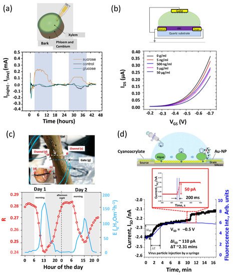
  24. Berto, M.; Vecchi, E.; Baiamonte, L.; Condò, C.; Sensi, M.; Di Lauro, M.; Sola, M.; De Stradis, A.; Biscarini, F.; Minafra, A.; et al. Label free detection of plant viruses with organic transistor biosensors. Sens. Actuators B Chem. 2019, 281, 150–156.
  25. Schöning, M.J.; Poghossian, A. Recent advances in biologically sensitive field-effect transistors (BioFETs). Analyst 2002, 127, 1137–1151.
  26. Pachauri, V.; Ingebrandt, S. Biologically sensitive field-effect transistors: From ISFETs to NanoFETs. Essays Biochem. 2016, 60, 81–90.
  27. Antonisse, M.M.; Reinhoudt, D.N. Potentiometric anion selective sensors. Electroanal. Int. J. Devoted Fundam. Pract. Asp. Electroanal. 1999, 11, 1035–1048.
  28. Jiao, Y.; Wang, X.; Chen, Y.; Castellano, M.J.; Schnable, J.C.; Schnable, P.S.; Dong, L. In-Planta Nitrate Detection Using Insertable Plant Microsensor. In Proceedings of the 2019 20th IEEE International Conference on Solid-State Sensors, Actuators and Microsystems & Eurosensors XXXIII (TRANSDUCERS & EUROSENSORS XXXIII), Berlin, Germany, 23–27 June 2019; pp. 37–40.
  29. Araújo, S.O.; Peres, R.S.; Barata, J.; Lidon, F.; Ramalho, J.C. Characterising the Agriculture 4.0 Landscape—Emerging Trends, Challenges and Opportunities. Agronomy 2021, 11, 667.
  30. Kalsoom, T.; Ramzan, N.; Ahmed, S.; Ur-Rehman, M. Advances in sensor technologies in the era of smart factory and industry 4.0. Sensors 2020, 20, 6783.
  31. Almalki, F.A.; Soufiene, B.O.; Alsamhi, S.H.; Sakli, H. A low-cost platform for environmental smart farming monitoring system based on IoT and UAVs. Sustainability 2021, 13, 5908.
  32. Munirathinam, S. Industry 4.0: Industrial internet of things (IIOT). In Advances in Computers; Elsevier: Amsterdam, The Netherlands, 2020; Volume 117, pp. 129–164.
  33. Dillon, T.; Wu, C.; Chang, E. Cloud computing: Issues and challenges. In Proceedings of the 2010 24th IEEE International Conference on Advanced Information Networking and Applications, Perth, Australia, 20–23 April 2010; pp. 27–33.
  34. Gröger, C. Building an industry 4.0 analytics platform. Datenbank-Spektrum 2018, 18, 5–14.
  35. Lichtenthaler, H.K. Vegetation stress: An introduction to the stress concept in plants. J. Plant Physiol. 1996, 148, 4–14.
  36. Brilli, F.; Loreto, F.; Baccelli, I. Exploiting plant volatile organic compounds (VOCs) in agriculture to improve sustainable defense strategies and productivity of crops. Front. Plant Sci. 2019, 10, 264.
  37. Zörb, C.; Geilfus, C.M.; Dietz, K.J. Salinity and crop yield. Plant Biol. 2019, 21, 31–38.
  38. Boyer, J.S. Plant productivity and environment. Science 1982, 218, 443–448.
  39. Coppedè, N.; Janni, M.; Bettelli, M.; Maida, C.L.; Gentile, F.; Villani, M.; Ruotolo, R.; Iannotta, S.; Marmiroli, N.; Marmiroli, M.; et al. An in vivo biosensing, biomimetic electrochemical transistor with applications in plant science and precision farming. Sci. Rep. 2017, 7, 16195.
  40. Amato, D.; Montanaro, G.; Vurro, F.; Coppedé, N.; Briglia, N.; Petrozza, A.; Janni, M.; Zappettini, A.; Cellini, F.; Nuzzo, V. Towards In Vivo Monitoring of Ions Accumulation in Trees: Response of an in Planta Organic Electrochemical Transistor Based Sensor to Water Flux Density, Light and Vapor Pressure Deficit Variation. Appl. Sci. 2021, 11, 4729.
  41. Janni, M.; Coppede, N.; Bettelli, M.; Briglia, N.; Petrozza, A.; Summerer, S.; Vurro, F.; Danzi, D.; Cellini, F.; Marmiroli, N.; et al. In Vivo Phenotyping for the Early Detection of Drought Stress in Tomato. Plant Phenom. 2019, 2019, 6168209.
  42. Diacci, C.; Abedi, T.; Lee, J.W.; Gabrielsson, E.O.; Berggren, M.; Simon, D.T.; Niittylä, T.; Stavrinidou, E. Diurnal in vivo xylem sap glucose and sucrose monitoring using implantable organic electrochemical transistor sensors. Iscience 2021, 24, 101966.
  43. Lee, S.W.; Lee, E.H.; Thiel, G.; Van Etten, J.L.; Saraf, R.F. Noninvasive measurement of electrical events associated with a single chlorovirus infection of a microalgal cell. ACS Nano 2016, 10, 5123–5130.
  44. Vurro, F.; Janni, M.; Coppedè, N.; Gentile, F.; Manfredi, R.; Bettelli, M.; Zappettini, A. Development of an In Vivo Sensor to Monitor the Effects of Vapour Pressure Deficit (VPD) Changes to Improve Water Productivity in Agriculture. Sensors 2019, 19, 4667.
  45. Michela, J.; Claudia, C.; Federico, B.; Sara, P.; Filippo, V.; Nicola, C.; Manuele, B.; Davide, C.; Loreto, F.; Zappettini, A. Real-time monitoring of Arundo donax response to saline stress through the application of in vivo sensing technology. Sci. Rep. 2021, 11, 18598.
  46. Takemoto, A.; Araki, T.; Uemura, T.; Noda, Y.; Yoshimoto, S.; Izumi, S.; Tsuruta, S.; Sekitani, T. Printable Transparent Microelectrodes toward Mechanically and Visually Imperceptible Electronics. Adv. Intell. Syst. 2020, 2, 2000093.
  47. Lee, K.; Park, J.; Lee, M.S.; Kim, J.; Hyun, B.G.; Kang, D.J.; Na, K.; Lee, C.Y.; Bien, F.; Park, J.U. In-situ synthesis of carbon nanotube–graphite electronic devices and their integrations onto surfaces of live plants and insects. Nano Lett. 2014, 14, 2647–2654.
  48. Bischak, C.G.; Flagg, L.Q.; Ginger, D.S. Ion exchange gels allow organic electrochemical transistor operation with hydrophobic polymers in aqueous solution. Adv. Mater. 2020, 32, 2002610.
  49. Tao, T.; Zhou, Y.; Ma, M.; He, H.; Gao, N.; Cai, Z.; Chang, G.; He, Y. Novel graphene electrochemical transistor with ZrO2/rGO nanocomposites functionalized gate electrode for ultrasensitive recognition of methyl parathion. Sens. Actuators B Chem. 2021, 328, 128936.
  50. Strand, E.J.; Bihar, E.; Gleason, S.M.; Han, S.; Schreiber, S.W.; Renny, M.N.; Malliaras, G.G.; McLeod, R.R.; Whiting, G.L. Printed Organic Electrochemical Transistors for Detecting Nutrients in Whole Plant Sap. Adv. Electron. Mater. 2021, 8, 2100853.
  51. Friederich, P.; León, S.; Perea, J.D.; Roch, L.M.; Aspuru-Guzik, A. The influence of sorbitol doping on aggregation and electronic properties of PEDOT: PSS: A theoretical study. Mach. Learn. Sci. Technol. 2020, 2, 01LT01.
  52. García, J.A.; Glasa, M.; Cambra, M.; Candresse, T. P lum pox virus and sharka: A model potyvirus and a major disease. Mol. Plant Pathol. 2014, 15, 226–241.
  53. Wang, H.; Wang, Y.; Hou, X.; Xiong, B. Bioelectronic Nose Based on Single-Stranded DNA and Single-Walled Carbon Nanotube to Identify a Major Plant Volatile Organic Compound (p-Ethylphenol) Released by Phytophthora Cactorum Infected Strawberries. Nanomaterials 2020, 10, 479.
  54. Wang, H.; Ramnani, P.; Pham, T.; Villarreal, C.C.; Yu, X.; Liu, G.; Mulchandani, A. Gas biosensor arrays based on single-stranded DNA-functionalized single-walled carbon nanotubes for the detection of volatile organic compound biomarkers released by huanglongbing disease-infected citrus trees. Sensors 2019, 19, 4795.
  55. Saraf, N.; Barkam, S.; Peppler, M.; Metke, A.; Vázquez-Guardado, A.; Singh, S.; Emile, C.; Bico, A.; Rodas, C.; Seal, S. Microsensor for limonin detection: An indicator of citrus greening disease. Sens. Actuators B Chem. 2019, 283, 724–730.
  56. Teklić, T.; Parađiković, N.; Špoljarević, M.; Zeljković, S.; Lončarić, Z.; Lisjak, M. Linking abiotic stress, plant metabolites, biostimulants and functional food. Ann. Appl. Biol. 2021, 178, 169–191.
  57. Diacci, C.; Lee, J.W.; Janson, P.; Dufil, G.; Méhes, G.; Berggren, M.; Simon, D.T.; Stavrinidou, E. Real-Time Monitoring of Glucose Export from Isolated Chloroplasts Using an Organic Electrochemical Transistor. Adv. Mater. Technol. 2020, 5, 1900262.
  58. Stitt, M.; Zeeman, S.C. Starch turnover: Pathways, regulation and role in growth. Curr. Opin. Plant Biol. 2012, 15, 282–292.
  59. Arkhypova, V.; Soldatkin, O.; Mozhylevska, L.; Konvalyuk, I.; Kunakh, V.; Dzyadevych, S. Enzyme biosensor based on pH-sensitive field-effect transistors for assessment of total indole alkaloids content in tissue culture of Rauwolfia serpentina. Electrochem. Sci. Adv. 2021, e2100152.
  60. Arkhypova, V.N.; Dzyadevych, S.V.; Soldatkin, A.P.; Anna, V.; Martelet, C.; Jaffrezic-Renault, N. Development and optimisation of biosensors based on pH-sensitive field effect transistors and cholinesterases for sensitive detection of solanaceous glycoalkaloids. Biosens. Bioelectron. 2003, 18, 1047–1053.
  61. Arkhypova, V.N.; Dzyadevych, S.V.; Soldatkin, A.P.; Korpan, Y.I.; Anna, V.; Gravoueille, J.M.; Martelet, C.; Jaffrezic-Renault, N. Application of enzyme field effect transistors for fast detection of total glycoalkaloids content in potatoes. Sens. Actuators B Chem. 2004, 103, 416–422.
  62. Korpan, Y.I.; Raushel, F.M.; Nazarenko, E.A.; Soldatkin, A.P.; Jaffrezic-Renault, N.; Martelet, C. Sensitivity and specificity improvement of an ion sensitive field effect transistors-based biosensor for potato glycoalkaloids detection. J. Agric. Food Chem. 2006, 54, 707–712.
  63. Herrmann, V.; Tesche, M. In vivo pH measurement in the xylem of broad-leaved trees using ion-sensitive field-effect transistors. Trees 1992, 6, 13–18.
  64. Izumi, R.; Ono, A.; Ishizuka, H.; Terao, K.; Takao, H.; Kobayashi, T.; Kataoka, I.; Shimokawa, F. Biological information (pH/EC) sensor device for quantitatively monitoring plant health conditions. In Proceedings of the 2017 IEEE SENSORS, Scotland, UK, 29 October–1 November 2017; pp. 1–3.
More
This entry is offline, you can click here to edit this entry!
Video Production Service