Human Heart Organoid Development: History
Please note this is an old version of this entry, which may differ significantly from the current revision.
Contributor: , , ,

The emergence of human-induced Pluripotent Stem Cells (hiPSCs) has dramatically improved the understanding of human developmental processes under normal and diseased conditions. The hiPSCs have been differentiated into various tissue-specific cells in vitro, and the advancement in three-dimensional (3D) culture has provided a possibility to generate those cells in an in vivo-like environment. Tissues with 3D structures can be generated using different approaches such as self-assembled organoids and tissue-engineering methods, such as bioprinting. Researchers are interested in studying the self-assembled organoids differentiated from hiPSCs, as they have the potential to recapitulate the in vivo developmental process and be used to model human development and congenital defects. Organoids of tissues such as those of the intestine and brain were developed many years ago, but heart organoids were not reported until recently.

  • heart development
  • congenital heart defect
  • organoid
  • anatomical structure
  • induction signal
  • molecular markers

1. Introduction

To model the in vivo developmental processes, 3D cell cultures were developed and categorized into different subtypes such as embryoid body (EB), gastruloid, spheroid, and organoid. EBs are aggregated differentiating embryonic stem cells (ESCs) or iPSCs. The cells in EBs are usually at early differentiation stages and do not organize into specific spatial patterns. In the cardiac stem cell field, EBs are used to generate lineage-specific cell types such as cardiomyocytes [1]. Gastruloids consist of three germ layers and are used to model the embryo gastrulation process. In theory, gastruloids can develop into an entire embryo if an appropriate differentiation environment is provided; in a recent study, a differentiation condition was designed to enable the cultured mouse embryos to develop to the hindlimb formation stage [2][3][4]. The spheroid is an aggregate of differentiated cell types or cancer cells. These cells do not self-assemble into the anatomical patterns observed in vivo and can be used to study the interactions among different cell types. Lastly, the organoid is a complicated cell aggregate in which the differentiated cells mostly belong to a specific organ and self-assemble into the in vivo-like anatomic patterns. Tissue-specific organoids, such as the cerebrum, liver, lung, and kidney, can be made with iPSCs or progenitor cells [5][6]. They usually have similar morphology and cell-type interactions to the in vivo organs, making organoids advantageous over other 3D models [7]. However, as the emergence of multi-lineage organoids and certain organoids can only recapitulate part of the organ-like features, the distinctions between the different types of 3D cell cultures are becoming less clear.
Multiple hiPSC-derived heart organoid systems were recently reported and claimed to have developed in vivo-like heart features. The heart is the first organ to develop in humans, and its defects affect about 1% of births [8]. Heart development originates from mesodermal cells, which later develop into cardiac progenitors in the two heart fields. First heart field (FHF) cells develop into the primitive heart tube, and second heart field (SHF) progenitors contribute to heart-tube elongation from both poles [9][10]. Meanwhile, the heart tube undergoes rightward looping, giving rise to the four-chambered heart. The heart chamber wall consists of three tissue layers: epicardium, myocardium, and endocardium. The heart also has other structures besides the chambers, including the inflow tract (IFT), the atrial ventricular canal (AVC), and the outflow tract (OFT) at the early stages, which contribute to the development of the chambers, valves, septum, and large vessels at later stages. The normal development of these structures is essential for generating a functional heart; if this process goes awry, it can cause congenital heart defects (CHDs) [10].
Although mouse models are highly valuable in studying certain CHDs such as hypoplastic left heart syndrome [11], many CHDs still do not have helpful animal models due to the potential genetic and pathological differences between species. Human-induced Pluripotent Stem Cells (hiPSCs) have a significant advantage in modeling these diseases, as the iPSCs can be directly reprogrammed from patient somatic cells, therefore preserving the genetic background of the patients. To model CHDs, the 3D differentiation condition (e.g., organoids) is essential as it can potentially develop features like those seen in the in vivo hearts. Besides modeling normal heart developmental processes and CHDs, heart organoids also have the potential to be used for drug screenings.

2. Heart fields Formation

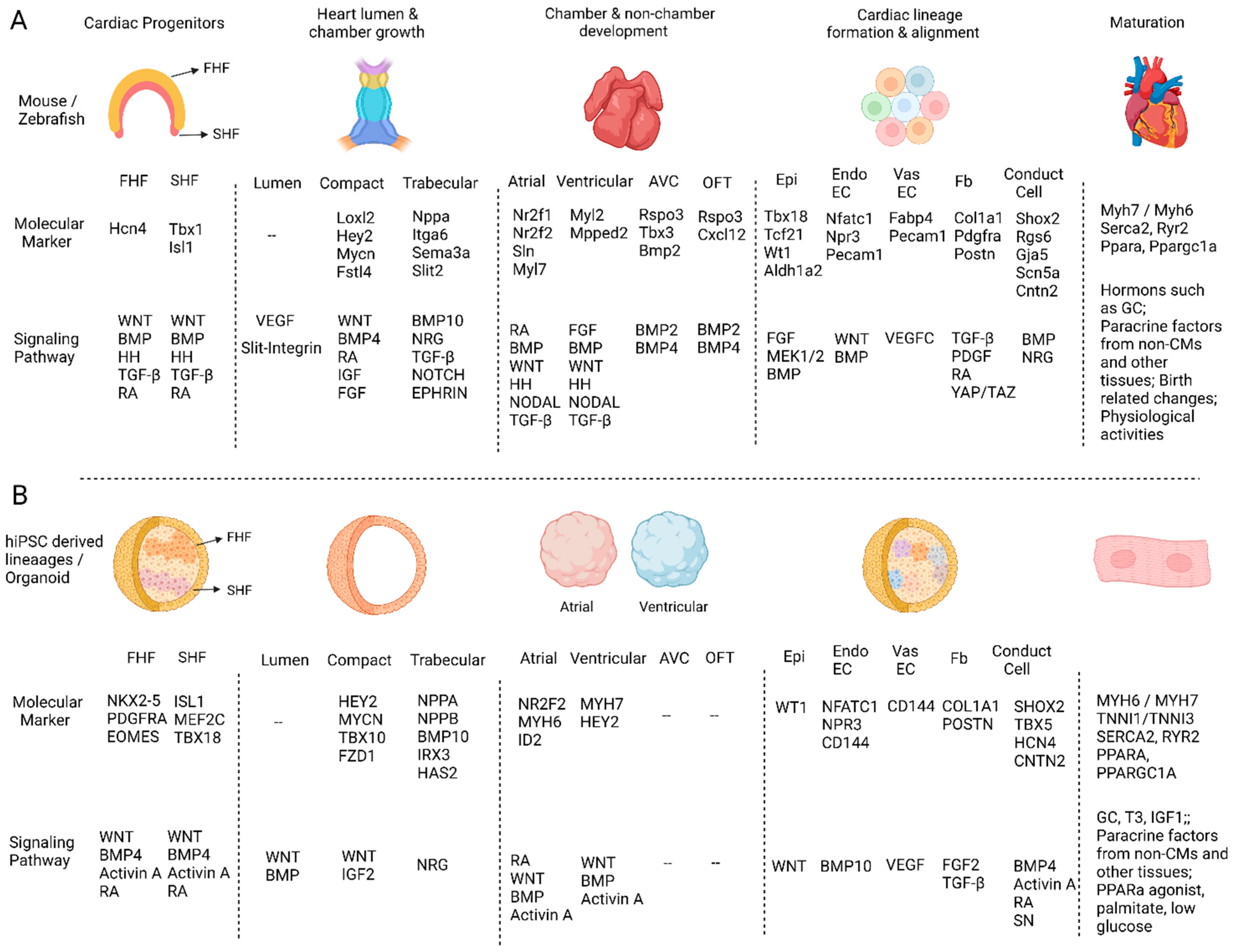
During mouse heart development, the two heart fields are specified sequentially and express marker genes such as Hcn4 (FHF), Isl1, and Tbx1 (SHF) [12][13]. The FHF originates from the lateral plate mesoderm and mainly contributes to linear heart-tube formation. FHF formation is induced by signals from the adjacent ectoderm, endoderm, embryonic midline, and posterior regions. Bone morphogenetic protein (BMP), Fibroblast growth factor (FGF), Transforming growth factor β (TGFβ), and WNT signaling pathways were reported to be involved in this process [14][15]. The SHF is derived from the pharyngeal mesoderm and contributes to heart development from both inflow and outflow poles after the linear heart-tube stage. Additionally, the SHF is specified by signals from the surrounding pharyngeal endoderm and neural crest cells. While SHF progenitor cell survival, proliferation, and deployment were found to be dependent upon WNT, BMP, Hedgehog (HH), and FGF signaling [15][16], the posterior limit of SHF was reported to be determined by retinoic acid (RA) [17]. Furthermore, TGF-β, HH, and FGF were reported to be important in zebrafish SHF progenitor cell proliferation and differentiation [18] (Figure 1A).
Jcdd 09 00125 g001
Figure 1. The comparison of molecular markers and regulatory signaling pathways in in vivo heart and human heart organoid development. (A) The major events during in vivo heart development and their molecular markers and regulatory signaling pathways. (B) The molecular markers and signaling pathways that have been reported to be important in regulating human iPSC-derived cardiac cell or cardiac organoid differentiations. “--” means non-applicable or no related studies have been reported.
The FHF and SHF were also discovered to develop in mouse 3D culture systems. Andersen et al. verified FHF/SHF-like cells in their mouse iPSC-derived organoids by showing similarities with embryonic FHF/SHF cells in their gene expression and differentiation potentials. They also found that the Bmp/Smad pathway and the Smad-independent BMP/WNT pathway specified FHF and SHF progenitors [12], respectively. Recently, Rossi et al. described mouse embryonic stem cells (mESCs)-derived gastruloids that were found to have cells expressing the FHF/SHF markers and had a spatial distribution like the in vivo progenitors [19]. Additionally, Lewis-Israeli et al. generated heart organoids from hiPSCs presenting characteristics of both heart fields in the same organoid. They identified NKX2-5, PDGFRA, and EOMES expression in the FHF progenitors and ISL1, MEF2C, and TBX18 expression in SHF cells (Figure 1B). These progenitors were described to respectively develop into left and right ventricular CMs based on the expression of HAND1 and HAND2 [20].
Next, it will be interesting to analyze the detailed structures in the organoid heart fields, as the FHF and SHF in mouse embryos are specified into small segments through the differential expression of HOX genes and RA signaling, and each segment respectively develops into related heart anatomical structures such as atrial and ventricular chambers [21]. Furthermore, these organoid heart fields do not seem to differentiate sequentially or form the specific spatial patterns seen in the heart fields in embryos, whose SHF locates dorsal and medial to the FHF to progressively contribute cells to both poles of the linear heart tube [10]. With further detailed analysis and appropriate manipulations, researchers expect the organoid heart fields to develop in a similar temporal and spatial manner as seen in embryonic heart field development.

3. Heart Lumen Development

Through genetic screening in Drosophila, the heart tube lumen formation was found to be regulated by a Slit-Integrin signaling pathway, which regulates actin cytoskeleton alignment to promote cardiac cell polarization in lumen development [22][23]. In mice, live imaging analysis revealed that the heart lumen developed from a split of two endocardial endothelial cell (EndoEC) layers at the cardiac crescent stage. It also found that cell rounding was unlikely to initiate lumen formation as the cardiac crescent cells are still columnar when the lumen begins to develop [24]. As part of the heart lumen developmental process, aorta lumen propagation initiates between stages 1S and 3S (E8.0), developing from adjacent endothelial cell (EC) contact after EC shape has changed. This process is regulated by VE-Cadherin and VEGF-A [25]. In humans, the heart lumen develops from the fusion of two endocardial tubes, each of which has a hollow lumen derived from the cardiogenic cords [26].
A recent study reported the induction of heart chamber-like structures in human heart organoids and found that Wnt-BMP signaling and transcription factor HAND1 were both critical in this process [27]. Further time-course analysis of organoid formation found that the lumen appeared after 2.5–3.5 days of differentiation at the cardiac mesoderm stage, which is earlier than when the mouse heart lumen develops in the cardiac crescent stage. Additionally, the study found that low WNT and Activin A levels can induce chamber formation with a partial inner lining of EndoECs. However, lumen formation does not seem to rely on the EndoECs, as the lumen can still form when the EndoECs developed on the outer organoid surface after VEGF treatment [27]. Lewis-Israeli et al. and the study also generated organoids with chamber-like structures, and these chambers also developed independently from the EndoECs [20][28]. As chamber formation in the current organoids does not go through the same process as in vivo heart lumen development, there is limited value in modeling human heart lumen formation under normal and diseased conditions using organoid cultures. However, heart organoids may still be valuable in studying other aspects of heart chamber development, such as heart pumping and looping.

4. Compact and Trabecular Myocardium Growth

Heart chamber growth was thought to balloon out from the looped hearts segmentally. The ventricular and atrial chambers were found to respectively expand from the linear heart tube on ventral and dorsal sides [29][30] and the expanded chambers to develop into two types of myocardium, with the compact myocardium on the outer surface and trabecular myocardium close to the lumen to increase cardiac output and oxygen uptake at early embryonic stages [31][32]. The CMs in compact myocardium highly express Loxl2, Hey2, Mycn, and Fstl4, while the CMs in trabecular myocardium express Nppa, Itga6, Sema3a, and Slit2 [32]. Compact and trabecular myocardium development was shown to be differentially regulated by signals from the epicardium and the endocardium [33][34][35]. Epicardium-derived signals such as BMP4, FGF, WNT, IGF, and RA were reported to promote CM proliferation in compact myocardium [35][36][37], and endocardium signals such as NOTCH, Neuregulin, Ephrin, and TGF-β were reported to promote trabecular myocardium development [31][38][39]. Furthermore, some signaling molecules expressed in the myocardium, such as BMP10, were also found to regulate the trabecular myocardium development [40] (Figure 1A).
To generate compact CMs from hiPSCs, WNT and IGF2 were added to the ventricular CM differentiation system on day 10 [41]. The CMs were shown to express typical compact myocardium marker genes such as HEY2, MYCN, TBX10, and FZD1. In contrast, the addition of Neuregulin to the differentiation system at day 10 can specify CMs into trabecular CMs expressing trabecular myocardium genes such as NPPA, NPPB, BMP10, IRX3, and HAS2 (Figure 1B). Similarly, the co-culture of EndoEC with CMs can promote the development of trabecular CMs, as EndoECs were known to be able to secrete Neuregulin in mice and zebrafish [42]. Next, it will be interesting to test if other EndoEC-derived growth factors such as TGF-β and NOTCH can also induce trabecular CM fate and if epicardium-derived factors such as RA, BMP, WNT, and FGF can promote compact CM development. Additionally, and most importantly, a test will be needed to determine if these factors can be applied locally to generate heart organoids with compact and trabecular myocardium at correct anatomical locations.

5. Heart Structure Development

The early stages of mouse heart development consist of the formation of the four chambers (left and right atrial; left and right ventricular) and two non-chamber structures—the atrial ventricular canal (AVC) and the outflow tract (OFT). While AVC at later developmental stages contributes to the development of the septum and atrioventricular valves, including the tricuspid and mitral valves, the OFT contributes to the formation of large vessels (aorta and pulmonary artery) and the semilunar valves, including the aortic and pulmonary valves [43]. The atrial CMs highly express Nr2f1, Nr2f2, Sln, and Myl7, while the ventricular CMs express Myl2 and Mpped2. Furthermore, while the left and right ventricular CMs differentially express Pcsk6, the left and right atrial CMs highly express Pitx2 and Shox2, respectively. In contrast, the AVC CMs express Rspo3, Tbx3, and Bmp2, and the OFT CMs express Rspo3 and Cxcl12 [32][44] (Figure 1A).
Atrial lineage specification is regulated by RA signaling in multiple species [45][46][47], while early dorsal-ventral patterning signals such as FGF and BMP also differentially promote atrial and ventricular lineage development in zebrafish [48]. As the left and right ventricular CMs develop from different heart fields, their lineage formation is primarily regulated by the heart field specification signals previously mentioned when discussing heart fields’ formation. The AVC and OFT share a structure named the endocardial cushion, which is induced by the interaction of BMP signaling, including BMP2 and BMP4 in myocardium and BMPR1A in EndoECs. Mouse endocardial cushion cells express marker genes such as Twist1, Msx1, and Snail. Endocardial cushion cells need to go through an endothelial-to-mesenchymal transition (EndoMT) process regulated by multiple signaling pathways, such as TGFβ, WNT/β-catenin, HIPPO, and NOTCH, to develop into valve cells [49].
Atrial and Ventricular CMs were found to co-exist in heart organoids but did not display in vivo-like spatial domains. RA signaling had been used to promote atrial CM lineage in monolayer and EB-based hiPSC differentiation, and the atrial and ventricular CM progenitors were distinguished based on the expression of CD235A and RALDH2 [50][51]. Researchers have also generated heart organoids with atrial or ventricular identities by adding (+) or omitting (−) RA at the cardiac mesoderm stage and found that the RA+ and RA- heart organoids had distinct membrane action potentials and Ca2+ transient activities. The chamber identity of these cells was further confirmed with immunofluorescence staining for chamber-specific marker genes such as MYH7, HEY2 (ventricular), NR2F2, MYH6, and ID2 (atrial) (Figure 1B). Researchers also performed single-cell mRNA sequencing (scRNA-seq) and random forest-based zone classification to analyze their cell identities systematically [28]. These analyses consistently support that the CMs in RA- heart organoids preferentially develop into ventricular CMs, while the CMs in RA+ organoids are more likely to develop into atrial CMs [28].
Interestingly, MYL2 is a robust ventricular CM marker gene in human fetal hearts but is barely expressed in the organoid ventricular CMs differentiated from the hiPSC line “WTC”. Considering that WTC and its derived transgenic lines have been broadly used in the cardiac stem cell field, researchers have investigated MYL2 expression in WTC-derived CMs from multiple labs based on their scRNA-seq results. They found that while MYL2 was barely detected in the WTC-derived CMs before differentiation day 30 in several studies, it was expressed in other hiPSC line-derived CMs with the same differentiation conditions [52][53][54]. However, there are some exceptions where MYL2 was found to be expressed in the WTC-derived CMs on day 30 in one study and day 90 in another. Both studies generated the CMs using a monolayer with small molecules protocol [53][55]. The cause of the differences in gene expression across the studies is still a mystery, but it will be important to investigate whether this expression variation also exists in other genes and other cell lines.

This entry is adapted from the peer-reviewed paper 10.3390/jcdd9050125

References

  1. Mummery, C.L.; Zhang, J.; Ng, E.S.; Elliott, D.A.; Elefanty, A.G.; Kamp, T.J. Differentiation of human embryonic stem cells and induced pluripotent stem cells to cardiomyocytes: A methods overview. Circ. Res. 2012, 111, 344–358.
  2. van den Brink, S.C.; Alemany, A.; van Batenburg, V.; Moris, N.; Blotenburg, M.; Vivié, J.; Baillie-Johnson, P.; Nichols, J.; Sonnen, K.F.; Martinez Arias, A. Single-cell and spatial transcriptomics reveal somitogenesis in gastruloids. Nature 2020, 582, 405–409.
  3. Aguilera-Castrejon, A.; Oldak, B.; Shani, T.; Ghanem, N.; Itzkovich, C.; Slomovich, S.; Tarazi, S.; Bayerl, J.; Chugaeva, V.; Ayyash, M. Ex utero mouse embryogenesis from pre-gastrulation to late organogenesis. Nature 2021, 593, 119–124.
  4. Simunovic, M.; Brivanlou, A.H. Embryoids, organoids and gastruloids: New approaches to understanding embryogenesis. Development 2017, 144, 976–985.
  5. Sutherland, R.M.; McCredie, J.A.; Inch, W.R. Growth of multicell spheroids in tissue culture as a model of nodular carcinomas. J. Natl. Cancer Inst. 1971, 46, 113–120.
  6. Lancaster, M.A.; Knoblich, J.A. Organogenesis in a dish: Modeling development and disease using organoid technologies. Science 2014, 345, 1247125.
  7. Sakalem, M.E.; De Sibio, M.T.; da Costa, F.A.d.S.; de Oliveira, M. Historical evolution of spheroids and organoids, and possibilities of use in life sciences and medicine. Biotechnol. J. 2021, 16, 2000463.
  8. Gilboa, S.M.; Devine, O.J.; Kucik, J.E.; Oster, M.E.; Riehle-Colarusso, T.; Nembhard, W.N.; Xu, P.; Correa, A.; Jenkins, K.; Marelli, A.J. Congenital heart defects in the United States: Estimating the magnitude of the affected population in 2010. Circulation 2016, 134, 101–109.
  9. Zou, Y.; Evans, S.; Chen, J.; Kuo, H.-C.; Harvey, R.P.; Chien, K.R. CARP, a cardiac ankyrin repeat protein, is downstream in the Nkx2-5 homeobox gene pathway. Development 1997, 124, 793–804.
  10. Buckingham, M.; Meilhac, S.; Zaffran, S. Building the mammalian heart from two sources of myocardial cells. Nat. Rev. Genet. 2005, 6, 826–835.
  11. Liu, X.; Yagi, H.; Saeed, S.; Bais, A.S.; Gabriel, G.C.; Chen, Z.; Peterson, K.A.; Li, Y.; Schwartz, M.C.; Reynolds, W.T. The complex genetics of hypoplastic left heart syndrome. Nat. Genet. 2017, 49, 1152–1159.
  12. Andersen, P.; Tampakakis, E.; Jimenez, D.V.; Kannan, S.; Miyamoto, M.; Shin, H.K.; Saberi, A.; Murphy, S.; Sulistio, E.; Chelko, S.P. Precardiac organoids form two heart fields via Bmp/Wnt signaling. Nat. Commun. 2018, 9, 3140.
  13. Cai, C.-L.; Liang, X.; Shi, Y.; Chu, P.-H.; Pfaff, S.L.; Chen, J.; Evans, S. Isl1 identifies a cardiac progenitor population that proliferates prior to differentiation and contributes a majority of cells to the heart. Dev. Cell 2003, 5, 877–889.
  14. Azhar, M.; Schultz, J.E.J.; Grupp, I.; Dorn, I.I.G.W.; Meneton, P.; Molin, D.G.; Gittenberger-de Groot, A.C.; Doetschman, T. Transforming growth factor beta in cardiovascular development and function. Cytokine Growth Factor Rev. 2003, 14, 391–407.
  15. Kelly, R.G.; Buckingham, M.E.; Moorman, A.F. Heart fields and cardiac morphogenesis. Cold Spring Harb. Perspect. Med. 2014, 4, a015750.
  16. Rochais, F.; Mesbah, K.; Kelly, R.G. Signaling pathways controlling second heart field development. Circ. Res. 2009, 104, 933–942.
  17. Ryckebusch, L.; Wang, Z.; Bertrand, N.; Lin, S.-C.; Chi, X.; Schwartz, R.; Zaffran, S.; Niederreither, K. Retinoic acid deficiency alters second heart field formation. Proc. Natl. Acad. Sci. USA 2008, 105, 2913–2918.
  18. Knight, H.; Yelon, D. Utilizing zebrafish to understand second heart field development. Etiol. Morphog. Congenit. Heart Dis. 2016, 25, 193–199.
  19. Rossi, G.; Broguiere, N.; Miyamoto, M.; Boni, A.; Guiet, R.; Girgin, M.; Kelly, R.G.; Kwon, C.; Lutolf, M.P. Capturing cardiogenesis in gastruloids. Cell Stem Cell 2021, 28, 230–240.e6.
  20. Lewis-Israeli, Y.R.; Wasserman, A.H.; Gabalski, M.A.; Volmert, B.D.; Ming, Y.; Ball, K.A.; Yang, W.; Zou, J.; Ni, G.; Pajares, N. Self-assembling human heart organoids for the modeling of cardiac development and congenital heart disease. Nat. Commun. 2021, 12, 5142.
  21. Bertrand, N.; Roux, M.; Ryckebüsch, L.; Niederreither, K.; Dollé, P.; Moon, A.; Capecchi, M.; Zaffran, S. Hox genes define distinct progenitor sub-domains within the second heart field. Dev. Biol. 2011, 353, 266–274.
  22. Jammrath, J.; Reim, I.; Saumweber, H. Cbl-Associated Protein CAP contributes to correct formation and robust function of the Drosophila heart tube. PLoS ONE 2020, 15, e0233719.
  23. Knox, J.; Moyer, K.; Yacoub, N.; Soldaat, C.; Komosa, M.; Vassilieva, K.; Wilk, R.; Hu, J.; Paz, L.d.L.V.; Syed, Q. Syndecan contributes to heart cell specification and lumen formation during Drosophila cardiogenesis. Dev. Biol. 2011, 356, 279–290.
  24. Ivanovitch, K.; Temiño, S.; Torres, M. Live imaging of heart tube development in mouse reveals alternating phases of cardiac differentiation and morphogenesis. eLife 2017, 6, e30668.
  25. Strilić, B.; Kučera, T.; Eglinger, J.; Hughes, M.R.; McNagny, K.M.; Tsukita, S.; Dejana, E.; Ferrara, N.; Lammert, E. The molecular basis of vascular lumen formation in the developing mouse aorta. Dev. Cell 2009, 17, 505–515.
  26. Sydelko, B.S. Anatomy & Physiology Online. J. Med. Libr. Assoc. JMLA 2013, 101, 163.
  27. Hofbauer, P.; Jahnel, S.M.; Papai, N.; Giesshammer, M.; Deyett, A.; Schmidt, C.; Penc, M.; Tavernini, K.; Grdseloff, N.; Meledeth, C. Cardioids reveal self-organizing principles of human cardiogenesis. Cell 2021, 184, 3299–3317.e22.
  28. Feng, W.; Schriever, H.; Jiang, S.; Bais, A.; Kostka, D.; Li, G. Computational profiling of hiPSC-derived heart organoids reveals chamber defects associated with Ebstein’s anomaly. bioRxiv 2020.
  29. Moorman, A.F.; Christoffels, V.M. Cardiac chamber formation: Development, genes, and evolution. Physiol. Rev. 2003, 83, 1223–1267.
  30. Schleich, J.-M.; Abdulla, T.; Summers, R.; Houyel, L. An overview of cardiac morphogenesis. Arch. Cardiovasc. Dis. 2013, 106, 612–623.
  31. Luxán, G.; D’Amato, G.; MacGrogan, D.; de la Pompa, J.L. Endocardial notch signaling in cardiac development and disease. Circ. Res. 2016, 118, e1–e18.
  32. Li, G.; Xu, A.; Sim, S.; Priest, J.R.; Tian, X.; Khan, T.; Quertermous, T.; Zhou, B.; Tsao, P.S.; Quake, S.R. Transcriptomic profiling maps anatomically patterned subpopulations among single embryonic cardiac cells. Dev. Cell 2016, 39, 491–507.
  33. Li, G.; Tian, L.; Goodyer, W.; Kort, E.J.; Buikema, J.W.; Xu, A.; Wu, J.C.; Jovinge, S.; Wu, S.M. Single cell expression analysis reveals anatomical and cell cycle-dependent transcriptional shifts during heart development. Development 2019, 146, dev173476.
  34. Smith, T.K.; Bader, D.M. Signals from both sides: Control of cardiac development by the endocardium and epicardium. Semin. Cell Dev. Biol. 2007, 18, 84–89.
  35. Kang, J.-O.; Sucov, H.M. Convergent proliferative response and divergent morphogenic pathways induced by epicardial and endocardial signaling in fetal heart development. Mech. Dev. 2005, 122, 57–65.
  36. Díaz del Moral, S.; Benaouicha, M.; Muñoz-Chápuli, R.; Carmona, R. The Insulin-like Growth Factor Signalling Pathway in Cardiac Development and Regeneration. Int. J. Mol. Sci. 2022, 23, 234.
  37. Buikema, J.W.; Mady, A.S.; Mittal, N.V.; Atmanli, A.; Caron, L.; Doevendans, P.A.; Sluijter, J.P.; Domian, I.J. Wnt/β-catenin signaling directs the regional expansion of first and second heart field-derived ventricular cardiomyocytes. Development 2013, 140, 4165–4176.
  38. Bruneau, B.G. Signaling and transcriptional networks in heart development and regeneration. Cold Spring Harb. Perspect. Biol. 2013, 5, a008292.
  39. Kodo, K.O.S.; Jahanbani, F.; Termglinchan, V.; Hirono, K.; InanlooRahatloo, K.; Ebert, A.D.; Shukla, P.; Abilez, O.J.; Churko, J.M.; Karakikes, I. iPSC-derived cardiomyocytes reveal abnormal TGF-β signalling in left ventricular non-compaction cardiomyopathy. Nat. Cell Biol. 2016, 18, 1031–1042.
  40. Chen, H.; Shi, S.; Acosta, L.; Li, W.; Lu, J.; Bao, S.; Chen, Z.; Yang, Z.; Schneider, M.D.; Chien, K.R. BMP10 is essential for maintaining cardiac growth during murine cardiogenesis. Development 2004, 131, 2219–2231.
  41. Funakoshi, S.; Fernandes, I.; Mastikhina, O.; Wilkinson, D.; Tran, T.; Dhahri, W.; Mazine, A.; Yang, D.; Burnett, B.; Lee, J. Generation of mature compact ventricular cardiomyocytes from human pluripotent stem cells. Nat. Commun. 2021, 12, 3155.
  42. Mikryukov, A.A.; Mazine, A.; Wei, B.; Yang, D.; Miao, Y.; Gu, M.; Keller, G.M. BMP10 signaling promotes the development of endocardial cells from human pluripotent stem cell-derived cardiovascular progenitors. Cell Stem Cell 2021, 28, 96–111.e7.
  43. Lin, C.-J.; Lin, C.-Y.; Chen, C.-H.; Zhou, B.; Chang, C.-P. Partitioning the heart: Mechanisms of cardiac septation and valve development. Development 2012, 139, 3277–3299.
  44. Consortium, T.M. Single-cell transcriptomics of 20 mouse organs creates a Tabula Muris. Nature 2018, 562, 367–372.
  45. Hochgreb, T.; Linhares, V.L.; Menezes, D.C.; Sampaio, A.C.; Yan, C.Y.; Cardoso, W.V.; Rosenthal, N.; Xavier-Neto, J. A caudorostral wave of RALDH2 conveys anteroposterior information to the cardiac field. Development 2003, 130, 5363–5374.
  46. Xavier-Neto, J.; Neville, C.M.; Shapiro, M.D.; Houghton, L.; Wang, G.F.; Nikovits, W.; Stockdale, F.E.; Rosenthal, N. A retinoic acid-inducible transgenic marker of sino-atrial development in the mouse heart. Development 1999, 126, 2677–2687.
  47. Yutzey, K.E.; Gannon, M.; Bader, D. Diversification of cardiomyogenic cell lineages in vitro. Dev. Biol. 1995, 170, 531–541.
  48. Martin, K.E.; Waxman, J.S. Atrial and sinoatrial node development in the zebrafish heart. J. Cardiovasc. Dev. Dis. 2021, 8, 15.
  49. O’Donnell, A.; Yutzey, K.E. Mechanisms of heart valve development and disease. Development 2020, 147, dev183020.
  50. Lee, J.H.; Protze, S.I.; Laksman, Z.; Backx, P.H.; Keller, G.M. Human pluripotent stem cell-derived atrial and ventricular cardiomyocytes develop from distinct mesoderm populations. Cell Stem Cell 2017, 21, 179–194.e4.
  51. Cyganek, L.; Tiburcy, M.; Sekeres, K.; Gerstenberg, K.; Bohnenberger, H.; Lenz, C.; Henze, S.; Stauske, M.; Salinas, G.; Zimmermann, W.-H. Deep phenotyping of human induced pluripotent stem cell–derived atrial and ventricular cardiomyocytes. JCI Insight 2018, 3, e99941.
  52. Friedman, C.E.; Nguyen, Q.; Lukowski, S.W.; Helfer, A.; Chiu, H.S.; Miklas, J.; Levy, S.; Suo, S.; Han, J.-D.J.; Osteil, P. Single-cell transcriptomic analysis of cardiac differentiation from human PSCs reveals HOPX-dependent cardiomyocyte maturation. Cell Stem Cell 2018, 23, 586–598.e8.
  53. Grancharova, T.; Gerbin, K.A.; Rosenberg, A.B.; Roco, C.M.; Arakaki, J.E.; DeLizo, C.M.; Dinh, S.Q.; Donovan-Maiye, R.M.; Hirano, M.; Nelson, A.M. A comprehensive analysis of gene expression changes in a high replicate and open-source dataset of differentiating hiPSC-derived cardiomyocytes. Sci. Rep. 2021, 11, 15845.
  54. Kathiriya, I.S.; Rao, K.S.; Iacono, G.; Devine, W.P.; Blair, A.P.; Hota, S.K.; Lai, M.H.; Garay, B.I.; Thomas, R.; Gong, H.Z. Modeling human TBX5 haploinsufficiency predicts regulatory networks for congenital heart disease. Dev. Cell 2021, 56, 292–309.e9.
  55. Paige, S.L.; Galdos, F.X.; Lee, S.; Chin, E.T.; Ranjbarvaziri, S.; Feyen, D.A.; Darsha, A.K.; Xu, S.; Ryan, J.A.; Beck, A.L. Patient-specific induced pluripotent stem cells implicate intrinsic impaired contractility in hypoplastic left heart syndrome. Circulation 2020, 142, 1605–1608.
More
This entry is offline, you can click here to edit this entry!
ScholarVision Creations