Nucleolar and Ribosomal Dysfunction: History
Please note this is an old version of this entry, which may differ significantly from the current revision.
Contributor: ,

Ageing is a complex and unavoidable process that can be defined as the functional decline after a period of maturity. In this respect, maturity is understood as the endpoint of development and the condition of maximal functional performance capability.  The nucleolus organizes around the sites of transcription by RNA polymerase I (RNA Pol I). rDNA transcription by this enzyme is the key step of ribosome biogenesis and most of the assembly and maturation processes of the ribosome occur co-transcriptionally. Therefore, disturbances in rRNA transcription and processing translate to ribosomal malfunction. Nucleolar malfunction has recently been described in the classical progeria of childhood, Hutchinson–Gilford syndrome (HGPS), which is characterized by severe signs of premature aging, including atherosclerosis, alopecia, and osteoporosis. A deregulated ribosomal biogenesis with enlarged nucleoli is not only characteristic for HGPS patients, but it is also found in the fibroblasts of “normal” aging individuals. Cockayne syndrome (CS) is also characterized by signs of premature aging, including the loss of subcutaneous fat, alopecia, and cataracts. It has been shown that all genes in which a mutation causes CS, are involved in rDNA transcription by RNA Pol I. 

  • nucleolus
  • aging
  • RNA Pol I
  • ribosome
  • loss of proteostasis
  • neurodegeneration

1. Aging and the Nucleolus

One question is whether the nucleolus, the site of ribosomal transcription and synthesis, is involved in the aging pathomechanisms or even a driver of age-associated physiological decline. The nucleolus is a non-membrane-bound organelle that organizes around the sites of rDNA transcription by RNA polymerase I (RNA Pol I) and the co-transcriptional pre-ribosomal assembly and processing [1]. The nucleolus is a dynamic organelle that dis- and reassembles during the cell cycle and contains a multitude of proteins involved in ribosomal biogenesis, cell cycle regulation, and stress responses [2][3].
In yeast cells, it has been shown that the redistribution of the silence information regulator, Sir2, from the telomeres to an AGE locus prolongs lifespan. The nucleolus has been identified as the AGE locus [4] and the accumulation of extrachromosomal rDNA circles resulting in nucleolar enlargement and fragmentation is one cause of aging in yeast [5]. This aging mechanism could not be confirmed in mammals, however the nucleolus attracted the attention of the aging research community.
The size of the nucleolus, during early microscopy, was recognized as, and is still valid as, a prognostic factor in tumor pathology and an indicator of the activity of the ribosomal synthesis machinery [6]. The nucleolar size, the activity of ribosomal biogenesis, and subsequent protein synthesis inversely correlate with lifespan in model organisms like Caenorhabditis elegans [7]. This implies that individuals with a high protein synthesis rate and turnover as well as large nucleoli might age more rapidly than individuals with a slow ribosome synthesis/protein synthesis activity and small nucleoli. This observation is further supported by the fact that inhibition of a central sensor/regulator of nutrient availability, mammalian target of rapamycin (mTOR), also regulates nucleolar size and activity and inhibition of mTOR by rapamycin which results in a reduced nucleolar size, alleviates age-related pathology, and even extends the lifespan of model organisms [8]. The metabolic theory of aging states that the metabolic rate of an organism, which is also mirrored in nucleolar size and activity, is one denominator of the aging process and lifespan [9][10]. Therefore, the nucleolar size and activity reflect metabolic conditions, and thereby the rate of cell and tissue aging, however, whether this is mere correlation or a causal association is currently not established. Consequently, the investigation of the pathomechanisms of progerias may help to determine whether the nucleolus is a downstream effector or a causal player in the chain of events that leads to the demise of the cellular, tissue, and organismal integrity during the aging process.

2. Nucleolar Size as a Hallmark of Aging

A study in C. elegans by the Antebi group [11] identified the nucleolus as a central effector in longevity pathways. Asking whether all lifespan-extending signaling pathways converge on the same downstream process, they discovered that nucleolar size and function are predictive markers of life expectancy. Starting from the identification of nucleolin (NCL1) as a downstream effector of caloric-restriction mediated lifespan extension, they demonstrated that NCL1 knockdown abrogated all the tested life-extending manipulations. NCL1 is a protein that inhibits rRNA transcription and protein synthesis [12]. Knockdown of ncl-1 in C. elegans led to enlarged nucleoli in several tissues [13]. Knockdown of ncl-1 blocked the lifespan-extending effects of the manipulations of a genetic caloric-restriction model on the TOR pathway, the insulin/insulin-like growth factor pathway, the germline-less mutants, the mitochondrial effectors, the translation-initiation factors, and the ribosomal S6kinase. Overexpression of ncl-1 reduced nucleolar size and increased lifespan. All lifespan-extending models exhibited small nucleoli and the size of the nucleoli in wild-type C. elegans was a predictor of life expectancy. Reduced nucleolar size was associated with a reduced ribosomal RNA and protein content, suggesting that the downregulation of ribosomal synthesis is an indicator of long life. Finally, the authors analyzed nucleolar size in long-lived Drosophila melanogaster and observed reduced nucleolar size in these insects as well as in muscle biopsies of dietary-restricted, exercising humans. Thereby, identified nucleolin and its nucleolar function as a central player in lifespan regulation and is in agreement with the metabolic theory of aging which proposes that interventions that dampen high-energy demanding processes, including ribosomal biogenesis and translation, prolong the lifespan. Moreover, the findings that small nucleoli are related to longevity [11] and large nucleoli to premature aging [14], could, when confirmed in larger human cohorts, be used as predictive parameters of metabolic health, biological aging, and life expectancy.

3. Cockayne Syndrome (CS)

The CS is a rare autosomal recessive disease that can be caused by six different genes, which are all involved in nucleotide excision repair (NER) of UV lesions [15]. It is characterized by a high skin UV sensitivity and severe developmental and degenerative disturbances with a mean life expectancy of 12 years [16][17]. CS patients present with a failure to thrive and, depending on the severity, degeneration of different organ systems, including the loss of subcutaneous fat tissue (“cachectic dwarfs”), alopecia, cataracts, and neurodegeneration. Neurodegeneration affects the peripheral system (hearing loss) as well as the central nervous system, with intellectual, gait, and speech impairments. Most of the symptoms found in this syndrome are normally associated with advanced age (cachexia, alopecia, cataracts, neurodegeneration), thus the CS presents as a progeroid disease. The CS is of segmental nature, because it does not display an elevated cancer incidence that is typical for xeroderma pigmentosum (XP), a high cancer-prone skin disease that is also caused by mutations in NER genes. Because mutations in the NER factors, which completely inactivate NER, are not followed by childhood degeneration but rather by XP, which is not characterized by a severe developmental failure [18], alternative explanations for the pathogenesis of premature aging are discussed [19]. One particular cellular feature that distinguishes the CS cells from cells of non-progeroid NER syndromes is the hypersensitivity to oxidizing agents. The CS cells undergo apoptosis when challenged with low doses of reactive oxygen species (ROS), interpreted as a consequence of non-repaired oxidative DNA damage [20][21]. If this is the case, then knockdown of the enzymes that repair oxidative DNA-damage should provoke a CS-phenotype in mice. However, the knockdown of base-excision repair glycosylases [22], even in combination with the protein responsible for most CS cases, i.e., Cockayne syndrome B protein (CSB), does not result in premature aging [23].
The CS phenotype in humans is induced by mutations in the Cockayne syndrome B protein (CSB) (70%), the Cockayne syndrome A protein (CSA) (20%), the subunits of the transcription/DNA repair factor TFIIH, XPB and XPD, and XPG and XPF, and two NER proteins. All of these proteins play central roles in the repair of helix-distorting lesions in DNA provoked by UV light. The total failure of this DNA repair pathway results in the cancer-prone skin disease XP that is characterized by the development of different cancers on UV-exposed skin. Early diagnosis and consequent UV protection reduces skin-cancer risk and allows almost normal development of XP children [24].
The CS cases that do not suffer from a combination with XP are cancer free, indicating that developmental impairment and premature aging are not caused by non-repaired mutagenic UV lesions, although CS patients are frequently highly UV sensitive. The same type of UV sensitivity can also be found in UV-sensitivity syndrome (UVsS), a rarely diagnosed hypersensitivity of the skin to UV light [25][26]. UVsS can be caused by mutations in the CS factors CSA or CSB, but does not display the severe phenotype of CS. The cells from UVsS patients display the same DNA-repair defect as the CS cells [27], thus the DNA-repair defect cannot be responsible for the severe phenotype of CS patients. One difference between the UVsS and CS cells is that the latter are hypersensitive to oxidizing agents, and thereby undergo apoptosis when challenged with elevated ROS [26]. The hypersensitivity to oxidation also differentiates the combined XP/CS patient cells from the XP patient cells, indicating that this feature might explain the severe developmental and premature aging symptoms found in CS [28]. The NER proteins are involved in the repair of oxidative DNA lesions [29], however, an oxidative DNA-repair pathway that is specific for CS but not for XP has currently not been identified [19]. Either DNA [20], proteins [30] or lipids [29] might be oxidized and cause the chain of events that result in childhood degeneration and premature aging. Therefore, alternative hypotheses for the premature aging phenotype of CS are discussed [19][30]. One striking feature of all CS-proteins is that they are involved in the rate-limiting step of ribosomal biogenesis, transcription by RNA Pol I in the nucleolus [31][32][33][34][35].

4. Loss of RNA Pol I Transcription Leads to Mitochondrial Dysfunction

In a series of knockdown experiments, Scheibye-Knudsen et al. demonstrated that there is a close association between ribosomal transcription by RNA Pol I and mitochondrial function [36]. Loss of the CS proteins, CSA or CSB, inhibited rRNA transcription in neuroblastoma cell lines and subsequent microarray analyses revealed a pronounced upregulation of translational and mitochondrial pathways. The specific inhibition of RNA Pol I transcription activity in different cell lines markedly raised mitochondrial membrane potential and superoxide production dependent on poly-ADP ribose polymerase 1 (PARP1). Ruling out that the mitochondrial phenotype is a consequence of translational deficiencies, the authors showed that transcriptional stalling at G4 quadruplex structures is enhanced in the absence of CSA or CSB and that CSB is able to resolve these transcription-obstacles. Interestingly, CSB overexpression in CSA-mutant patient cells could overcome mitochondrial phenotype and PARP1 activation. By avoiding replication effects through the usage of non-dividing cells, the authors demonstrated that treatment with the G4-stabilizing drug pyridostatin, or the RNA Pol I inhibitor CX5461, led to the PARP1 activation. This was followed by the loss of NAD+ and an increase of nicotinamide. Reassessing this chain of events in C. elegans, the authors demonstrated that inhibition of RNA Pol I transcription activity and stabilization of G4 quadruplex structures lead to accelerated aging and a shortened lifespan that could partially be rescued by a NAD+ precursor. Therefore, premature aging in CS is described as a consequence of a failure in the resolution of secondary DNA structures that are predominantly localized in the rDNA.

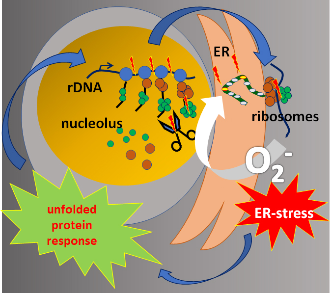
5. Loss of Proteostasis in CS

In considering the cellular consequences of a disturbance in RNA Pol I transcription, as described in the publications above, Alupei et al. investigated ribosomal function and cellular signaling in CS-patient cells in comparison to reconstituted and UVsS cells [30]. The authors revealed a circulus vitiosus originating from a disturbed ribosomal transcription leading to the repression of this vital pathway as depicted in Figure 1. A reduced rate of the 47S pre-rRNA transcription, indicating disturbed RNA Pol I transcription, and a decreased protein synthesis were detected in CSA- and CSB-mutated CS cells. Interestingly the number of ribosomes was not reduced, suggesting a disturbance in the translation process. Indeed, they found increased translation infidelity in both CSA- and CSB-mutated cells, indicating a malfunction of the ribosomes. The translation accuracy of the ribosome was analyzed by transfection experiments using a mutant luciferase plasmid with a point mutation inactivating the enzyme [37]. With erroneous incorporation of the correct amino acid, the luciferase activity was restored, whereas with accurate translation, the point mutation was translated, and the enzyme remained inactive. Increased luminescence, and thus increased translational infidelity, were observed in CSA- and CSB-mutated CS cells. By protein unfolding using urea and labelling the exposed hydrophobic residue with the fluorescent dye 4,4′-Dianilino-1,1′-binaphthyl-5, (BisANS), the stability of the proteome was determined. The authors observed increased proteome instability in both CSA- and CSB-mutated CS cells. In the presence of elevated ROS levels, an increase of carbonylated proteins in both CSA- and CSB-mutated cells was observed. Moreover, elevated levels of unstable and misfolded proteins led to endoplasmic reticulum (ER) stress, as indicated by an increased protein level of the ER-stress marker GRP78. The ER stress activates the unfolded proteins response (UPR), as observed by increased phosphorylation of eIF2α. The eIF2α phosphorylation leads to apoptosis in both CSA- and CSB-mutated cells. The oxidative hypersensitivity of CS cells was overcome by the addition of different chaperones, indicating that this particular feature of CS cells might not be due to oxidative DNA damage, but rather because of misfolded proteins susceptible to oxidation. Furthermore, it was shown that decreased RNA Pol I transcription activity is not only caused by the mutation of CSA and CSB, but also by ER stress, because treatment with the chaperone tauroursodeoxycholic acid (TUDCA) de-repressed the transcription by RNA Pol I and protein synthesis. Therefore, decreased RNA Pol I transcription is followed by ribosomal malfunction, loss of proteostasis, and ER stress-induced inhibition of rRNA synthesis, which together lead to a vicious cycle and cell death in CS cells. This pathomechanism might explain developmental defects and neurological degeneration observed in CS [30].
Cells 08 00534 g002 550
Figure 1. Graphical illustration of the hypothesis: Disturbances in RNA polymerase I transcription result in rRNA processing defects, altered subunit composition, and reduced translational fidelity of the ribosomes. Misfolded and oxidised proteins provoke ER stress and an UPR that affects ribosomal biogenesis.

This entry is adapted from the peer-reviewed paper 10.3390/cells8060534

References

  1. Bassler, J.; Hurt, E. Eukaryotic Ribosome Assembly. Annu. Rev. Biochem. 2018, 88.
  2. Andersen, J.S.; Lyon, C.E.; Fox, A.H.; Leung, A.K.; Lam, Y.W.; Steen, H.; Mann, M.; Lamond, A.I. Directed proteomic analysis of the human nucleolus. Curr. Biol. 2002, 12, 1–11.
  3. Bensaddek, D.; Nicolas, A.; Lamond, A.I. Quantitative proteomic analysis of the human nucleolus. In The Nucleolus; Humana Press: New York, NY, USA, 2016.
  4. Kennedy, B.K.; Gotta, M.; Sinclair, D.A.; Mills, K.; McNabb, D.S.; Murthy, M.; Pak, S.M.; Laroche, T.; Gasser, S.M.; Guarente, L. Redistribution of silencing proteins from telomeres to the nucleolus is associated with extension of life span in S. cerevisiae. Cell 1997, 89, 381–391.
  5. Sinclair, D.A.; Guarente, L. Extrachromosomal rDNA circles—A cause of aging in yeast. Cell 1997, 91, 1033–1042.
  6. Penzo, M.; Montanaro, L.; Trere, D.; Derenzini, M. The Ribosome Biogenesis-Cancer Connection. Cells 2019, 8, 55.
  7. Tiku, V.; Antebi, A. Nucleolar Function in Lifespan Regulation. Trends Cell Biol. 2018, 28, 662–672.
  8. Antikainen, H.; Driscoll, M.; Haspel, G.; Dobrowolski, R. TOR-mediated regulation of metabolism in aging. Aging Cell 2017, 16, 1219–1233.
  9. Finkel, T. The metabolic regulation of aging. Nat. Med. 2015, 21, 1416–1423.
  10. Lopez-Otin, C.; Galluzzi, L.; Freije, J.M.P.; Madeo, F.; Kroemer, G. Metabolic Control of Longevity. Cell 2016, 166, 802–821.
  11. Tiku, V.; Jain, C.; Raz, Y.; Nakamura, S.; Heestand, B.; Liu, W.; Spath, M.; Suchiman, H.E.D.; Muller, R.U.; Slagboom, P.E.; et al. Small nucleoli are a cellular hallmark of longevity. Nat. Commun. 2017, 8, 16083.
  12. Frank, D.J.; Roth, M.B. ncl-1 is required for the regulation of cell size and ribosomal RNA synthesis in Caenorhabditis elegans. J. Cell Biol. 1998, 140, 1321–1329.
  13. Hedgecock, E.M.; Herman, R.K. The ncl-1 gene and genetic mosaics of Caenorhabditis elegans. Genetics 1995, 141, 989–1006.
  14. Buchwalter, A.; Hetzer, M.W. Nucleolar expansion and elevated protein translation in premature aging. Nat. Commun. 2017, 8, 328.
  15. Ferri, D.; Orioli, D.; Botta, E. Heterogeneity and overlaps in nucleotide excision repair (NER) disorders. Clin. Genet. 2019.
  16. Nance, M.A.; Berry, S.A. Cockayne syndrome: Review of 140 cases. Am. J. Med Genet. 1992, 42, 68–84.
  17. Laugel, V. Cockayne syndrome: The expanding clinical and mutational spectrum. Mech. Ageing Dev. 2013, 134, 161–170.
  18. Theil, A.F.; Hoeijmakers, J.H.; Vermeulen, W. TTDA: Big impact of a small protein. Exp. Cell Res. 2014, 329, 61–68.
  19. Brooks, P.J. Blinded by the UV light: how the focus on transcription-coupled NER has distracted from understanding the mechanisms of Cockayne syndrome neurologic disease. DNA Repair 2013, 12, 656–671.
  20. Bohr, V.; Anson, R.M.; Mazur, S.; Dianov, G. Oxidative DNA damage processing and changes with aging. Toxicol. Lett. 1998, 102–103, 47–52.
  21. D’Errico, M.; Pascucci, B.; Iorio, E.; Van Houten, B.; Dogliotti, E. The role of CSA and CSB protein in the oxidative stress response. Mech. Ageing Dev. 2013, 134, 261–269.
  22. Sampath, H. Oxidative DNA damage in disease--insights gained from base excision repair glycosylase-deficient mouse models. Environ. Mol. Mutagenesis 2014, 55, 689–703.
  23. Khobta, A.; Epe, B. Repair of oxidatively generated DNA damage in Cockayne syndrome. Mech. Ageing Dev. 2013, 134, 253–260.
  24. Tamura, D.; DiGiovanna, J.J.; Khan, S.G.; Kraemer, K.H. Living with xeroderma pigmentosum: comprehensive photoprotection for highly photosensitive patients. Photodermatol. Photoimmunol. Photomed. 2014, 30, 146–152.
  25. Horibata, K.; Iwamoto, Y.; Kuraoka, I.; Jaspers, N.G.; Kurimasa, A.; Oshimura, M.; Ichihashi, M.; Tanaka, K. Complete absence of Cockayne syndrome group B gene product gives rise to UV-sensitive syndrome but not Cockayne syndrome. Proc. Natl. Acad. Sci. USA 2004, 101, 15410–15415.
  26. Nardo, T.; Oneda, R.; Spivak, G.; Vaz, B.; Mortier, L.; Thomas, P.; Orioli, D.; Laugel, V.; Stary, A.; Hanawalt, P.C.; et al. A UV-sensitive syndrome patient with a specific CSA mutation reveals separable roles for CSA in response to UV and oxidative DNA damage. Proc. Natl. Acad. Sci. USA 2009, 106, 6209–6214.
  27. Itoh, T.; Fujiwara, Y.; Ono, T.; Yamaizumi, M. UVs syndrome, a new general category of photosensitive disorder with defective DNA repair, is distinct from xeroderma pigmentosum variant and rodent complementation group I. Am. J. Hum. Genet. 1995, 56, 1267–1276.
  28. Soltys, D.T.; Rocha, C.R.; Lerner, L.K.; de Souza, T.A.; Munford, V.; Cabral, F.; Nardo, T.; Stefanini, M.; Sarasin, A.; Cabral-Neto, J.B.; et al. Novel XPG (ERCC5) mutations affect DNA repair and cell survival after ultraviolet but not oxidative stress. Hum. Mutat. 2013, 34, 481–489.
  29. Yu, Y.; Cui, Y.; Niedernhofer, L.J.; Wang, Y. Occurrence, Biological Consequences, and Human Health Relevance of Oxidative Stress-Induced DNA Damage. Chem. Res. Toxicol. 2016, 29, 2008–2039.
  30. Alupei, M.C.; Maity, P.; Esser, P.R.; Krikki, I.; Tuorto, F.; Parlato, R.; Penzo, M.; Schelling, A.; Laugel, V.; Montanaro, L.; et al. Loss of Proteostasis Is a Pathomechanism in Cockayne Syndrome. Cell Rep. 2018, 23, 1612–1619.
  31. Iben, S.; Tschochner, H.; Bier, M.; Hoogstraten, D.; Hozak, P.; Egly, J.M.; Grummt, I. TFIIH plays an essential role in RNA polymerase I transcription. Cell 2002, 109, 297–306.
  32. Hoogstraten, D.; Nigg, A.L.; Heath, H.; Mullenders, L.H.; van Driel, R.; Hoeijmakers, J.H.; Vermeulen, W.; Houtsmuller, A.B. Rapid switching of TFIIH between RNA polymerase I and II transcription and DNA repair in vivo. Mol. Cell 2002, 10, 1163–1174.
  33. Bradsher, J.; Auriol, J.; Proietti de Santis, L.; Iben, S.; Vonesch, J.L.; Grummt, I.; Egly, J.M. CSB is a component of RNA pol I transcription. Mol. Cell 2002, 10, 819–829.
  34. Schmitz, K.M.; Schmitt, N.; Hoffmann-Rohrer, U.; Schafer, A.; Grummt, I.; Mayer, C. TAF12 recruits Gadd45a and the nucleotide excision repair complex to the promoter of rRNA genes leading to active DNA demethylation. Mol. Cell 2009, 33, 344–353.
  35. Koch, S.; Garcia Gonzalez, O.; Assfalg, R.; Schelling, A.; Schafer, P.; Scharffetter-Kochanek, K.; Iben, S. Cockayne syndrome protein A is a transcription factor of RNA polymerase I and stimulates ribosomal biogenesis and growth. Cell Cycle 2014, 13, 2029–2037.
  36. Scheibye-Knudsen, M.; Tseng, A.; Borch Jensen, M.; Scheibye-Alsing, K.; Fang, E.F.; Iyama, T.; Bharti, S.K.; Marosi, K.; Froetscher, L.; Kassahun, H.; et al. Cockayne syndrome group A and B proteins converge on transcription-linked resolution of non-B DNA. Proc. Natl. Acad. Sci. USA 2016, 113, 12502–12507.
  37. Azpurua, J.; Ke, Z.; Chen, I.X.; Zhang, Q.; Ermolenko, D.N.; Zhang, Z.D.; Gorbunova, V.; Seluanov, A. Naked mole-rat has increased translational fidelity compared with the mouse, as well as a unique 28S ribosomal RNA cleavage. Proc. Natl. Acad. Sci. USA 2013, 110, 17350–17355.
More
This entry is offline, you can click here to edit this entry!
Video Production Service