In Vitro Placental Models of Human Trophoblast: History
Please note this is an old version of this entry, which may differ significantly from the current revision.

The complex process of placental implantation and development affects trophoblast progenitors and uterine cells through the regulation of transcription factors, cytokines, adhesion receptors and their ligands. Differentiation of trophoblast precursors in the trophectoderm of early ontogenesis, caused by the transcription factors, such as CDX2, TEAD4, Eomes and GATA3, leads to the formation of cytotrophoblast and syncytiotrophoblast populations. The molecular mechanisms involved in placental formation inside the human body along with the specification and differentiation of trophoblast cell lines are, mostly due to the lack of suitable cell models, not sufficiently elucidated. This research is an evaluation of current technologies, which are used to study the behavior of human trophoblasts and other placental cells, as well as their ability to represent physiological conditions both in vivo and in vitro. An in vitro 3D model with a characteristic phenotype is of great benefit for the study of placental physiology. At the same time, it provides great support for future modeling of placental disease. 

  • trophoblast stem cells
  • trophoblast invasion
  • organoids

1. Primoculture Trophoblasts Monolayer Cells

The placenta’s in vitro model allows the modeling of various cellular and metabolic processes inside the human body. However, the ability to detect early processes in placentation and the mechanisms by which human TE function is specific, is currently poorly understood. Several authors cultured trophoblast cells in the past. Traditionally, cell cultures have been performed using two-dimensional (2D) systems where cells grow in a monolayer. Researchers isolated trophoblast cells from the scattered placental tissue that was not contaminated with blood elements, macrophages and mesenchymal cells using Percoll gradients [1]. The populations of mononuclear cells of placental tissue were obtained in the samples, which showed structural properties, as well as some biochemical properties of trophoblasts. Cells harvested from the middle gradient band appeared round, with the majority having a diameter of about 10–20 μm [1]. Scientists still use the isolation of primary cells from placental tissue. From the terminal placental tissue following the enzymatic digestion, Percoll centrifugation makes it possible to isolate CTBs, which, following in vitro culturing, form multinucleated structures characterized by upregulated markers of STB identity, such as hCG [2]. The growth of placental cells from the explants is provided as another possibility. Explant cultures have grown from smaller pieces of placental tissue, several millimeters in size. Individual tissue sections were immersed in the culture medium and the cells proliferated from the explanted tissue. The growth of placental cells via explants is facilitated in hypoxic conditions, where cultivation in closed flasks promotes rapid growth. Under the above-mentioned conditions, cytotrophoblast proliferation increases. Throughout this procedure, it was possible to prepare cultures of trophoblastic cells that replicate, sometimes in restricted mode, for two to three cycles, excluding chorionic gonadotropins. The best results were achieved when the original placental explants were obtained from the CTBs of the first-trimester placental column, in contrast to the wounds where the villi pieces are located [3].

2. Human Cancer Cell Lines

Human cell lines are used in experiments for extended periods. The human choriocarcinoma cell line JAGs are known cell lines that have endocrine production, secreting gonadotropin, human chorionic somatomammotropin, progesterone, estrogen and estradiol. The JEG-3 line is partially similar in functionality to the placental tissue for trophoblast invasion in vitro [4]. Human choriocarcinoma cell lines, BeWo cells, began to be used in the 1980s as an in vitro model for the placenta. The b30 subclone BeWo cells can grow on a membrane system to form confluent cell layers. These layers allow testing of metabolism in the placenta [5]. HTR-8/SVneo was developed in the first trimester using an EVTs infected with simian virus T antigen (SV40). HTR-8/SVneo cell lines are widely used to study trophoblast functions, including cell fusion, migration and invasion. The purity of each cell line is therefore crucial so that it can be used as a model of recapitulating trophoblast cells [6]. The JEG-3 human choriocarcinoma cell line is grown in the form of a single cell suspension cultured without serum. In addition, JEG-3 and JAR, cancer cell lines with an abnormal number of chromosomes have, in some cases, distinctly different transcriptomic profile compared to EVT [7]. In an in vitro invasion assay, authors demonstrated the significant invasive capacity of the JEG-3 cell line, which was compared to the JAR cell line. It was further found that the expression of heparanase mRNA protein in human choriocarcinoma cells JEG-3 and JAR was clearly higher than in normal chorion [8]. The JEG-3 cell line expresses endogenous HLA-G, which is useful as a positive control for the EVT layer [9][10][11]. JEG-3, BeWo, JAR, HTR-8/Svneo and Swan-71 cell lines are the most commonly used for experimental testing of trophoblast migration [7].

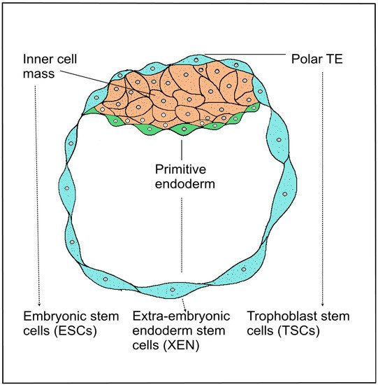
3. Trophoblast Stem Cells of the Blastocyst

Human stem cells can currently be isolated by several methods. Mammalian blastocyst consists of two types of cell layers, the outer TE surrounded by pluripotent cells, forming the inner cell mass (ICM). Blastocysts give rise to three stem cell entities—the pluripotent ESCs, which are derived from ICM developed from the epiblast, and two types of extraembryonic stem cells, primitive eXtraembryonic ENdoderm-derived (XEN) cells and TSCs derived from extraembryonic ectoderm. Human embryonic stem (hESCs) cells are usually derived from the ICM of blastocysts. The derivation of hESCs is therefore considered ethically controversial, and the embryo is destroyed after isolation. Multipotent polar TE cells later develop into the embryonic part of the placenta. Stem cells of all three lines, ESCs, XEN and TSCs, self-renew and retain their fate-specific development potential in vitro [12]. TSCs were derived from TE blastocysts or extraembryonic ectoderm following the implantation [13]. Several authors describe successful cultivation of the TSC line, which was derived from human embryonic blastomeres and chorionic mesenchymal cells and cultured with 10% fetal bovine serum and fibroblast growth factor 2 (FGF2) [14][15][16]. Recently described TSCs from the blastocyst are bipotential. These cells are capable of differentiating into vCTBs and STBs and are suitable for the formation of 3D epithelial organoids, closely resembling the structure and physiology of the original organ (Figure 1).
Biomedicines 10 00904 g004 550
Figure 1. Schematic development of a blastocyst. Mammalian blastocyst consists of two types of cell layers, the outer trophectoderm surrounded by pluripotent cells, forming the inner cell mass (ICM). Blastocysts give rise to three stem cell entities—the pluripotent embryonic stem cells (ESCs), which are derived from ICM developed from the epiblast, and two types of extraembryonic stem cells, primitive eXtraembryonic ENdoderm-derived (XEN) cells and trophoblast stem cells (TSCs) derived from extraembryonic ectoderm. Solid lines—description of blastocyst cell layers. Dotted lines—three types of blastocyst-derived stem cells.

4. Induced Stem Cell Engineering Cell Fate

In mammals, cell fate segregation takes place shortly after fertilization, when the outer morula cells specialize in the future trophoblasts. Embryonal cell fate is controlled by several stimuli, including cell polarity and position, cell–cell signaling and differential expression of TFs. Unlike the in vivo environment, the in vitro conditions are different. Embryonic cell does not spontaneously differentiate into extraembryonic cell types. In an in vitro environment, it is possible to target the fate of cells with specific TFs. Induced PSCs can be generated from somatic cells or adult fibroblasts after forced expression of pluripotent TFs. Similarly, direct reprogramming to multipotent trophoblastic stem cells can be induced. It has been shown that hTSCs can be generated by somatic cells by two methods of reprograming—either adult fibroblasts with the TFs SOX2, Octamer-binding transcription factor 4 (Oct4), Krüppel-like factor 4 (KLF4) or the transformation of PSCs. Human-induced PSCs, which have been generated by reprograming somatic cells, are capable of differentiating into a wide range of body tissue types [13]. It has been shown that a reduced Oct4 expression induces trophoblastic morphology. In the overexpression of the Oct4 antagonist, TF CDX2 is able to induce a trophoblast, its morphology and upregulation of trophoblast markers. Eomes, a factor behind CDX2, is overexpressed and affected by differentiation toward TE/TSC, making both CDX2 and Eomes strong candidates for key TE regulators. Decreased expression of Oct4 in ESCs resulted in the loss of pluripotency and the formation of a monolayer by trophoblast-like cells. CDX2 and Eomes have a key effect on the regulation of differentiation into the trophoblast line. TEAD4 acts on CDX2 during preimplantation, leading to the initiation of TE formation [12]. Furthermore, TFs were identified as inevitable for the successful induction of TSCs, GATA3, Eomes and the transcription factor TFAP2C. Eomes and TFAP2C bind to the TEAD4, GATA3 and E74-like ETS transcription factor 5 (ELF5) genes and positively regulate the expression of ELF5, a protective gene in trophoblastic line differentiation in the early blastocyst. The triple casts of ELF5, Eomes and TFAP2C were enriched for TSC proliferation and potency of TSCs. GATA3 binds to expression and upregulates CDX2, which also increases self-expression as ELF5 and Eomes. GATA3 also induces Eomes expression independently of CDX2 [17]. When it comes to comparing the molecular differences in humans and rodents, known molecular differences include the timing of the CDX2 expression, a key TE TF that is detected in humans only after blastocyst formation. GATA3 expression is more pronounced in human TE, presumably to compensate for the late CDX2 expression. Differences in expression patterns also exist between human and mouse blastocysts. For example, Oct4 is ubiquitously expressed in all human blastocyst cells on days 5–7, while in mice, it is restricted to the ICM [18]. TSCs are characterized by the morphological, transcriptional and functional characteristics of the human cytotrophoblast in vitro. TSC self-renewal could be maintained on a layer of inactive mouse embryonic fibroblast feeder cells that are provided with factors related to the NODAL pathway and supplemented with FGF4, heparin and fetal bovine serum. The induction of human trophoblast progenitors is an important factor. TSCs could be maintained under these conditions for several passages [19]. A technique has recently been described for inducing human-induced trophoblast progenitor (iTP) cells by directly reprogramming fibroblasts with murine trophoblast line-specific transcription factors consisting of CDX2, Eomes and ELF5. Human iTP cells were given epithelial morphology and could be maintained in vitro for more than 2 months [20]. The authors proposed the recommended criteria for the TSC phenotype of GATA3, KRT7 and TFAPC2 markers that lack the expression of HLA class I, ELF5 promoter hypomethylation and C19MC expression. In addition, DNA methylation deficient progenitor cells can efficiently differentiate into trophoblast-type cells [2][7][21].

5. Trophoblast Organoids and Spheroids as Placental Model

5.1. Placental Tissue Culture

More sophisticated in vitro cultivation methods are currently available. In 3D cultures, cells can proliferate and differentiate into a networked environment of biological or synthetic materials that simulate natural ECM. Human TSCs were successfully derived from the blastocyst in the first trimester VCT. Authors analyzed primary transcriptomes of human trophoblast cells in order to deduce how CTBs are maintained in their undifferentiated form in an in vivo condition. Culture conditions and derived human TSCs from CTBs and blastocysts were optimized. Line markers, such as alpha 6 integrin subunit (ITGA6) and TP63, were confirmed on cytotrophoblasts, including ITGA5, HLA-G extravillous trophoblasts, placental lactogen (CSH1) and chorionic gonadotropin (CGB) syncytiotrophoblasts. A 3D model of human TS cells from the blastocyst using a suitable Matrigel was created, and growth of EVT cells from the placental explants was induced. TS cells were cultured for a long time, and the cells expressed the markers KRT7, TP63, GATA3, TEAD4. The markers CGB and SDC1 were highly expressed on syncytiotrophoblasts. On the contrary, HLA-G and vimentin were weakly expressed [2]. Placental organoids mimicking the human trophoblast include cells of extraembryonic origin. Primary culture can be used to create isolated organoids. It was shown that a suspension of trophoblast-enriched cells was obtained in the first trimester by enzymatic cleavage of villi from the placental tissue. Placental villous stromal cells (PVSCs) were isolated by digestion of the tissue remaining, following the initial trypsin and collagenase digestion. The formation of small organoid clusters that formed after 7–10 days was observed. Trophoblast organoids were passaged and placed on differentiation plates. Then, they were maintained in the trophoblast organoid differentiation medium [22]. TSCs with the ability to differentiate into STBs and EVTs are ideal for generating organoids. An in vitro generation of human cytotrophoblastic organoid cultures (CTB-ORGs) from a purified first trimester capable of self-renewal and expansion under defined culture has been shown. Purified first-trimester CTBs were applied to human trophoblast organoids from placental tissues and subsequently integrated into a Matrigel matrix containing a mixture of growth factors and signaling inhibitors [14][21]. Matrigel is widely used to induce the growth of EVT cells from the perinatal tissue [14][23]. Trophoblast culture medium (TOM) composed of EGF, FGF2, CHIR99021 (WNT activator), A83-01 (TGFβ/SMAD inhibitor) and R-spondin-1 was used to culture EVT cells [24]. Early placental proliferative 3D structures expressed markers of the human CTB strain in the epithelial cell layers and spontaneously underwent cell fusion toward the center [14]. Self-renewing trophoblast organoids grow as complex structures that accurately recapitulate the structure of the placental villi in vivo, where vCTBs expresses the markers EpCAM (epithelial cell adhesion molecule) and cadherin-1 (CDH1) [14]. Organoids have been shown to express CDX2, TP63, TFAP2C, TFAP2A, TEAD4 and GATA3 in the outer layers of CTB-ORG [14][25]. Lee’s authors report a phenotype characteristic of human cytotrophoblastic organoid cultures (CTB-ORG) and primary first-trimester trophoblasts, HLA class I profile, ELF5 methylation and microRNA (miRNA) expression from the miRNA cluster chromosome 19 (C19MC) [21]. Integrin P1 proliferative markers are characteristic of human TP niches in the first trimester of the placenta [26]. The differentiation process of early extraembryonic tissues is important in monitoring the processes of placentation in vitro. Human PSCs are used in the study of trophoblast differentiation [27]. In vitro techniques mimic in vivo ones, where TSC also differentiates into EVT, a process crucial for proper placentation. The advantage of the 3D model is the arrangement into complex structures generating both STBs and EVT. The formation of the simulated natural structure of the placental villi can be closely monitored histologically. The lacunae present in the syncytial regions are similar to those found in vivo [14][24]. Authors’ data now provide functional evidence that naive hPSCs indeed have enhanced potential to access trophoblast fates during both spontaneous differentiation assays and upon treatment with recently devised conditions for hTSC isolation [2][28]. Other authors describe a sheep placenta model in a series of studies analyzing the dynamic interrelationships between the trophoblast and the uterine epithelium. Trophoblastic epithelium consists mostly of typical trophoblast cells and minimal binucleate trophoblast cells derived from them. Binucleate cells synthesize and accumulate placental lactogen [29][30].

5.2. Organoids Mimic EVT

Trophoblast migration through the maternal decidua and remodeling spiral arteries affects the extracellular environment. Matrix-integrin binding induces a signal transduction cascade. Such process plays a key role in controlling the cellular behavior and expression of MMP2 and MMP9, which facilitate collagen skeletal degradation. The degradation of ECM EVTs can be reproduced in vitro, where the cells degrade the Matrigel layer on which they are cultured. Authors used an EVT-like cell line HTR8/SVneo, which in combination with Matrigel induces differentiation to the EVT phenotype [31]. The phenotype with EVT differentiation potential includes the following markers: cytokeratin 7 (KRT7), CD9 trophoblast stem cell lines EVT proteases MMP2, MMP9, ECM degradation inhibitors TGFB1, adhesion molecules TGFB2, ITG extracellular EGA and extracellular ITHk, cadherin CDH5 [31].

5.3. Spheroids of Placenta-Derived Mesenchymal Stem Cells

Placenta-derived mesenchymal stem cells (MSCs) are used for regenerative medicine thanks to their multilineage potential and regenerative properties. MSCs can be isolated from extraembryonic tissues, umbilical cord, placental tissue, amniotic and chorionic membranes, where they are involved in maintaining stem cell and tissue niches [32][33][34][35][36]. PMSCs have a characteristic viability and proliferation [37]. MSCs preparation as spheroids is a method used for optimizing the improvement in the efficacy of MSC-based therapeutics. Culturing MSCs in the form of three-dimensional spheroids is a simple and reproducible method and has several advantages over the 2D monolayer culture [38]. Human amniotic mesenchymal stem cells (hAMSCs) were cultured in a serum-free DMEM medium. Mesenchymal phenotype of CD90+, CD73+, CD13+, CD45—and HLA-DR—was confirmed in the tested cells [39]. The tested hAMSC spheroids had increased expression of angiogenic and growth factors HGF, PDGF, VEGF, FGF1, EGF, as well as immunosuppressive factors IL6, TGF-β, COX2. It has been detected that culturing MSCs in the form of spheroids was suitable for maintaining multipotency and paracrine production of hAMSCs [39]. MSCs have a therapeutic effect either due to their requirement for replacement cells or their immunomodulatory and paracrine activities, which promote tissue regeneration. MSCs appear to be the most effective treatment option for patients suffering from musculoskeletal diseases [40][41].

5.4. Characteristics Phenotype of Trophoblast Organoids

Specific trophoblast markers make it possible to recognize the degree of cell development and cell differentiation. Phenotyping of organoid markers of trophoblasts is very important to determine their defined phenotype. The authors Turco et al. recommend the characterization of the following markers: HLA-ABC, HLA-G, ITGA2 [22]. Transcription factor GATA3 affects the embryonic development of various tissues and acts in inflammatory and humoral immune responses. KRT7 stimulates DNA synthesis in cells. KRT7 is expressed by EVT, specifically STBs and vCTBs, which express HLA class I. HLA-G positivity is characteristic of EVT cells, thus it is an accepted specific marker detected for EVT identification [9]. Trophoblast cells from animal placenta express cytokeratins II (KRTII). Trophoblast cells are distinguishable from syncytia cells, which had a very low expression on KRTII [30]. Interestingly, the inhibitors of placental differentiation have been described. A study investigated the effect of transforming growth factor β1 (TGFβ1) on cytotrophoblast differentiation. It has been proven that TGFβ1 acts as a major inhibitor of trophoblast differentiation and concomitant peptide hormone secretion [42]. Invasive trophoblasts have a capability to provide chemotactic signals to uterine leukocytes and affect decidual angiogenesis and apoptosis by secreting hCG [43]. CDX2 is essential for trophoblastic development, vasculogenesis in the mesoderm of the yolk sac, allantoic growth and chorioallantoic fusion [44]. CDX2 is expressed in a mouse model for 3.5 days post-coitum in the trophectoderm region. Along with CDX1 and CDX4, CDX2 is one of three caudal-related genes in the human genome. Genes are also present in most vertebrates’ genomes. CDX2 is thought to play an important role in the pathways controlling the embryonic axial elongation and anterior-posterior patterning [44]. TP63 is a member of the p53 family of transcription factors, and TP63 is involved in skin development and regulation of ASCs. TP63 encodes a transcript isoform that plays a role in ovarian germ cell survival [45]. It has been found that human stem cells induce trophoblasts by induction of BMP 4, leading to the formation of cells with the p63+/KRT7+ phenotype, which is a stem CTB. It has also been observed that p63 expression in CTB cultures had an inhibitory effect on the secretion of hCG [46]. The presence of transcription factors involved in the regulation of trophoblast development has been noted. TFAP2A, TFAP2C and GATA3 in CTB organoids were also expressed in STB and CTB placentas in the first trimester. The observed mRNAs were also detectable in CTB-ORG [14][47][48].

This entry is adapted from the peer-reviewed paper 10.3390/biomedicines10040904

References

  1. Kliman, H.J. Uteroplacental blood flow. The story of decidualization, menstruation, and trophoblast invasion. Am. J. Pathol. 2000, 157, 1759–1768.
  2. Okae, H.; Toh, H.; Sato, T.; Hiura, H.; Takahashi, S.; Shirane, K. Derivation of human trophoblast stem cells. Cell Stem Cell 2018, 22, 50–63.
  3. Ringler, G.E.; Strauss, J.F., III. In vitro systems for the study of human placental endocrine function. Endocr. Rev. 1990, 11, 105–123.
  4. Bangma, J.; Szilagyi, J.; Blake, B.E.; Plazas, C.; Kepper, S.; Fenton, S.E. An assessment of serum-dependent impacts on intracellular accumulation and genomic response of per-and polyfluoroalkyl substances in a placental trophoblast model. Environ. Toxicol. 2020, 35, 1395–1405.
  5. Heaton, S.J.; Eady, J.J.; Parker, M.L.; Gotts, K.L.; Dainty, J.R. The use of BeWo cells as an in vitro model for placental iron transport. Am. J. Physiol.-Cell Physiol. 2008, 295, 1445–1453.
  6. Abou-Kheir, W.; Barrak, J.; Hadadeh, O.; Daoud, G. HTR-8/SVneo cell line contains a mixed population of cells. Placenta 2017, 50, 1–7.
  7. Abbas, Y.; Turco, M.Y.; Burton, G.J.; Moffett, A. Investigation of human trophoblast invasion in vitro. Hum. Reprod. Update 2020, 26, 501–513.
  8. Jingting, C.; Yangde, Z.; Yi, Z.; Huining, L.; Rong, Y. Heparanase expression correlates with metastatic capability in human choriocarcinoma. Gynecol. Oncol. 2007, 107, 22–29.
  9. Apps, R.; Murphy, S.P.; Fernando, R.; Gardner, L.; Ahad, T.; Moffett, A. Human leucocyte antigen (HLA) expression of primary trophoblast cells and placental cell lines, determined using single antigen beads to characterize allotype specificities of anti-HLA antibodies. Immunology 2009, 127, 26–39.
  10. Apps, R.; Sharkey, A.; Gardner, L.; Male, V.; Trotter, M.; Miller, N.; Moffett, A. Genome-wide expression profile of first trimester villous and extravillous human trophoblast cells. Placenta 2011, 32, 33–43.
  11. Manaster, I.; Goldman-Wohl, D.; Greenfield, C.; Nachmani, D.; Tsukerman, P.; Hamani, Y. MiRNA-mediated control of HLA-G expression and function. PLoS ONE 2012, 7, e33395.
  12. Kubaczka, C.; Kaiser, F.; Schorle, H. Breaking the first lineage barrier–many roads to trophoblast stem cell fate. Placenta 2017, 60, 52–56.
  13. Castel, G.; Meistermann, D.; Bretin, B.; Firmin, J.; Blin, J.; Loubersac, S. Induction of Human Trophoblast Stem Cells from Somatic Cells and Pluripotent Stem Cells. Cell Rep. 2020, 33, 108419.
  14. Haider, S.; Meinhardt, G.; Saleh, L.; Kunihs, V.; Gamperl, M.; Kaindl, U. Self-renewing trophoblast organoids recapitulate the developmental program of the early human placenta. Stem Cell Rep. 2018, 11, 537–551.
  15. Genbacev, O.D.; Prakobphol, A.; Foulk, R.A.; Krtolica, A.R.; Ilic, D. Trophoblast L-selectin-mediated adhesion at the maternal-fetal interface. Science 2003, 299, 405–408.
  16. Zdravkovic, T.; Nazor, K.L.; Larocque, N.; Gormley, M.; Donne, M.; Giritharan, G.; Fisher, S.J. Human stem cells from single blastomeres reveal pathways of embryonic or trophoblast fate specification. Development 2015, 142, 4010–4025.
  17. Yang, Y.; Parker, G.C.; Puscheck, E.E.; Rappolee, D.A. Direct reprogramming to multipotent trophoblast stem cells, and is pluripotency needed for regenerative medicine either? Stem Cell Investig. 2016, 3, 24.
  18. Deglincerti, A.; Croft, G.F.; Pietila, L.N.; Zernicka-Goetz, M.; Siggia, E.D.; Brivanlou, A.H. Self-organization of the in vitro attached human embryo. Nature 2016, 533, 251–254.
  19. Roberts, R.M.; Fisher, S.J. Trophoblast stem cells. Biol. Reprod. 2011, 84, 412–421.
  20. Chen, Y.; Wang, K.; Gong, Y.G.; Khoo, S.K.; Leach, R. Roles of CDX2 and EOMES in human induced trophoblast progenitor cells. Biochem. Biophys. Res. Commun. 2013, 431, 197–202.
  21. Lee, C.Q.; Gardner, L.; Turco, M.; Zhao, N.; Murray, M.J.; Coleman, N. What is trophoblast? A combination of criteria define human first-trimester trophoblast. Stem Cell Rep. 2016, 6, 257–272.
  22. Turco, M.Y.; Moffett, A. Development of the human placenta. Development 2019, 146, 163428.
  23. Miller, R.K.; Genbacev, O.; Turner, M.A.; Aplin, J.D.; Caniggia, I. Human placental explants in culture: Approaches and assessments. Placenta 2005, 26, 439–448.
  24. Turco, M.Y.; Gardner, L.; Kay, R.G.; Hamilton, R.S.; Prater, M.; Hollinshead, M.S. Trophoblast organoids as a model for maternal–fetal interactions during human placentation. Nature 2018, 564, 263–267.
  25. Kretzschmar, K.; Clevers, H. Organoids: Modeling development and the stem cell niche in a dish. Dev. Cell 2016, 38, 590–600.
  26. Lee, C.Q.; Turco, M.Y.; Gardner, L.; Simons, B.D.; Hemberger, M.; Moffett, A. Integrin α2 marks a niche of trophoblast progenitor cells in first trimester human placenta. Development 2018, 145, 16.
  27. Horii, M.; Touma, O.; Bui, T.; Parast, M.M. Modeling human trophoblast, the placental epithelium at the maternal fetal interface. Reproduction 2020, 160, R1–R11.
  28. Dong, C.; Beltcheva, M.; Gontarz, P.; Zhang, B.; Popli, P.; Fischer, L.A.; Theunissen, T.W. Derivation of trophoblast stem cells from naïve human pluripotent stem cells. eLife 2020, 9, 52504.
  29. Wooding, F.B.P. The synepitheliochorial placenta of ruminants: Binucleate cell fusions and hormone production. Placenta 1992, 13, 101–113.
  30. Kadyrov, M.; Garnier, Y.; Gantert, M.; Kramer, B.W.; Kaufmann, P.; Huppertz, B. Cytokeratin antibodies as differential markers of trophoblast and fetomaternal syncytial plaques in the sheep placentome. Placenta 2007, 28, 1107–1109.
  31. Highet, A.R.; Zhang, V.J. Use of Matrigel in culture affects cell phenotype and gene expression in the first trimester trophoblast cell line HTR8/SVneo. Placenta 2012, 33, 586–588.
  32. Bárcia, R.N.; Santos, J.M.; Teixeira, M.; Filipe, M.; Pereira, A.R.S.; Ministro, A. Umbilical cord tissue–derived mesenchymal stromal cells maintain immunomodulatory and angiogenic potencies after cryopreservation and subsequent thawing. Cytotherapy 2017, 19, 360–370.
  33. Parolini, O.; Alviano, F.; Bagnara, G.P.; Bilic, G.; Bühring, H.J.; Evangelista, M. Concise review: Isolation and characterization of cells from human term placenta: Outcome of the first international Workshop on Placenta Derived Stem Cells. Stem Cells 2008, 26, 300–311.
  34. Bačenková, D.; Rosocha, J.; Tóthová, T.; Rosocha, L.; Šarisský, M. Isolation and basic characterization of human term amnion and chorion mesenchymal stromal cells. Cytotherapy 2011, 13, 1047–1056.
  35. Bačenková, D.; Trebuňová, M.; Zachar, L.; Hudák, R.; Ižaríková, G.; Šurínová, K.; Živčák, J. Analysis of same selected immunomodulatory properties of chorionic mesenchymal stem cells. Appl. Sci. 2020, 10, 9040.
  36. Janockova, J.; Slovinska, L.; Harvanova, D.; Spakova, T.; Rosocha, J. New therapeutic approaches of mesenchymal stem cells-derived exosomes. J. Biomed. Sci. 2021, 28, 39.
  37. Lankford, L.; Chen, Y.J.; Saenz, Z.; Kumar, P.; Long, C.; Farmer, D.; Wang, A. Manufacture and preparation of human placenta-derived mesenchymal stromal cells for local tissue delivery. Cytotherapy 2017, 19, 680–688.
  38. Rettinger, C.L.; Fourcaudot, A.B.; Hong, S.J.; Mustoe, T.A.; Hale, R.G.; Leung, K.P. In vitro characterization of scaffold-free three-dimensional mesenchymal stem cell aggregates. Cell Tissue Res. 2014, 358, 395–405.
  39. Miceli, V.; Pampalone, M.; Vella, S.; Carreca, A.P.; Amico, G.; Conaldi, P.G. Comparison of immunosuppressive and angiogenic properties of human amnion-derived mesenchymal stem cells between 2D and 3D culture systems. Stem Cells Int. 2019, 2019, 7486279.
  40. Vikartovska, Z.; Humenik, F.; Maloveska, M.; Farbakova, J.; Hornakova, L.; Murgoci, A.N.; Cizkova, D. Adult Stem Cells Based Therapies in Veterinary Medicine. Arch. Vet. Sci. Med. 2020, 3, 40–50.
  41. Trebuňova, M.; Gromošová, S.; Bačenková, D.; Rosocha, J.; Živčák, J. Biocompatibility of the human mesenchymal stem cells with bovine bone tissue at the cellular level in vitro. Lékař A Tech. Clin. Technol. 2018, 48, 59–65.
  42. Morrish, D.W.; Bhardwaj, D.; Paras, M.T. Transforming growth factor β1 inhibits placental differentiation and human chorionic gonadotropin and human placental lactogen secretion. Endocrinology 1991, 129, 22–26.
  43. Knöfler, M.; Pollheimer, J. IFPA Award in Placentology lecture: Molecular regulation of human trophoblast invasion. Placenta 2012, 33, 55–62.
  44. Chawengsaksophak, K.; de Graaff, W.; Rossant, J.; Deschamps, J.; Beck, F. Cdx2 is essential for axial elongation in mouse development. Proc. Natl. Acad. Sci. USA 2004, 101, 7641–7645.
  45. Crum, C.P.; McKeon, F.D. p63 in epithelial survival, germ cell surveillance, and neoplasia. Annu. Rev. Pathol. Mech. Dis. 2010, 5, 349–371.
  46. Li, Y.; Moretto-Zita, M.; Leon-Garcia, S.; Parast, M.M. p63 inhibits extravillous trophoblast migration and maintains cells in a cytotrophoblast stem cell-like state. Am. J. Pathol. 2014, 184, 3332–3343.
  47. Biadasiewicz, K.; Sonderegger, S.; Haslinger, P.; Haider, S.; Saleh, L.; Fiala, C. Transcription factor AP-2α promotes EGF-dependent invasion of human trophoblast. Endocrinology 2011, 152, 1458–1469.
  48. Plessl, K.; Haider, S.; Fiala, C.; Pollheimer, J.; Knöfler, M. Expression pattern and function of Notch2 in different subtypes of first trimester cytotrophoblast. Placenta 2015, 36, 365–371.
More
This entry is offline, you can click here to edit this entry!
Video Production Service