As countries face the challenges of aging populations, there is a need to promote healthy aging and provide adequate social protection. To promote a healthy aging process and prevent aging-related health problems, correctly understanding aging mechanisms and developing effective and affordable intervention strategies for anti-aging have great social significance and huge economic benefits. During this process, an animal model of aging is a powerful tool for us to study the mechanism of aging.
2. Systemic-Induced Accelerated Aging Mouse Model
In this section, we will review some commonly used mouse models of accelerated systemic aging including drug treatment, genetic engineered models, irradiation induction, etc. They are characterized by an aging phenotype in multiple tissues or organs, reflecting systemic aging. The systemic-induced accelerated aging mouse models are summarized in Table 1.
Table 1. Summary of systemic-induced accelerated aging mouse models.
| Type |
Subdivision |
Phenotypes |
| D-galactose-induced senescence model |
Brain |
Cognitive impairment
Mitochondrial dysfunction
Neuronal degeneration
Apoptosis
Depressive and anxious |
| Heart |
Cardiac fibrosis
Collagen accumulation
Fibroblasts disordered arrangement |
| Kidney |
Kidney index ↓
Uric acid & Cys-C ↑
Glomerular and tubular damage ↑ |
| Liver |
Liver fibrosis
Glycogen levels ↓
Lipid deposition ↑ |
Reproductive
system |
Estrogen and progesterone ↓
Ovarian follicle regression
Uterine wall endometrial gland atrophy
Disrupt estrous cycles |
| Intestinal flora |
Disturbance |
| Lung |
Oxidative stress ↑
Fibrotic status
Chronic inflammation |
| SAMP mice |
SAMP 1 |
Aging amyloidosis
Immune dysfunction
Renal atrophy
Hearing loss
Senile pulmonary hyperinflation |
| SAMP 6 |
Senile osteoporosis
Myeloid progenitor cell senescence |
| SAMP 8 |
Astrogliosis
Microgliosis
Neurodegeneration
Amyloid accumulation
MAPT hyperphosphorylation |
| SAMP 10 |
Learning and memory impairment
Cerebral cortex and limbic system atrophy |
| Rps9 D95N mouse |
|
Altered fur
Cataracts
Hunched posture
Body composition function & body weight ↓
Fat mass & muscle strength ↓
Shortened lifespan
Mouse urinary syndrome
Extramedullary hematopoiesis |
| |
Lama−/− |
Short lifespan
Growth retardation
Muscular dystrophy
Altered lipid metabolism |
| |
Wrn∆hel/∆hel |
Short lifespan
Abnormal hyaluronic acid excretion
Metabolic abnormalities
Increased genomic instability and cancer incidence |
| |
Csa−/−, Csb−/− |
Short lifespan
Reduced fat mass
Photoreceptor cell loss
Neural pathology |
| Progeria syndrome mouse |
XpdTTD/TTD |
Short lifespan
Trichothiodystrophy |
| Bub1bH/H, Xpg−/− |
Brain atrophy
Neuronal loss
Neurofibrillary deposition of Aβ or senile plaques |
| Bub1bH/H, Bub1b+/GTTA |
Mean muscle fiber diameter ↓
Muscle fiber size variation ↑
Intermuscular fibrosis
Regenerative capacity of skeletal muscles ↓ |
| Mitochondrial DNA polymerase mutant mouse |
|
Lifespan ↓
Weight loss
Subcutaneous fat ↓
Hair loss
Kyphosis
Osteoporosis
Anemia
Fertility ↓
Spermatogonia depletion
Heart enlargement |
| Total body irradiation (TBI) model |
|
Progressive premature frailty
Cognitive decline
Whole blood antioxidant capacity ↓
RBC glutathione ↓
Thymic involution
Articular cartilage and bone degeneration
Ovarian environment damage |
| Ozone-induced senescence model |
|
Cognitive decline
Memory impairment
AD symptoms
Lung tumor growth ↑ |
| Chronic jet-lag mouse |
|
Accelerated initial tumor growth
Shortened mouse survival
Induce spontaneous hepatocellular carcinoma
Obesity
Depression
Addiction
Abnormal cardiac structure
Impaired cardiac function |
2.1. The D-Galactose-Induced Senescence Model
D-galactose is a common aldohexose that exists naturally in the body and in daily foods [
8]. After ingestion, a healthy adult can metabolize and eliminate a maximum daily dose of 50 g of galactose from the body within about eight hours [
9]. However, when galactose accumulates to high levels, reactive oxygen species (ROS) are generated by mitochondrial respiratory chain enzymes, xanthine oxidase, lipoxygenase, cyclooxygenase, nitric oxide synthase, and peroxidase. Increased ROS can subsequently lead to elevated oxidative stress and inflammation, inducing mitochondrial dysfunction and apoptosis [
10]. Meanwhile, the elevated mitochondrial ROS level can lead to the activation of many biochemical pathways, such as the polyol pathway, the formation of advanced glycation end products (AGEs), the activation of protein kinase C, and the hexosamine pathway [
11,
12]. Overall, D-galactose-induced methods can increase aging markers such as AGEs, receptors for advanced glycation end products (RAGEs), aldose reductase (AR), sorbitol dehydrogenase (SDH), decreased telomerase activity, shortened telomere, β-site amyloid precursor protein cleaving enzyme 1 (BACE-1), amyloid β (Aβ), aging-related pathways (p16, p21, p53, etc.), and positive senescence-associated β-galactosidase (SA-β-gal) staining [
13]. Multiple tissues and organs, including the brain, heart, lung, liver, kidney, reproductive system, gastrointestinal system, and so on, manifest aging phenotypes after D-galactose treatment [
14].
Previous works have shown that D-galactose induces brain aging by increasing mitochondrial dysfunction, oxidative stress, inflammation, apoptosis, and decreasing the expression of brain-derived neurotrophic factor. D-galactose injections may induce brain aging similar to human brain aging in many ways, including mitochondrial dysfunction, increased oxidative stress, decreased ATP production, neuronal degeneration and apoptosis, and cognitive deficits [
13,
15,
16,
17]. D-galactose increases the neuro-inflammation markers via activating NF-κB, leading to memory impairment [
18,
19]. Besides learning and memory inhibition, D-galactose-treated mice also exhibit depressive and anxious behaviors [
20].
The leading cause of death in elderly people worldwide is cardiovascular disease [
21]. D-galactose treatment increases the risk of cardiovascular disease, which is associated with excess ROS and oxidative stress. Persistent oxidative stress has been revealed to be related to decreased ferric reducing antioxidant power and lower activity of Cu-Zn superoxide dismutase, leading to myocardial damage [
22]. Studies have shown that galactose reduces endogenous hydrogen sulphide producing enzyme cystathionine γ-lyase (CSE) [
23] and antioxidant enzymes such as catalase (CAT), haem oxygenase-1 (HO-1), superoxide dismutase (SOD), glutathione peroxidase (GSH-Px), and nitric oxide synthase (NOS), leading to decreased total antioxidant capacity and inducing lipid peroxidation markers including malondialdehyde (MDA), lipid hydroperoxides (L-OOH), and conjugated dienes (CD) in cardiac tissue [
24,
25]. D-galactose increases whole heart weight and left ventricle weight, which is associated with hypertension and aging. At the same time, the heart tissue showed enlarged myocardial fibers, blurred structure, shortened distortion, widening of the interval, and obvious capillaries of myocardial interstitial congestion [
26]. D-galactose treatment resulted in cardiac fibrosis, significant accumulated collagen, and disordered arrangement of fibroblasts compared with the control. D-galactose also increased cardiac apoptosis markers [
27]. Excessive D-galactose can be transformed to advanced AGEs via the Maillard reaction [
28]. AGEs bind to the receptors, RAGE, increasing ROS production via NADPH oxidase. NADPH oxidase further activates p38 MAP kinases, causing transcription factor NF-κB translocating into the nucleus, where transcription of inflammatory cascades like tumor necrosis factor alpha (TNF-α) are enhanced [
29]. D-galactose treatment can also increase fibrotic markers such as connective tissue growth factor (CTGF), transforming growth factor β1 (TGF-β1), phosphorylated mitogen-activated protein kinase 1/2 (p-MEK1/2), phosphorylated extracellular signal-regulated kinase 1/2 (p-ERK1/2), matrix metalloproteinase (MMP), and pathological specific protein 1 (SP1) [
30].
D-galactose treatment also increases oxidative markers (e.g., MDA) and decreases antioxidant enzymes (e.g., SOD, GSH-Px and NOS) in lung, liver, and renal tissues [
26,
31,
32,
33,
34,
35]. D-galactose treatment can affect lung elastic constitution. The primary effects of treatment on the lungs are increased alveolar size and reduced elastic recoil, which may facilitate airway closure [
36]. D-galactose treatment is considered to successfully mimic the natural aging process by increasing oxidative stress, fibrotic status, and chronic inflammation in the lungs.
The liver is the main site where D-galactose is metabolized, thus excess D-galactose in the body may significantly affect the liver. As previously mentioned, high levels of D-galactose can react with free amines in the amino acids to form AGEs, which is found to be involved in the progression of various liver diseases [
12]. High levels of D-galactose lead to the accumulation of its final metabolite, galactitol, which will eventually lead to ROS accumulation through the p38 MAPK/NRF2/HO-1 signaling pathway and cause cellular osmotic stress in the liver [
37]. D-galactose treatment significantly increased apoptotic proteins including Bax, procaspase-3, and caspase-3, and raised the ratio of Bax/Bcl-2 in the liver tissue [
34,
38,
39]. Various changes similar to natural aging were also observed in the D-galactose-treated livers. Compared with controls, the livers of D-galactose-treated animals had lower levels of glycogen and more lipid deposition. Masson’s trichrome staining showed obvious collagen fibers around blood vessels and in the interphase of liver tissue, and irregular pseudo-lobules around liver tissue, indicating a state of liver fibrosis [
40,
41].
D-galactose treatment significantly reduced the renal index of animals, and markers of acute kidney injury such as uric acid and cystatin C (Cys-C) were also increased [
42]. In the kidney, D-galactose administration resulted in an increase in the TUNEL-positive cells and DNA fragmentation. In addition, p21 expression and the staining intensity of SA-β-gal were also increased in kidney cells [
43]. Extensive glomerular and tubular damages were detected in D-galactose-treated animals, as the number of tubules with cellular necrosis from the renal cortices and outer medulla were significantly increased [
43,
44].
In the male reproductive system, D-galactose induced oxidative stress, marked by an increase in MDA levels in the prostate, testis, epididymis, and decrease in SOD activity in the testis. Peroxidation in the testicular and epididymal mitochondrial fractions was also significantly increased after D-galactose treatment [
45]. Female reproductive aging is characterized by decreased levels of estrogen, progesterone, inhibin B, anti-Müllerian hormone, and androgens, which include free testosterone, dehydroepiandrosterone (DHEAS), and androstenedione [
46]. Compared to the control groups, D-galactose administration produced aging-associated changes like reduced estrogen and progesterone levels and increased FSH and luteinizing hormone (LH) levels. Ovarian follicle regression and atrophy on the uterine wall and endometrial gland were observed after D-galactose treatment, which indicates disrupted estrous cycles and damaged uterine and ovarian tissues [
47].
D-galactose injection can lead to changes in the level of oxidative stress that affect the microbial environment in the intestine and lead to intestinal flora disorder [
48]. The ecology of intestinal flora is closely related to aging, and intestinal ecological disturbance can lead to accelerated aging and a shortened lifespan [
49]. Transferring gut microbiota from aged to young germ-free mice triggered innate immune and inflammatory responses. Effects of aging include increased differentiation of CD4
+ T cells in the spleen, upregulation of intestinal inflammatory cytokine such as TNF-α, and increased cycling of bacterial-derived inflammatory cytokines [
50,
51]. In addition, D-galactose administration significantly decreased the small intestine propulsion rates and prolonged gastrointestinal transit time [
49]. The aging gut triggers chronic inflammation, leading to gut dysplasia and intestinal dysplasia in turn leads to defective epithelial function, predisposing the host to infection, neoplasia, and increased mortality [
52].
2.2. Senescence-Accelerated Mouse/Prone
Researchers from Kyoto University found an aging phenotype from a subset of pups while maintaining an inbred line of AKR/J mice. Characteristics of these mice include hair loss, reduced activity, and a shortened lifespan. These aging traits are thought to develop as a result of elevated oxidative stress and are inherited by their offspring. Further, the accelerated aging mice were grouped into several distinct subtypes according to their phenotypes [
53].
Senescence-accelerated mouse/prone (SAMP) is a group of inbred mouse strains that typically exhibit accelerated aging [
54]. Meanwhile, since it shows various aging-related diseases similar to humans, such as aging amyloidosis, senile osteoporosis, learning/memory impairment, etc., in specific lines, it is widely used for aging research. Cellular senescence in various cell types, including astrocytes, endothelial cells, progenitor cells, retinal epithelial cells, and fibroblasts, was found in the aging SAMP mice [
55,
56,
57]. SAMP mice exhibit an increase in ROS generated by mitochondria or other cellular sites, which not only causes damage to mitochondria, but also triggers degradation that leads to the aging outcome [
58].
SAMP1 mice are characterized by aging amyloidosis, immune dysfunction, renal atrophy, hearing loss, and senile pulmonary hyperinflation [
59,
60]. The lifespan of the SAMP1 mice is about 40% shorter than senescence-accelerated resistant mice (SAMR1), and various signs of aging appear early in appearance.
The SAMP6 mice model is a senile osteoporosis model animal that tends to develop osteoporosis at an early stage with aging due to its low bone density in instar [
61]. Bone marrow transplantation experiments have revealed that the cause of SAMP6 osteoporosis is abnormalities in bone marrow stem cells [
62]. The incidence of spontaneous leg fractures due to osteoporosis is high in adult SAMP6 mice. Cellular senescence in myeloid progenitors disrupts their differentiation in favor of adipogenesis over osteogenesis. This mechanism is thought to contribute to low osteoblast activity and osteoporosis in SAMP6 mice and the elderly [
61,
63,
64].
The SAMP8 mice develop age-associated deficits in learning and memory and also exhibit various age-related neuropathological changes similar to aging humans [
65,
66]. Neuropathological changes including astrogliosis, microgliosis, and neurodegeneration occur as early as five months of age [
67]. SAMP8 mice also showed accumulated amyloid and age-related microtubule-associated protein tau (MAPT) hyperphosphorylation, as well as increased nitric oxide synthase activity, further demonstrating their feasibility as a brain aging model [
68,
69]. In addition, decreased activities in SOD, CAT, glutathione reductase, and GSH-Px, and increased activity in acyl-CoA oxidase were detected in SAMP8 mice at 1–12 months of age [
70]. SAMP6 and SAMP8 mice also develop many other age-related diseases, including retinal degeneration, testosterone deficiency [
71], myocardial fibrosis [
72], and hepatic lipid deposition [
73].
The SAMP10 mice exhibit learning and memory impairment with aging, and atrophy is observed in the cerebral cortex and limbic system, so it is considered to be a spontaneous model animal for aging and brain degeneration. The atrophy of the frontal cerebral cortex and olfactory bulb is marked in SAMP10 mice [
74,
75].
So far, more than ten strains of SAMP mice have been identified and widely used in aging studies, each of which can develop various age-related diseases such as renal fibrosis (shrinking of the kidneys), immune dysfunction, and degenerative joint disease like osteoarthritis (OA), etc.
2.3. Rps9 D95N Mouse
Rps9 D95N is a ribosome ambiguity mutation that causes error-prone protein synthesis in mammalian ribosomes, resulting in increased error-prone translation. Rps9 D95N mutant mice exhibit features of accelerated aging, including morphological (altered fur, cataracts, and hunched posture), physiological (body composition and function, body weight, fat mass, and muscle strength), and pathological (shortened lifespan, mouse urinary syndrome and extramedullary hematopoiesis), which also complements and explains the link between accumulation of erroneous proteins resulting from protein mistranslation and individual aging [
76].
2.4. Progeria Syndrome Mouse
Mouse models of progeria syndrome have emerged as an attractive tool for evaluating intervention strategies for unhealthy aging due to their short lifespan, relatively simple strategies by single gene deletion or mutation, and their strong phenotypic similarities to normal aging [
7]. To understand important mechanisms of the aging process, progeria mice
(Lmna−/−,
Wrn∆hel/∆hel,
Csa−/− or
Csb−/−) from common progeria types such as Hutchinson-Gilford Progeria, Werner Syndrome, and Cockayne Syndrome are being widely used.
The most common phenotypes in mouse models of progeria can be observed in bones, joints, skin, nervous system, adipose tissue, skeletal muscle, cardiovascular system, liver, kidney, and the hematopoietic system. Less common lesions occur in the gonads, eyes, and occasionally in the gastrointestinal tract [
77].
Some human progeria syndromes like Werner Syndrome exhibit osteoporosis. Current data from a mouse model of progeria indicate that senile osteoporosis is the result of reduced bone turnover and loss of bone mass due to defects in osteoblast progenitor cells, osteoblast differentiation, or osteoblast function [
77]. Degenerative joint disease is another major symptom that affects the elderly. Mutation in the
Xpd gene of nucleotide excision repair (NER) leads to a short lifespan, causing trichothiodystrophy (TTD) in humans. As expected,
XpdTTD/TTD mice exhibited a significant decrease in subchondral bone plate thickness compared to that observed in wild-type mice. Surprisingly, female
XpdTTD/TTD mice exhibited less cartilage damage and fewer lost articular cartilage, compared to WT females [
78].
During the aging process, skeletal muscle was inevitably accompanied with a reduction in muscle mass, known as sarcopenia or age-related muscle atrophy, and macroscopic examination of sarcopenic mice will show weight loss and marked reduction in muscle mass. Sarcopenia is characterized by the loss of muscle fibers and smaller fiber cross-sectional area that is defined as fiber atrophy [
79]. The progeria mouse model
Bub1bH/H and
Bub1b+/GTTA mice showed decreased mean muscle fiber diameter, increased myofiber size variation, increased intermuscular fibrosis, and impaired regenerative capacity in skeletal muscles [
80].
Common brain aging diseases include Alzheimer’s disease (AD) and cognitive dysfunction syndrome. Brain atrophy, neuronal loss, neurofibrillary deposition of Aβ or senile plaques, intraneuronal tauopathies (neurofibrillary tangles, NFTs), cerebrovascular amyloid angiopathy, neuronal lipofuscinosis, vascular and meningeal calcification, decreased white matter integrity, and astrogliosis are common age-related neurological pathologies [
81]. The brains of several mouse models of progeria show neurodegenerative changes at an early stage. Similar to age-related gliosis,
Bub1bH/H mice have increased numbers of astrocytes and microglia at one and five months of age, respectively. Additionally,
Xpg−/− mice developed more astrocytes and increased activation of microglia in the brain and spinal cord [
82].
3. Tissue-, Organ-, or System-Specific Mouse Models of Aging-Related Diseases
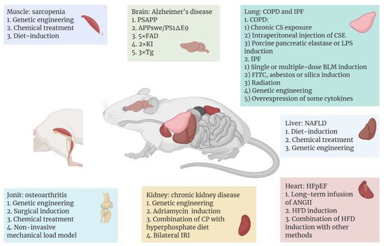
Among the leading effects of ageing is the heightened incidence of various aging-related diseases, and mouse models continue to serve an essential part in the study of the pathogenesis and treatment of these illnesses. A variety of commonly used and emerging mouse models have been developed for different aging-related diseases, with the aim of reproducing as closely as possible the progression of the diseases in humans (Figure 1).
Figure 1. Aging-related diseases include Alzheimer’s disease (AD), sarcopenia, heart failure with preserved ejection fraction (HFpEF), non-alcoholic fatty liver disease (NAFLD), chronic kidney disease (CKD), osteoarthritis, chronic obstructive pulmonary disease (COPD), and idiopathic pulmonary fibrosis (IPF), which occur in different tissues or organs. With the help of mice as model organisms, researchers have used different methods to establish disease models in different tissues or organs of mice. In recent years, some common or emerging methods of modeling aging-related diseases are shown above, and these methods are classified according to the organ to which the disease mainly affects.
Figure 1 was created with BioRender software (
https://biorender.com/ (accessed on 20 April 2022)).
3.1. Model of Aging Brain or Nerve System
Although there are some genetic similarities between mice and humans, the two still diverge in the expression levels of certain age-related genes, which leads to potentially different aging process in the central nervous system of humans and mice [
122]. Therefore, changing the expression levels of those specific genes that contribute to the aging process of the brain is the main strategy for modeling the aging brain in mice. For now, to ensure that experimental results can be extrapolated to humans, research on such models has mainly concentrated on evolutionarily conserved mechanisms that modulate aging. These conserved mechanisms include genomic instability, epigenetic changes, telomere attrition, mitochondrial dysfunction, loss of proteostasis, and dysregulated nutrient sensing [
122,
123].
For instance, as a neurodegenerative disease, AD represents one of the most common neurological disorders. The number of people affected by the disease has been recorded as over tens of millions worldwide, and the number will continue to rise. Also, it is the most common cause of dementia [
124]. Over the past decades of research, with the help of animal models, substantial advances have been made, expanding our understanding of this disease.
So far, the etiology of AD is not yet completely understood. It is thought that genetic elements play key roles in the onset of such disease [
125]. The typical histopathological features of AD are Aβ deposits and NFTs in the brain. However, wild-type mice do not spontaneously exhibit symptoms of AD [
126]. Because AD-related proteins differ in sequence, pathogenicity, and number of isoforms between rodents and humans [
127], the main strategy for modeling AD in mice is to construct transgenic mice that cause amyloid deposition or NFTs in the brain.
Common AD-associated mutant proteins in humans include Aβ, presenilin 1 (PS1), apolipoprotein E (ApoE), and MAPT. In the 1990s, the first transgenic AD model mice stably expressing the mutant human Aβ precursor protein (APP) were constructed [
128]. After this, transgenic mice carrying multiple human AD-related mutant proteins emerged. One of the most commonly used AD models today is hAPP/PS1 lines, which carry both mutated human APP and PSEN1, including transgenic strains PSAPP, APPswe/PS1ΔE9, 5XFAD, and 2xKI [
129]. Compared to monogenic lines containing only mutated APP or PS1, these transgenic mice exhibit earlier and faster onset of amyloid accumulation and cognitive impairment [
130]. However, such AD models do not exhibit signs of NFTs, which can be imitated by mouse models that express human MAPT. Based on this, Oddo et al. constructed a 3xTg model combining the human APP, PS1, and MAPT mutations [
131]. Since this model can show both Aβ deposition and NFTs in the brain, it is considered to be the well-established transgenic model of AD. Consistent with patients with AD, some of these transgenic mouse models (e.g., PS19, APPswe/PS1ΔE9) showed increased levels of NF-κB pathway-related proteins (IKKβ, p65, and COX-2) in the brain, indicating upregulation of brain inflammation in these mice. Therefore, the use of such mouse models will also help us to further clarify the relationship between neuroinflammation and the pathogenesis of AD [
132,
133].
3.2. Model of Aging Muscle
The aging of the body is accompanied by the aging of the skeletal muscles. Among other things, sarcopenia, which is a widespread progressive skeletal muscle disorder, is associated with an increased probability of adverse consequences like falls, fractures, physical disability, and death, and its risk increases with age [
134]. Modeling of sarcopenia in mice is divided into two main categories: models of genetic engineering and chemical or dietary-induced models.
For genetic engineering models, the majority of research has employed knockout (KO) mice. For example, mitofusion2 (Mfn2) is one of the important protein components mediating mitochondrial fusion, and Mfn2 KO mice exhibit mitochondrial dysfunction in skeletal muscle cells and specific atrophy of type IIb glycolytic fibers [
135]. Collagen VI, an extracellular matrix (ECM) protein, has a critical role in skeletal muscle. Six-month-old
Col6α1−/− mice exhibit alterations of the diaphragm consistent with aged wild-type mice, such as abnormal tricarboxylic acid (TCA) cycle and decreased autophagy in diaphragm cells, indicating the
Col6α1−/− mouse could be considered as a premature model of skeletal muscle aging [
136]. Additionally, interleukin 10 (IL-10) [
137], SOD1 [
138], and NOD-like receptor protein 3 (NLRP3) [
139] deficient mice have also been employed in studying the pathogenesis of sarcopenia as well as the intervention of therapeutically targeting such a disease. Overexpression of certain proteins can also lead to sarcopenia, such as TNF-α transgenic mice that exhibit reduced muscle mass, muscle fiber diameter, and Pax7
+ muscle stem-cell content [
140].
For chemical or diet-induced sarcopenia models, dexamethasone is a common inducer, which is essentially a glucocorticoid that triggers muscle atrophy in mice. It is shown that dexamethasone induces upregulation of ubiquitin ligases in muscle, including muscle atrophy F-box (MAFbx) and muscle ring finger 1 (MuRF1), which may further mediate the degradation of muscle atrophy-associated proteins [
141,
142]. In addition, diet-induced sarcopenia mouse models allow us to investigate the co-occurrence of sarcopenia with other disorders, e.g., sarcopenia can also develop from the reduction in muscle mass and strength caused by certain chronic diseases. Fabián et al. [
143] treated mice with hepatotoxin 5-diethoxycarbonyl-1,4-dihydrocollidine (DDC), thereby inducing sarcopenia secondary to chronic liver disease (CLD), as evidenced by reduced muscle strength and motility, as well as the reduction in muscle fiber size and its type of transformation in mice.
3.3. Model of Aging Heart
Heart failure is a complex disease that can eventually result from almost all cardiovascular disorders, like myocardial infarction, atherosclerosis, and hypertension. Based on the left ventricular ejection fraction, heart failure is clinically classified into two major categories: heart failure with reduced ejection fraction or preserved ejection fraction (HFpEF) [
144]. Among them, due to the increasing morbidity and mortality of HFpEF in recent years and the lack of effective therapeutic options for this disease, research on HFpEF has received increasing attention and as a result, some mouse models of HFpEF have been developed.
Long-term infusion of angiotensin II (ANGII) into mice based on a mini-osmotic pump is one of the most common methods of modeling HFpEF. Elevated levels of ANGII in the mouse circulatory system can trigger vasoconstriction, hypertension, aldosterone secretion, TGF-β-mediated inflammation, and fibrosis, and ultimately cardiac hypertrophy [
144]. These symptoms closely resemble those exhibited by HFpEF in humans. Furthermore, in addition to causing obesity, a high-fat diet (HFD) is also known to induce a host of cardiac-related symptoms, including left ventricular hypertrophy, HFpEF, and diastolic dysfunction [
145,
146,
147,
148]. In addition, Withaar et al. [
149] constructed a model with HFD and ANGII treatment, which exhibited higher levels of cardiac fibrosis as well as more severe diastolic dysfunction and cardiac hypertrophy compared to the single-treatment group with HFD or ANGII. Also, Schiattarella et al. [
150] developed a model in which both HFD and the constitutive NO synthase inhibitor N
ω-nitro-L-arginine methyl ester (L-NAME) were imposed. Although L-NAME caused an increase in diastolic and systolic blood pressure, the HFD-L-NAME group exhibited more significant cardiomyocyte hypertrophy and a reduction of myocardial capillary density.