Your browser does not fully support modern features. Please upgrade for a smoother experience.
Please note this is an old version of this entry, which may differ significantly from the current revision.
Subjects:
Cell Biology
Extracellular vesicles (EVs) are small cargo-containing structures with a lipid bilayer but do not have the cellular machinery required to replicate. They have been shown to play a role in cell-to-cell communication, as they can be found to transport biological material including proteins, lipids, ribonucleic acid (RNA), and deoxyribonucleic acid (DNA) between cells, leading to cellular changes within multi-cellular organisms. It is a process that has been conserved through evolution, found both in prokaryotes and eukaryotes.
- extracellular vesicle
- exosome
- transfer RNA
- tRNA fragment
- tRNA half
- cancer
1. Introduction
Extracellular vesicles (EVs) are an umbrella term for a heterogenous array of secreted membrane vesicles and can be further distinguished by cell source, biogenesis pathways, and size ranges, thus contributing to diverse nomenclature [2]. Two broad categories that are often used distinguish microvesicles (MVs) and exosomes on the basis of cellular biogenesis [2]. Microvesicles are generated by the outward budding of the phospholipid bilayer generating vesicles made directly from the plasma membrane. These vesicles tend to be larger with MVs having a size range of up to 1000 nm or larger in some cases [2,3]. Exosomes, on the other hand, involve intracellular mechanisms forming multivesicular endosomes (MVEs) which contain intraluminal vesicles (ILVs). ILVs are formed from the budding of MVEs which then fuse with the plasma membrane and get released extracellularly as exosomes. Exosomes are usually smaller in size (<150 nm) closely reflecting that of ILVs [2,3] and therefore also express specific markers that relate to their endosomal origin [2].
EVs have been found to be present in virtually all types of biofluids, including cerebrospinal fluid (CSF) [4], urine [5], blood [6], bile [7], and breast milk [8] as well as being released by other organisms and even plants [9]. Numerous studies focus on their carrier material which contains lipids, protein, and RNA in an endeavour to link them to the pathophysiology of a wide range of diseases [1]. With the current advances in RNA sequencing technologies and bioinformatic strategies, this has drawn attention to the wide range of small RNA (sRNA) content in EVs.
Whilst much of the literature has focused on studying micro RNAs (miRNAs) [10], short 16–22 nucleotide (nt) single-stranded molecules with a role in canonical transcription and translational pathways, less is known about the role of transfer RNAs (tRNA) despite being the most abundant RNA in the human genome [11], and the first non-coding RNA to have been discovered [12]. The canonical cytoplasmic tRNA molecule is 76 nt long and well-conserved, with a cloverleaf secondary structure and a tertiary L-shaped structure that separates the anticodon triplet for recognition of template mRNA from the amino acid attachment site. Whilst tRNAs are present in all species for protein translation, tRNA genetic expression is species-specific and with over 270 different tRNA sequences present among approximately 450 tRNA human genes, highly complex in eukaryotes [13]. Furthermore, the genetic expression of tRNAs is subject to modifications such as base-specific methylation, altering function, and fragmentation which can generate whole new subspecies of RNA molecules. This has led to, over the past decade [14,15], a wide expansion in tRNA biology with regulatory functions affecting gene expression, protein synthesis, and the stress response as significant downstream functions [11].
The fragmentation products of tRNAs are a potential source of new functionality away from conventional protein synthesis. tRNA-halves and tRNA-derived fragments (tRFs) are two such fragmentation products of tRNAs and have distinct biogenesis pathways (Figure 1). tRNA-halves are thought to be formed by nucleases such as angiogenin as well as Dicer and RNase Z which are upregulated under varying cellular conditions including stress [11,14,16]. The simplest fragmentation is a cleavage of the anti-codon loop forming either 5′ or 3′ tRNA-halves with lengths corresponding to half the full-length of mature tRNAs [14]. tRFs on the other hand, includes 5′ and 3′-tRFs with shorter nucleotide sequences and are named based on where the cleavage occurred, and are thought to be produced from the cleavage occurring in or around the D-loop or T-loop structure of mature tRNAs, respectively [16]. However, tRFs can also arise from 3′ nuclease cleavage of 5′ tRNA-halves and as such, the enzymes responsible for the production of smaller tRFs remain not well understood [16]. There are also tRFs that do not correspond to the 5′ or 3′ end and are called internal tRFs. Downstream effects of tRFs are thought to be related to RNA machinery, associating with argonaute proteins to exhibit miRNA-like regulatory and silencing activity on mRNA to facilitate post-transcriptional repression [16]. This represents a new spectrum of molecular pathways that converge to modify gene expression and thus influence cells in health and disease.
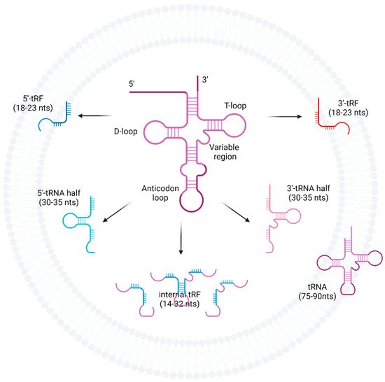
Figure 1. Fragmentation of tRNAs into tRFs, tRNA-halves, and internal tRFs. tRNAs are cleaved at the anti-codon loop by enzymes such as angiogenin, Dicer, or RNase Z into either 5′ or 3′-tRNA-halves. tRFs can arise from mature tRNA, pre-tRNA as well as tRNA-halves, and are formed when cleavage occurs at either the D-loop or T-loop. However, the enzymes responsible for tRF production are less well understood.
2. Current Insights
The researchers have focussed on the tRNAs, tRNA-halves, and tRFs associated with human EVs, in health and pathological disease states. They have described the various biofluids used for EV capture and characterised the different EV isolation methods used in protocols for the subsequent identification of EV tRNAs contained within. Importantly, we have shown that multiple pathologies and especially malignancies can be distinguished by differential EV tRNA expression and may thus be of clinical significance for both diagnosis and prognosis of the disease.
The abundance of tRNAs contained within EVs is highly variable and may depend on tissue and disease type, ranging from as low as 0.04% in the blood plasma EVs of gastric cancer patients [41] to as high as >95% from EVs obtained from healthy placental syncytiotrophoblasts [32]. However, tRNAs appear to be a consistent finding in EVs, occurring in sequencing samples with as high a prevalence (and sometimes higher) as miRNAs. Additionally, EV isolation methods will need to be tailored to the requirements of the experiment, with highly selective methods such as density gradient preferable when contamination with RBP is to be avoided. We found that 46.5% of the protocols used precipitation as their EV isolation method, and this has been recognised as having relatively low purity [77,78]. Moreover, the technological limitations of sequencing methods may further add to the complexity of interpreting the EV tRNAs found. Our review highlighted a few limitations of current sequencing technologies. In many studies, the library preparation is limited to specific size ranges [40] and low abundance species can lead to selective biases in enrichment. [21] Moreover, conventional sequencing methods may not effectively capture RNAs that are post-transcriptionally modified [23]. Finally, Tosar et al. [79] highlighted how piRNA transcripts differ from some tRNA-halves by only one nucleotide. Individual piRNA and as well as other small cytoplasmic RNA species are highly homologous to major tRNA fragments within commonly used databases; thus, mapping of reads may lead to an under- or over-representation of tRNA fragments. The limitations encountered with sequencing technologies could be addressed through novel pipelines such as that reported by Amorim et al. [22]. Ongoing collaborative work such as the Extracellular RNA Communication Consortium has made efforts to improve this and work towards strategies such as deconvolution to determine tRNA sources [80].
It is increasingly recognised that tRNA-derived fragments have a role in mammalian cells, with 5′-tRNA-halves having been associated with actions on the ribosome leading to stress granule formation [81] and translational inhibition. Smaller tRFs share a size similarity to miRNAs and there is significant evidence in the wider (non-EV) literature that they can have actions in conjunction with Argonaute [82], the Piwi subfamily, and other RNA-binding proteins [82,83].
tRNAs and their fragmentation products have been found as an EV biomarker in circulating fluid and tissue for a number of malignancies including leukaemia, prostate, ovarian, pancreatic, and colorectal cancers [84,85]. In our study, breast cancer showed particular promise with several differentially expressed tRNAs (tRF-Arg-CCT-017, tRF-Gly-CCC-001, and tiRNA-Phe-GAA-003) associated with key regulatory pathways such as Wnt signalling, classically involved in cancer initiation and progression. [86] In fact, several other malignancies such as colorectal cancers [87], gastric cancers [88], and glioblastomas [89] also secrete cancer-stem-cell-associated EVs, which act on the β-catenin/Wnt-signalling pathways to increase stemness, in turn increasing their tumorigenic potential.
In addition, the majority of the studies in our review showed that the fragmentation products of tRNAs, namely 5′-tRFs and 5′ tRNA-halves, were the most abundant sub-species of tRNAs identified in EVs. A possible reason for the observation of 5′ products was shown to be potentially due to reverse transcription failure due to post-transcriptional modifications in otherwise full-length tRNAs by Shurtleff et al. who used an alternative thermostable sequencing system. What is also clear is that simple analysis of tRNA molecules by broad amino acid (isoacceptor types) may not be sophisticated enough to determine potentially clinically relevant changes in EV tRNA profiles. Further validation of sequencing by qRT-PCR is necessary to prove true differential expression and this means additionally incorporating the standardised use of tRNA molecular terminology.
We have also highlighted that several biological mechanisms have been clearly associated with changes in tRNA-halves levels. For instance, osteoblastogenesis revealed that BMSCs alter their cargo content in EVs from full-length tRNAs to tRNA-halves as their differentiation progresses [62]. We have noted the evidence that there are time-dependent changes in the EV tRNA profile of cancer patients and may perhaps reflect clinical changes such as disease burden [36]. In addition, tRFs have even been shown to possess potential diagnostic potential in a multitude of diseases.
tRNA-sorting mechanisms in EVs have also been the focus of various studies, with RNA-binding proteins [23], surface markers [26], and tRNA enzymatic stability as some of the mechanisms identified [25]. Potential therapeutic uses of EV tRNA include the loading of synthetic oligonucleotides into EVs via parent cells to alter downstream gene expression in donor cells [25]. This particular paper by Gambaro et al. [25] demonstrated key mechanisms for tRNA sorting, such as the concentration-dependent transfection and selection of stable synthetic oligonucleotides against enzyme degradation, through well-controlled transfection experiments.
Due to the heterogeneity of the studies included in our review, a meta-analysis could not be conducted; thus, a narrative synthesis of the literature was undertaken. Many studies may include tRNA data but not explicitly mention this and will have been excluded. We also restricted our search to articles involving human cell models or patient-derived biofluids which may exclude important studies using animal EV models that could have shed some light on tRNA markers across other species.
This entry is adapted from the peer-reviewed paper 10.3390/ijms23073692
This entry is offline, you can click here to edit this entry!