You're using an outdated browser. Please upgrade to a modern browser for the best experience.
Role of Epicardial Adipose Tissue in Cardiovascular Diseases: History
Please note this is an old version of this entry, which may differ significantly from the current revision.

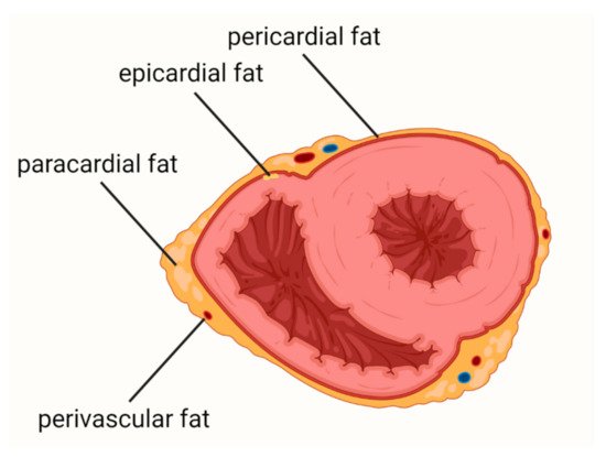
Cardiovascular diseases (CVDs) are the leading causes of death worldwide. Epicardial adipose tissue (EAT) is defined as a fat depot localized between the myocardial surface and the visceral layer of the pericardium and is a type of visceral fat. EAT is one of the most important risk factors for atherosclerosis and cardiovascular events and a promising new therapeutic target in CVDs. In health conditions, EAT has a protective function, including protection against hypothermia or mechanical stress, providing myocardial energy supply from free fatty acid and release of adiponectin. In patients with obesity, metabolic syndrome, or diabetes mellitus, EAT becomes a deleterious tissue promoting the development of CVDs. 

  • atherosclerosis
  • cardiovascular diseases
  • epicardial adipose tissue
  • EAT
  • inflammation

1. Introduction

Cardiovascular diseases (CVDs) remain one of the leading causes of death worldwide [1,2], entailing enormous costs for healthcare systems [3]. Many of them could be avoided, as cardiovascular risk factors are largely reversible.
Epicardial adipose tissue (EAT) is defined as a fat depot localized between the myocardial surface and the visceral layer of the pericardium, and is a type of visceral fat [4]. Therefore, as is the case with abdominal obesity, EAT is one of the most important risk factors for atherosclerosis and cardiovascular events [5,6,7]. Moreover, the volume and thickness of EAT correlate with intra-abdominal fat mass and severity of obesity [8,9] and are independently associated with cardiovascular events [10]. Additionally, there are reports that EAT may be a new therapeutic target in CVDs [11,12].
The most common classification of the adipose tissue surrounding the heart includes (i) epicardial adipose tissue, (ii) pericardial adipose tissue, (iii) paracardial adipose tissue, and (iv) perivascular adipose tissue (Figure 1). Epicardial fat is located below the visceral pericardium. Pericardial fat consists of adipose tissues between the visceral and parietal pericardial layers and the fat depot on the external surface of the parietal pericardium. Paracardial fat involves fat deposits outside the parietal pericardium. The perivascular adipose tissue is a fat around the coronary arteries, irrespective of location [13].
 
Figure 1. Adipose tissue surrounding the heart.
There are many differences between EAT and other types of adipose tissue, including anatomical, histological, embryological, and genetic differences [14,15]. EAT is located between the pericardium and the myocardium [5] and is not separated from the myocardium and vessels by fascia, allowing paracrine or vasocrine effects [16] via cytokines and chemokines [17]. In health conditions, EAT has a protective function, including protection against hypothermia [18] or mechanical protection for the coronary circulation [19]. Additionally, EAT has an important role in energy supply to the myocardium [20]. Thanks to the ability to use free fatty acid (FFA) quickly, EAT may protect the myocardium from the cardiotoxic effect of a large amount of FFA [21]. The secretion of adiponectin from epicardial adipocytes is also an important function of EAT. Adiponectin protects coronary circulation, improves endothelial function, reduces oxidative stress, and indirectly decreases the level of interleukin-6 (IL-6) and C-reactive protein (CRP) [22,23,24]. However, under specific conditions such as obesity, metabolic syndrome, or diabetes mellitus, the protective properties may be destroyed and EAT becomes a deleterious tissue promoting the development of CVDs.

2. Coronary Artery Disease

It has been shown that EAT and CAD are closely related at different levels: (i) in patients with CAD, the secretion of adipokines from EAT is altered; (ii) EAT has a proinflammatory features in patients with CVD risk factors and/or CAD; (iii) the amount of EAT and/or its proinflammatory state correlate with the severity of CAD and the instability of the atherosclerotic plaques; (iv) there is a relationship between EAT’s function and coronary microvascular dysfunction and artery spasm [14,17,26,27,28,29,30,31,32,33,34,35,36,37,38,39,40,41,42,43,44,45,46,47,48,49].
In patients with obesity, metabolic syndrome, or CAD, the epicardial adipocytes secrete less adiponectin and more leptin than in healthy people [26,27]. The decreased adiponectin expression attenuates endothelial function and leads to increased tumor necrosis factor-α (TNF-α) production, which increases inflammation and oxidative stress. The increased leptin level promotes adhesion of monocytes, macrophage-to-foam cell transformation, and unfavorable changes in lipid and inflammatory cytokine levels in adipose tissue [28]. All these processes result in the development and destabilization of atherosclerotic plaques [29].
Inflammation plays a crucial role in atherosclerosis, and EAT as a tissue with proinflammatory properties provides a huge contribution to coronary plaque formation [14,17,30,31].
Wang et al. assessed EAT in computed tomography (CT) in patients with and without diabetes, showing that EAT volume (EATV) is higher in diabetic patients and is associated with components of metabolic syndrome, coronary atherosclerosis, and coronary calcium scores [33]. In the Framingham Study, a significant association was found between epicardial fat and coronary artery calcification, which was significant after adjustment for traditional cardiovascular risk factors [34]. In women with chest pain and angiographically normal coronary arteries, there was a correlation between EAT thickness and reduced coronary flow reserve [46]. Kanaji et al. showed that in CAD patients with a single de novo lesion, PCAT attenuation is significantly associated with global coronary flow reserve [47].
Finally, attention should be paid to the current tendency to study the relationship of CAD not only with the thickness and volume of EAT, but also with its structure and size of adipocytes [50,51]. One study has found that the size and degree of hypertrophy of the epicardial adipocytes are related to CAD severity [51].

3. Heart Failure

Among patients with HF, approximately 50% have preserved ejection fraction (HFpEF). HFpEF is a heterogeneous disease with a complex pathogenesis which is not fully understood. This complexity is due to the fact that it can be caused or exacerbated by a variety of comorbidities, including cardiac and extracardiac abnormalities. Thus, the group of patients with HFpEF is very diverse [52,53]. HFpEF is the most common myocardium disorder among obese patients [54].

Based on the hitherto studies, it can be concluded that: (i) there is an association between EATV and the development of HfpEF; (ii) patients with HFpEF and obesity represent a distinct phenotype of the disease; (iii) EAT thickness or volume may have a greater impact on HFpEF than obesity per se; (iv) EAT participates in the pathogenesis of HfpEF due to EAT’s proinflammatory properties, intensification of fibrosis, and influence on myocardial substrate utilization.

There is a correlation between the severity of left ventricle (LV) diastolic dysfunction and the volume of EAT [56,57,58]. 

Patients with coexisting obesity and HFpEF had a different clinical phenotype than patients with HFpEF without obesity [60]. Obokata et al. compared patients with HFpEF and obesity, HPpEF without obesity, and a non-obese control group without HF [60]. Among obese HFpEF patients, diabetes and sleep apnea were more prevalent, whereas in the non-obese HFpEF patients, atrial fibrillation was more common. Additionally, the obese HFpEF patients had lower concentrations of N-terminal prohormone of brain natriuretic peptide (NT-proBNP), compared to the non-obese cohort. Furthermore, subjects with the obese HFpEF phenotype had increased plasma volume, a higher rate of concentric LV remodeling, greater right ventricular (RV) dilatation, and a higher rate of RV dysfunction. Obese patients also displayed worse exercise capacity, more pronounced hemodynamic abnormalities during exercise, and impaired pulmonary vasodilation. EAT thickness assessed by echocardiography was 20% higher in the obese HF group compared to non-obese HF, and 50% higher compared to the control group [60].

In obese patients with increased plasma volume, the ability of LV to dilate is insufficient, leading to cardiac overfilling and disproportionate increases in cardiac filling pressures. It seems that the inadequate ventricular distensibility is caused by cardiac fibrosis and microvascular rarefaction [65,66]. Moreover, the quantity of fibrosis assessed in CMR is associated with prognosis and outcomes in HFpEF [67]. Obese patients displayed more EAT [8,9] and therefore it seems likely that they were more exposed to cytokines released from the EAT reservoir.

4. Atrial Fibrillation

AF is the most common arrythmia in the adult population in the world, and its prevalence is increasing. It is estimated that AF affects up to 4% of the population in Australia, Europe, and the USA [76]. The involvement of hemodynamic stress in the pathogenesis of AF is well-documented, and hypertension is the most common risk factor [77]. Valvular diseases also significantly contribute to the development of this arrhythmia [78]. These disorders cause the remodeling of heart chambers, including enlargement of the left atrium (LA) and an increase in LA pressure. Alleviation of hemodynamic stresses can reduce AF’s burden [77,78]. However, there is a large group of patients with AF who do not have hypertension or valvular disease but do have the features of atrial myopathy (LA enlargement, increased LA pressure), as observed in imaging studies [79]. It is known that inflammation is associated with the development of atrial myopathy [80], including both inflammation in course of systemic inflammatory diseases [81,82,83,84] and metabolic disorders accompanied by adipose tissue inflammation [85,86]. The risk of developing AF is especially increased in rheumatoid arthritis [81] and psoriasis [82]. Among the metabolic diseases, special attention should be paid to obesity [85] and diabetes mellitus [86]. In these states, AF’s burden was proportional to the severity of metabolic disorders, such as glycemic control [85,86,87,88].
Several potential mechanisms linking EAT with AF, including: (i) proinflammatory status of EAT; (ii) reactive oxygen species (ROS) released from EAT; (iii) fatty infiltration of the atrium; (iv) dysfunction of the autonomic nervous system in EAT.
AF and inflammation are closely associated [80,81,82,83,84,85,86]. EAT can release inflammatory factors and contribute to inflammation and fibrosis in the adjacent myocardium via paracrine signaling. It should be emphasized that EAT has some features of brown adipose tissue, such as the presence of the uncoupling protein-1, which is a thermogenic protein specific to brown adipocytes [89]. These properties are mainly expressed in conditions of health and low oxidative stress [90,91,92]. The healthy EAT is a source of adiponectin, which may reduce inflammation and fibrosis [91,92,93,94]. In obesity, EAT loses its protective properties and becomes a tissue with a proinflammatory profile, subsequently increasing the risk of atrial myopathy and AF [95,96,97,98,99]. Mazurek et al. showed that inflammatory activity of EAT reflected by maximal standardized uptake value of FDG in PET/CT was higher in patients with AF than in the control group and it was not related to BMI [100].
It has been suggested that ROS play an important role in the pathogenesis of AF [101,102]. EAT has been shown to be richer in ROS than other fat depots [103], but at the same time, this effect was reduced by adiponectin [93].
It also seems that fatty infiltration into atrial myocardium plays an important role in the pathogenesis of AF, as demonstrated by histological examinations [104]. Fatty infiltration was more pronounced in persistent AF, compared with paroxysmal AF [105]. EATV and fatty infiltration were associated with cardiac conduction abnormalities [106]. It was postulated that EAT can change electrophysiological features and ion currents by cytokine, adipokine, and adipocyte infiltration, causing electrical substrate formation for AF [107].
It should be noted that EAT contains significant amounts of ganglionated plexi which are a part of the autonomic nervous system (ANS), which may play a role in the pathogenesis of AF [109,110]. The thickness of the EAT was related to ANS dysfunction [111], and catheter ablation of epicardial fat-reduced cardiac ANS activity, which makes it an interesting therapeutic perspective [112].
There are also several other less-understood potential mechanisms which may explain the involvement of EAT in the pathogenesis of AF, such as the local aromatase effect [113,114,115]. A significant positive correlation was determined between the total aromatase content of EAT and the occurrence/duration of triggered atrial arrhythmias [114]. Further mechanisms are pending investigation.
 

This entry is adapted from the peer-reviewed paper 10.3390/biology11030355

This entry is offline, you can click here to edit this entry!
Academic Video Service