Your browser does not fully support modern features. Please upgrade for a smoother experience.
Please note this is an old version of this entry, which may differ significantly from the current revision.
Subjects:
Neurosciences
In 2012, Iliff et al., for the first time, identified a novel structure in the brain called the glymphatic system. This system is considered as a crucial fluid-clearance system in the brain.
- photobiomodulation
- near-infrared light
- glymphatic system
- meningeal lymphatic vessels
1. The System, Its Components, and Pathways
Based on physiological findings of communication among different parts of the brain, the existence of a specific lymphatic drainage system in the brain of vertebrates has been suggested [18,19]. In 2012, Iliff et al., for the first time, identified a novel structure in the brain called the glymphatic system [20]. This system is considered as a crucial fluid-clearance system in the brain [21,22]. Studies on mouse models using different fluorescent tracers constructed this glymphatic drainage pathway in the brain [23,24].
This system consists of five main functional components, each facilitating the movement of CSF and ISF (Figure 1). The first compartment of the glymphatic system consists of the production of CSF by epithelial cells of the choroid plexus in the cerebral ventricles and circulation of CSF in the subarachnoid space, followed by the second, periarterial influx of CSF into the brain parenchyma. In fact, periarterial influx refers to the entrance of CSF into the periarterial spaces surrounding the arteries and its penetration deep into the brain tissue. Arterial pulsation caused by smooth muscle cells intensifies CSF movement inward along the periarterial space [25]. Exchange of CSF and ISF is the third component of this system, which occurs in the interstitial space of the brain parenchyma (Figure 2).
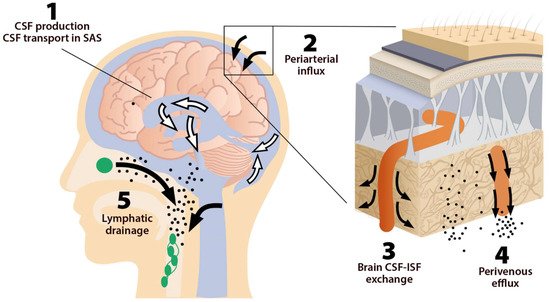
Figure 1. The five components of the glymphatic system. The fluid transport pathway is divided into five distinct segments: (1) cerebrospinal fluid (CSF) is produced by the choroid plexus and likely by extrachoroidal sources (capillary influx and metabolic water production); (2) arterial wall pulsatility drives CSF deep into brain along perivascular spaces; (3) CSF enters the brain parenchyma supported by aquaporin-4 (AQP4) water channels and disperses within the neuropil; interstitial fluid (ISF) mixes with CSF, (4) accumulates in the perivenous space, and drains out of the brain via (5) meningeal and cervical lymphatic vessels, as well as along cranial and spinal nerves Fluids from both the brain and the cribriform plate drain into the cervical lymphatic vessels, which then empty into the venous system at the level of the subclavian veins. The olfactory/cervical lymphatic drainage route is the primary bulk flow pathway.
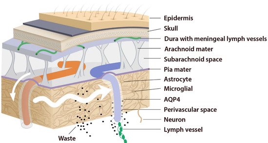
Figure 2. Periarterial influx of CSF into the brain tissue (small white arrow). CSF–ISF exchange supported by AQP4 channels in the vascular end feet plastered along the arterioles. From here, the fluid leaves the axons and moves towards the perivenous space in a path supported by astrocytes. Astrocytic AQP4 water channels facilitate this perivenous efflux of interstitial fluid, which drains to the dural lymphatic vessels.
Astrocytes are believed to facilitate the fluid movement between periarterial spaces and the interstitium through water channels such as aquaporins-4 (AQP4) [20,26]. The fourth component is the glymphatic efflux, which consists of drainage of ISF into the perivenous spaces. The meningeal lymphatic system is the fifth component and final downstream clearance of the glymphatic system. MLVs drain waste products and other solutes from the CNS [9]. This ISF then flows towards the leptomeningeal arteries located at the cortical surface (sulci) and ultimately moves into the cervical lymphatics [20].
Indeed, this system was named “glymphatic” based on the involvement of glial cells “gl” and its similar function with the “lymphatic system” [27,28]. The brain glymphatic system has several essential physiological functions such as drainage of ISF from the parenchymal section of the brain to nearby lymph nodes. It is also involved in communication with the immune system, which regulates and monitors brain responses to neuroinflammation [29]. Moreover, the glymphatic system possesses numerous physiological functions in addition to solute clearance [30]. It is hypothesized that the glymphatic system has a role in rapid lipid transportation across the blood–brain barrier (BBB) and promote glial signaling [31]. Additionally, CSF is involved in the transportation of apolipoprotein E, essential for cholesterol transport, and most notably, synaptic plasticity [32]. CSF influx is also a vehicle for glucose and other vital nutrients that are necessary for the metabolism of astrocytes and neurons [30].
Lifestyle factors, genetics, and pathological conditions can modulate brain clearance and influence the risk of developing neurodegenerative diseases [33]. Several factors such as genetic phenotypes, body posture, aging, and the sleep–wake cycle could influence these physiological functions [23] so that an impaired cerebral lymphatic system is counted as a risk factor for neurodegenerative [34], neuroinflammatory [12], and neurovascular diseases [13] and tumors, as well as impaired recovery from brain injuries [12] (Figure 3). Pathological conditions can strongly affect the brain lymphatic systems. In various vascular disorders including hypertension, atherosclerosis, and small vessel diseases [35], any alteration in the composition of the constituent proteins can result in a significant decline in vascular plasticity and decrease cerebral blood flow (CBF) into the perivascular pathways. In arterial stenosis (either cervical or intracranial), blockage of CBF and obstruction of perivascular or paravascular channels are observed [13], leading to reduced ISF flow resulting in loss of CSF clearance from the brain. Glymphatic system dysfunction has been demonstrated to be associated with many neurological diseases such as AD and PD [14,28]. The glymphatic system has been described as the “final common pathway” for neurodegenerative diseases [36].
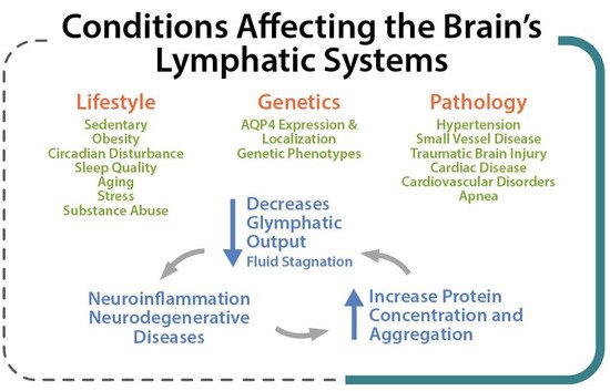
Figure 3. Lifestyle, Genetic and Pathological conditions that can strongly affect the brain lymphatic systems.
2. MLVs, Olfactory/Cervical Lymphatic Drainage Route, and Their Association with CSF Circulation
The absence of a conventional lymphatic vasculature in the CNS prompted a series of studies on rodents and human brains to identify MLVs as the lymphatic system of the CNS [37,38]. MLVs seem to provide a critical route for drainage of ISF and CSF. Various macromolecules and immune cells pass from CNS into the lymph nodes located in the deep cervical area [39,40,41]. More recently, strong evidence demonstrated that MLVs might be associated with the regulation of immune responses and also involved in the pathogenesis of neuroinflammatory diseases [42]. Animal studies have also shown impaired meningeal lymphatic function in AD [9] and PD [14]. In a neuroimaging study using a dynamic contrast-enhanced MRI, patients with idiopathic PD exhibited markedly decreased flow through the MLVs along the superior sagittal sinus and sigmoid sinus, as well as a significant delay in deep cLNs (dcLNs) perfusion [43].
Under normal physiological conditions, the olfactory/cervical lymphatic drainage route serves the primary bulk flow drainage pathway. The ethmoid bone and particularly the cribriform plate located at the anterior aspect of the brain (between the anterior cranial fossa and the nasal cavity) is considered a critical extracranial site of CSF outflow [44]. CSF in subarachnoid space passes through the cribriform plate along the olfactory nerves to the nasal lymphatics and cLNs. At the end of the route, CSF is deposited into the extracranial lymphatic system [13]. The continuous circulation and drainage of CSF are critical for removing CSF metabolic products and maintaining normal neural functions. The outflow routes of CSF are the arachnoid villi of the dural superior sagittal sinus [45], olfactory nerves, across the cribriform plate, and into the cervical lymphatic pathway [46].
The cribriform plate is a fenestrated bony plate of the ethmoid that separates the cranial and nasal cavities (Figure 4). Even though there are lymphatic vessels in the meninges [47], it has been demonstrated that CSF can drain through the cribriform plate in both humans and other mammals [48]. The main pathway by which CSF is removed from the skull is through the cribriform plate associated with the olfactory nerves [49]. The CSF is absorbed by lymphatic vessels located in the submucosa of the olfactory epithelium, in the nasal mucosa after passing the cribriform plate, and then drained into the cLNs. Any damage to the cribriform plate (by traumatic brain injuries or surgical methods) can lead to acute blockage of CSF outflow and, as a result, increase in resting intracranial pressure (ICP) and outflow resistance, emphasizing that the olfactory pathway represents the leading site for the CSF drainage [50]. There is a space between the olfactory sensory axons that provides a conduit for the outflow of CSF. Any damage to these nerves can also diminish the outflow of CSF through the cribriform plate [49] (Figure 4).
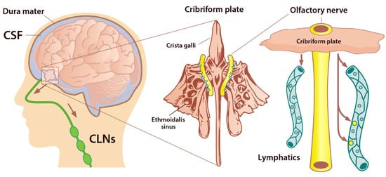
Figure 4. Perineural space surrounding olfactory nerve penetrates the nasal mucosa through the cribriform plate. The cribriform plate of the ethmoid bone is considered the key extracranial site of CSF outflow. CSF in SAS passes across the cribriform plate along olfactory nerves to nasal lymphatics and enters cervical lymph nodes (adapted from Semyachkina-Glushkovskaya et al. 2021).
AQP are a family of small integral membrane proteins that significantly boost the permeability of cells to water and facilitate the movement of fluid down the pressure gradient in various tissues, including the brain [51]. So far, 13 AQPs have been found in mammals (AQP0–12). AQP1 maintains CSF production by the choroid plexus, and it is also expressed along the periphery of the olfactory bulb, nerve junction, and lining the foramina of the cribriform plate. Moreover, there are high levels of AQP1, 3, and 5 within the nasal cavity. These AQPs facilitate the flow of fluid out of the olfactory bulb and subarachnoid space into the nasal cavity via the extensive network of lymphatic vessels, which play an essential role in moving fluid throughout the body. AQPs are found in the meninges and at the cribriform plate and olfactory bulb junction [11]. These vessels crossing the cribriform plate play a key role in transporting fluid from the cranial cavity to the nasal cavity olfactory sensory nerves. Following CSF absorption by lymphatics, it is conveyed in larger ducts through numerous lymph nodes and eventually is deposited into the body’s lymphatic system. Evidence has also shown that aging decreases the elimination rate of CSF from the nasal/cribriform plate region [52,53].
3. Sleep and Clearance of the Brain
The glymphatic system uses convective flow between the CSF and ISF to remove toxic metabolites in/from the brain. CSF enters the brain parenchyma (functional parts) along a paraarterial route and exchanges with the ISF [54]. The ISF carries extracellular solutes from the interstitial (extracellular) space in the brain along paravenous drainage pathways (Figure 1). This activity is dramatically boosted during sleep and is related to increased interstitial volume, possibly by shrinkage of astroglial cells [55]. Emerging evidence shows that sleep is the primary driver of glymphatic clearance and is essential for the maintenance of brain function via the discharge of metabolites and neurotoxic wastes from the brain, which accumulates in the highly active brain during waking hours [36,56].
Comparing the brain ISF volume during deep sleep to wakefulness, the volume of the brain’s ISF increases by 40–60% [57]. Astrocytic AQP4 water channels that encircle the brain’s vasculature contribute to this increase in ISF. This increase in ISF is required for proper glymphatic function and facilitates the clearance of soluble proteins, waste products, and excess extracellular fluid. ISF increase leads to a 2-fold faster removal of neurotoxic waste products such as lactate and Aβ from the brain. This increase in the clearance of brain waste happens during non-rapid eye movement (NREM) sleep [58], and the majority of glymphatic activity occurs during deep, slow-wave sleep. Poor sleep quality and short sleep duration result in an increased amount of Aβ in the CSF as well as a risk of Aβ plaque formation [59]. In addition, tau levels have been shown to be increased in the ISF of the hippocampus following sleep deprivation [60]. Evidently, these neurobiological mechanisms can support the fact that neurodegenerative diseases such as AD, PD, Huntington disease, and frontotemporal dementias are strongly linked to sleep disturbances [61]. With the glymphatic system in mind, it is of interest to note that sleep quality decreases as a function of normal aging, and individuals over 60 years old rarely enter deep NREM (stages 3). The effectiveness of glymphatic fluid transport is directly linked to the prevalence of slow-wave activity. Therefore, the age-related impairment in sleep quality can cause a catastrophic drop in the clearance of brain waste and potentially increase the incidence risk of neurodegenerative diseases [36].
Evidence revealed that endocytosis occurs across the BBB during sleep, and inhibition of this process causes the need for more sleep [62]. In addition, several studies have reported that sleep deprivation can increase the activity of several pro-inflammatory mediators such as C-reactive protein, interleukin (IL)-1β, IL-6, IL-17, interferon-γ (IFN-γ), and tumor necrosis factor-alpha (TNF-α). These mediators suppress astrocytic maintenance of the BBB, causing an increase in its permeability [63,64]. Sleep deprivation has been shown to decrease influx efficiency along the perivascular space, thus impairing the function of the glymphatic system and disturbing AQP4 polarization in a mouse model [65].
This entry is adapted from the peer-reviewed paper 10.3390/ijms23062975
This entry is offline, you can click here to edit this entry!