Bête Noire of Chemotherapy and Targeted Therapy: History
Please note this is an old version of this entry, which may differ significantly from the current revision.
Contributor: , , ,

Tumor cells struggle to survive following treatment. The struggle ends in either of two ways. The drug combination used for the treatment blocks the proliferation of tumor cells and initiates apoptosis of cells, which is a win for the patient, or tumor cells resist the effect of the drug combination used for the treatment and continue to evade the effect of anti-tumor drugs, which is a bête noire of therapy. Cancer-associated fibroblasts are the most abundant non-transformed element of the microenvironment in solid tumors. Tumor cells play a direct role in establishing the cancer-associated fibroblasts’ population in its microenvironment.

  • cancer-associated fibroblasts
  • resistance
  • chemotherapy

1. Introduction

Cancer-associated fibroblasts (CAFs) within the tumor microenvironment (TME) are non-transformed, tumor-cell-activated heterogeneous populations of cells having multiple origins and functions [1,2]. Detailed descriptions of the origin, functions, interactions with tumor cells, and heterogeneity of CAFs were previously provided by us elsewhere [3,4]. CAFs are activated by tumor cells in their favor. Once activated in an established tumor, CAFs act as crucial supporters of tumor growth, progression, and response to treatment.
The functions of CAFs in an established tumor include the following: (1) ECM (extracellular matrix) remodeling via collagenolysis to promote invasion and EMT (endothelial–mesenchymal transition); (2) increasing tissue stiffness to initiate angiogenic resistance and immune suppression; (3) induction of tumor angiogenesis; (4) secretomic induction of EMT by TGFbeta; (5) increasing secretomic factors of tumor-promoting or immune-suppressing ligands such as hepatocyte growth factor; fibroblast growth factors 1 and 2; stromal cell-derived factor 1 (SDF1/CXCL12); chemokine (C-C motif) ligands (CCL) 2, 5, 7, and 16; interleukin 6/8; and platelet derived growth factor; (6) metabolic reversal of reverse Warburg effect (non-glycolysis in tumor cells, glycolysis in stroma cells) and ‘lactate shuttle’ effect; (7) immune evasion via activation of M2 macrophages (CD163 positive); (8) inhibition of apoptosis in tumor cells; (9) activation of many of pro-proliferative tumor cell signaling; (10) immune reprogramming and antigen presentation; (11) adaptation to oxidative stress and hypoxic response; (12) promotion of stemness-promoting signals; (13) promotion of metastasis-associated phenotypes; (14) attenuation of drug response [1,5,6,7,8,9,10,11,12,13,14,15,16,17,18].
The range of functions of CAFs is comprehensive, and the actions of CAFs are contextual. The interactions of CAFs with tumor cells and TME components change with the evolution of the tumor, its metastatic progression, and its response to therapy. In summary, the functions of CAFs are structured to assist and promote tumor cells via direct and indirect interactions. Thus, CAFs form a centralized communication network within the TME that favors tumor cell growth, metastasis, and resistance to drug treatment [19]. The versatility of the functions of CAFs’ make them abettors of drug resistance and identifies them as prospective anti-tumor therapy targets [20,21].

2. CAF Heterogeneity and Resistance to Chemotherapy in Solid Tumors

2.1. CAF Heterogeneity

CAFs are heterogeneous in terms of their origin in different organ-type cancers, as well as in the progression of the disease. The heterogeneous subpopulations of CAFs, such as myoblastic CAFs (myCAFs) and inflammatory CAFs (iCAFs), have been extensively studied in fibroinflammatory PDAC disease characterized by dense and highly proliferating desmoplastic stroma. In fact, Li et al. identified genes associated with the differentiation of myCAFs and iCAFs [22,23,24]. Adipose-derived MSCs (AD-MSCs) have been shown to possess a high multilineage potential and self-renewal capacity and were reported as the CAF sources in PDAC by Miyazaki et al. [24]. Their study identified that AD-MSCs could differentiate into distinct CAF subtypes, myCAFs and iCAFs, depending on the different co-culture conditions in vitro. The diverse functions of iCAFs and myCAFs have also been reported in cholangiocarcinoma; breast cancers; prostate, head, and neck squamous cell carcinoma; and bladder and colon cancers. The diversity of CAF subpopulations was also recently reported to promote the growth of cholangiocarcinoma, wherein hepatic stellate cells (HSC) are the primary cause of CAF differentiation into myCAFs and iCAFs [25]. The hyaluronan synthase 2 myCAFs, but not type I collagen-expressing myCAFs, promoted tumor progression, while HGF-expressing iCAFs enhanced tumor growth via tumor-expressed MET, thereby directly linking CAFs to tumor cells. Another subset of CAFs, FAP+CAFs, were identified by Kieffer et al. in breast cancers that mediated immunosuppression and immunotherapy resistance via a positive feedback loop between specific CAF-S1 clusters and Tregs [26]. In prostate cancer, a differential mode of activation of iCAFs and myCAFs has been reported [27]. IL-1a/ELF3/YAP pathways are involved in iCAF differentiation, while TGF-beta1 induces myCAFs. One of the ways CAFs classically interact with the tumor cell EMT function was reported by Goulet et al. in bladder cancer, where IL-6 cytokine was found to be highly expressed in iCAFs, and its receptor IL-6R was found on RT4 bladder cancer cells [28]. Perhaps the most intriguing functional heterogeneity of CAFs was reported by Pan et al. in PDAC-CAF-exhibited organ-specific metastatic potential leading to different levels of heterogeneity of CAFs in different metastatic niches [29]. Several cell signaling pathways have been reported to be involved in the functioning of iCAFs and myCAFs, including the Hedgehog pathway [30]; Wnt pathway [31]; integrin a11B1 signaling [32]; cMET-HGF pathway [25]; IL-6 signaling [28]; EMT signaling via transcription factors SNAIL1, TWIST1, and ZEB1 [28]; and IL1B-mediated crosstalk [33]. Recently, Steele et al. reported that the Hedgehog pathway acts in a paracrine manner in PDAC, with ligands secreted by tumor cells signaling to stromal CAFs. The Hedgehog pathway activation is higher in PDPN+ alphaSMA+ myCAFs compared with iCAFs, and its inhibition impairs tumor growth by altering the fibroblast compartment in PDAC. Hedgehog pathway inhibition resulted in a reduction in myCAF numbers and a significant expansion of iCAFs, leading to an increase in the iCAF/myCAF ratio. As iCAFs are a source of inflammatory signals, the authors observed an increase in iCAFs upon Hedgehog inhibition, which correlated with changes in immune infiltration (significantly decreased CD8+ T cells and increased CD4+ T cells and CD25+CD4+ T cells; abundant FOXP3+ regulatory T cells) that are consistent with a more immunosuppressive pancreatic cancer microenvironment. The paracrine activation differentially elevated myCAFs compared with iCAFs, leading to favorable alterations of cytotoxic T cells and Tregs, causing increased immunosuppression [30]. Wnt signaling in CAFs represents a non-cell-autonomous mechanism for colon cancer progression [31]. Mose et al. reported Sfrp1 epithelial–mesenchymal transition phenotype induction in tumor cells without affecting tumor-intrinsic Wnt signaling, suggesting involvement of non-immune stromal cells. Low levels of Wnt signals induced the iCAF subtype, which in co-culture with organoids induced EMT, whereas high levels induced contractile myCAFs to attenuate the EMT phenotype.
The tumors with (1) an accumulation of stromal CAFs, (2) the presence of fibrotic stroma, (3) a high expression level of stroma signature genes, or (4) a high tumor/stroma ratio in the primary tumor are associated with poor prognosis in various cancers, including colon, gastric, esophagus, breast, NSCLC (non-small cell lung cancer), and liver cancers [34,35,36,37,38,39,40]. It is understood that chemotherapy’s limited effect (benefit) and the progression or recurrence of disease through therapy in many solid tumors are attributed to the development of resistance within tumor cells in support of the stroma. As a dominant component of tumor stroma, CAFs interact with both a tumor cell and the TME. The versatility of CAF functions and their several modes of interaction with tumor cells and all components of stroma (ECM and cells of the TME) indicate that a metastasis or progression of disease following treatment is aided and abetted by CAFs. Once a therapy-resistive circuitry is established between tumor cells and the CAFs of the stroma, tumor-centric therapy alone essentially becomes insufficient. 
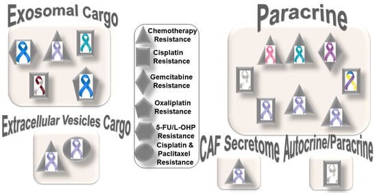
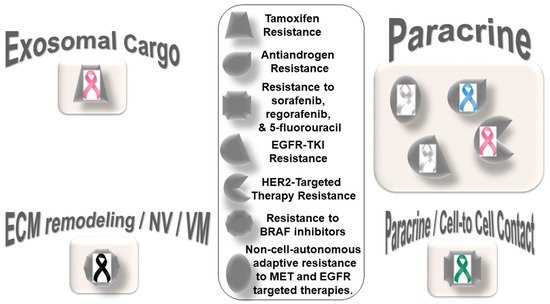
Figure 1 presents the distribution pattern of the types of resistance to chemotherapy based on specific mediators of CAF functions in solid tumors. The four types of mediators of action employed by CAFs to orchestrate the development of resistance to chemotherapy are presented in the cartoon. The most common mode of interaction is a paracrine, wherein CAFs signal to either tumor cells or other components of the TME via characteristic secretomes. In addition to the involvement of characteristic secretomes, exosomal cargos delivering different miRNAs that target various cell signaling proteins are common mediators of CAFs. The paracrine mode of action of CAFs is the predominant form of action, represented by six types of organ tumors (organ tumors are indicated by their respective ribbon colors, as presented in the figure legends). CAF crosstalk with tumor cells, and the TME occurs via exosomal cargo, imparting resistance to four organ cancers. The extracellular vesicle, secretome, and autocrine or paracrine modes are much less involved in the modes of action (Figure 1). The sizes of the boxes indicate the number of studies in each box. Among resistance to different types of chemotherapies, cisplatin resistance has been found to be very common, which is involved in both paracrine and exosomal cargo modes of action (the shapes in the inset indicate the types of resistances in different tumors).
Figure 1. Distribution pattern of types of resistance to chemotherapy based on specific mediators of CAF functions in solid tumors: The four mediators employed by CAFs to orchestrate the development of resistance to chemotherapy are presented in the cartoon. The most common mode of interaction is paracrine, wherein CAFs signal to either tumor cells or other components of the TME via characteristic secretome. In addition to the involvement of the characteristic secretome, exosomal cargos delivering different miRNAs that target various cell signaling proteins are common mediators of CAF actions. Among different organ cancers, gastric cancers have been reported to be the most common tumors in which CAFs are involved in the development of resistance to chemotherapy. The sizes of the boxes indicate the number of studies in each box. The shapes indicate the types of resistance in different tumors (inset). L-OHP is a new derivative of oxaliplatin; 5-FU is fluorouracil. Organ tumors are indicated by their respective ribbon colors. Head and neck cancer: white and burgundy; stomach cancer: periwinkle blue; colon cancer: dark blue; ovarian cancer: teal; lung cancer: white or pearl; breast cancer: pink; pancreatic cancer: purple; bladder cancer: blue, yellow, and purple.
Among the different types of solid tumors, gastric cancers have been reported to be the most common tumors exhibiting CAF-mediated resistance to chemotherapy, which involve paracrine, exosomal cargo, extracellular vesicle, and secretomic modes of action. Secretion of IL-11 from CAFs activated the IL-11/IL-11R/gp130/JAK/STAT3/Bcl anti-apoptosis signaling pathway in gastric cancer cells. Thus, CAF-derived IL-11 secretion caused resistance to chemotherapy regimens in gastric cancers [41]. In another study, CAF-induced activation of the JAK-STAT signaling has been proposed to confer chemoresistance in gastric cancer cells, while interleukin-6 (IL-6) was identified as a CAF-specific secretory protein that protects gastric cancer cells via paracrine signaling. Interestingly, clinical data have shown that IL-6 was differentially expressed in the stromal portion of cancer tissues, while IL-6 upregulation was positively correlated with poor responsiveness to chemotherapy [42]. In line with the above facts, several CAF-targeting agents have been tested in experimental models, as reviewed elsewhere [43]. Resistance to conventional chemotherapeutics in gastric cancers has been reported to be mediated by CAF-derived extracellular vesicles [44]. Annexin A6 initiated network formation and drug resistance within the ECM via activation of beta1 integrin-FAK-YAP signaling. Annexin A6 within CAF extracellular vesicles has been shown to stimulate FAK-YAP signaling by stabilizing beta1 integrin at the cell surface of gastric cancer cells, which subsequently induces drug resistance. In addition to extracellular vesicles, CAFs also communicate via exosomal cargos, which carry miRNAs and mediate resistance to specific chemotherapeutic agents, as presented in the following section.

2.2. CAFs and Specific Resistance to Cisplatin

Reports of CAF-mediated development of cisplatin resistance are more prevalent than any other chemotherapy agent. In certain solid tumors, the mechanism involved intracellular pathway signaling such as JNK or NF-κB, adhesion molecules such as annexin A3, or specific proteins such as plasminogen activator inhibitor-1. In lung cancers, CAFs have been reported to express a higher level of annexin A3 (ANXA3) than normal fibroblasts. The crosstalk was demonstrated using CAF-CM (CAF-conditioned media) incubation, which increased the ANXA3 level in lung cancer cells, which subsequently enhanced cisplatin resistance by inhibiting cisplatin-induced apoptosis involving ANXA3/JNK signaling [45]. In lung adenocarcinoma, cisplatin resistance was associated with the expression of SMAalpha expression [46]. In their study, Masuda et al. demonstrated that the inhibition of plasminogen activator inhibitor-1 increased the chemotherapeutic effect in lung cancer through suppressing the myofibroblast characteristics of CAFs. CAF-derived IL-8 promoted chemoresistance to cisplatin in gastric cancer via NF-κB activation and ABCB1 upregulation [47]. In bladder cancers, stromal CAFs enhanced cisplatin resistance via stimulating IGF-1/ERbeta/Bcl-2 signaling, wherein CAFs regulated ERbeta expression through IGF-1/AKT/c-Jun signaling following c-Jun phosphorylation and promoted ESR2 gene transcription [48]. In other cancers, exosomal cargo carried miRNA to mediate the CAFs’ effect. In ovarian cancer, CAF-mediated cisplatin resistance was reported to involve CAF-derived exosomes, which overexpressed miR-98-5p [49]. In immunocompromised mice, miR-98-5p targeted CDKN1A to inhibit CDKN1A expression and promoted cisplatin resistance by virtue of cell cycle progression. In head and neck cancer, cisplatin resistance is perpetrated by CAF-derived exosomal miR-196a targeting CDKN1B and ING6 [50]. Whether the nature of CAF mediators of cisplatin resistance is organ-specific or not needs to be concluded with more data in this field. From the current literature, it is evident that exosomal miRNA predominantly mediates platinum-based chemotherapy resistance (cisplatin and oxaliplatin), with a few exceptions such as tamoxifen resistance in breast [51] and radioresistance in colorectal cancers [52,53]. In the context of resistance to radiotherapy, CAFs are highly radio-resistant, even at high doses of radiation. CAFs resist apoptosis signals following radiation and become senescent, producing a distinct combination of immunoregulatory molecules. Hence, acquired radio resistance has been associated with CAF function [54,55]. A recent minireview summarized findings on the interactions between CAF, ionizing radiation, and immune cells in the tumor microenvironment [56]. Targeting CAFs, regulatory T cells, and tumor-associated macrophages in combination radio–immunotherapies has been reported to improve cancer treatment [57]. Future studies will also need to clarify the functional segregation of the two modes of events and whether it exists in the development of CAF-mediated resistance in solid tumors.

2.3. CAFs and Specific Resistance to Paclitaxel

CAF-mediated resistance to paclitaxel was reported in ovarian cancers. In ovarian cancers, the lipoma-preferred partner gene has been reported to mediate CAF–endothelial cell crosstalk in signaling chemoresistance [58]. CAFs upregulated the lipoma-preferred partner gene in microvascular endothelial cells via calcium-dependent signaling, and lipoma-preferred partner expression levels in intratumoral microvascular endothelial cells correlated with survival and chemoresistance in patients. Lipoma-preferred partners upregulated focal adhesion and stress fiber formation to promote endothelial cell motility and permeability. Experimental suppression of lipoma-preferred partners improved paclitaxel delivery to cancer cells by decreasing intratumoral microvessel leakiness.

2.4. CAFs and Specific Resistance to a Combination of Cisplatin and Paclitaxel

Specific resistance to a combination of cisplatin and paclitaxel aided by CAFs is encountered in gastric cancers. Exosomal miR-522 suppressed ferroptosis and promoted acquired chemoresistance (decreased chemosensitivity) by targeting ALOX15 and blocking lipid–ROS accumulation involving the intercellular pathway. Both cisplatin and paclitaxel treatment promoted miR-522 secretion from CAFs by activating the USP7/hnRNPA1 axis, leading to ALOX15 suppression and decreased lipid–ROS accumulation in gastric cancer cells [59].

2.5. CAFs and Specific Resistance to Oxaliplatin

CAFs orchestrate oxaliplatin resistance in colorectal cancers [60]. Colorectal cancer-associated lncRNA is transferred from CAFs to the cancer cells via exosomes, where it suppresses colorectal cancer (CRC) cell apoptosis, confers chemoresistance, and activates the Wnt/beta-catenin pathway. Long-non-coding RNA interacts directly with mRNA stabilizing protein (human antigen R) to increase beta-catenin mRNA and protein levels. Specific resistance to 5-FU/L-OHP (oxaliplatin) has been reported in colorectal cancers. In colorectal cancers, chemotherapy resistance was attributed to CAF-secreted exosomes [61]. A direct transfer of exosomes to colorectal tumor cells led to a significant increase in miR-92a-3p levels in cancer cells. An increased expression of miR-92a-3p activated the Wnt/beta-catenin pathway and inhibited mitochondrial apoptosis by directly inhibiting FBXW7 and MOAP1, contributing to stemness, EMT, metastasis, and 5-FU/L-OHP resistance.

2.6. CAFs and Specific Resistance to Gemcitabine

CAF-mediated resistance to gemcitabine involves CAF-derived SDF-1. SDF-1 stimulated malignant progression and gemcitabine resistance in pancreatic cancer due to paracrine induction of SATB-1 within tumor cells. SDF-1-mediated upregulation of SATB-1 expression in tumor cells contributed to the maintenance of CAF properties, forming a reciprocal feedback loop involving the SDF-1/SATB-1 pathway [62]. It is apparent from the results of the above studies that mediators of CAFs in the development of resistance to different chemotherapeutics are specific not only to organ cancers but also the particular drug. In an ideal world, we should be searching for an organ-specific blood-based marker that can correlate or indicate CAF-mediated development of resistance to chemotherapy.

3. CAFs and Resistance to Targeted Therapy in Solid Tumors

CAF-mediated resistance to targeted therapy in solid tumors can be categorized into (1) specific resistance to hormone-receptor-targeted anti-cancer drugs and (2) specific resistance to non-hormonal pathway-targeted anti-cancer drugs (Figure 2). One characteristic feature of this type of resistance is the lack of mediation via miRNA compared to resistance to chemotherapy. The only exception to this characteristic is a novel subset of CD63+ CAFs that mediated resistance to tamoxifen in breast cancers via exosomal miR-22 [51]. CD63+ CAFs have been reported to secrete miR-22-rich exosomes, which act through its targets, ERalpha and PTEN, to confer tamoxifen resistance in breast cancer cells. The details of the development of resistance to hormone receptor-targeted anti-cancer drugs mediated by CAFs in breast cancers have been reviewed elsewhere [63]. CAFs have been involved in mediating anti-androgen resistance in prostate cancers in a paracrine manner. Zhang et al. identified neuregulin 1 (NRG1) in the CAF supernatant [64]. CAF-derived NRG1 promoted resistance in tumor cells through the activation of HER3 involving the NRG1/HER3 axis, proving a paracrine mechanism of anti-androgen resistance in prostate cancer. In line with the above fact, an inadequate response to second-generation anti-androgen therapy was recorded in castration-resistant patients with NRG1 activity.
Figure 2. Distribution pattern of types of resistance to targeted therapy based on specific mediators of CAF functions in solid tumors: The four types of mediators of action employed by CAFs to orchestrate the development of resistance to targeted therapy are presented in the cartoon. The most common mode of interaction is paracrine, wherein CAFs signal to either tumor cells or other components of the TME via characteristic secretome. In addition to the involvement of characteristic secretome, exosomal cargos delivering different miRNAs that target various cell signaling proteins are common mediators of CAF action. The sizes of the boxes indicate the number of studies in each box. The shapes indicate the types of resistance in different tumors (inset). Organ tumors are indicated by their respective ribbon colors. Lung cancer: white or pearl; skin cancer: black. liver cancer: emerald green; breast cancer: pink; prostate cancer: light blue.
The role of the activation of EGFR, Wnt/beta-catenin, Hippo, TGF-beta, and JAK/STAT cascades in CAFs in relation to the chemoresistance and invasive or metastatic behavior of cancer cells [65] has strengthened the concept that CAFs should be included as a target for therapy in solid tumors. CAF-mediated resistance to non-hormonal pathway-targeted anti-cancer drugs has been observed in lung, breast, melanoma, and hepatocellular cancers. CAF-mediated non-cell-autonomous adaptive resistance to MET- and EGFR-targeted therapies in lung cancers via a metabolic shift involving paracrine crosstalk between tumor cells under drug exposure and their surrounding CAFs has been reported [66]. Apicella et al. demonstrated that with prolonged exposure to tyrosine kinase inhibitors (TKIs), EGFR- or MET-addicted cancer cells undergo a metabolic shift upregulating glycolysis and lactate production. High secreted levels of lactate stimulate CAFs to produce hepatocyte growth factor (HGF) in a nuclear factor kappa B (NFkB)-dependent manner. This HGF, in turn, activates MET-dependent signaling within cancer cells, counteracting the effects of tyrosine kinase inhibitors (TKIs). In tumor cells of lung adenocarcinoma with EGFR mutations, primary EGFR-TKI resistance was associated with high hepatocyte growth factor in CAFs [67]. Conditioned media from CAFs increased the resistance of PC-9 cells to EGFR-TKI, indicating that with the secretion of higher amounts of CAF-derived humoral factors, HGF is responsible for EGFR-TKI resistance [67]. Understandably, this kind of fail-safe metabolic reprogramming not only allows cellular resistance to the drug but also re-establishes a tumor–TME circuitry, which can also merge with the local immune signaling [68,69,70,71]. As with prostate cancers [64] and melanomas [72], CAFs have been involved in developing resistance to targeted therapies in breast cancers. CAFs participate in the HER2-targeted therapy resistance in breast cancers via the TAF/FGF5/FGFR2/c-Src/HER2 axis [73]. CAF-derived NRG1 (an HER3 ligand) causes resistance to trastuzumab [74,75], TKIs [76], and T-DM1 [77] in HER2-positive breast cancers. In the Neosphere trial, HER2-positive breast tumors with high NRG1 expression appeared to resist trastuzumab–docetaxel but not pertuzumab–trastuzumab–docetaxel [78]. Guardia et al. identified CAFs as the primary source of NRG1 in HER2-positive breast cancers. The study showed their role in mediating resistance to trastuzumab, which can be overcome by dual anti-HER2 blockade following pertuzumab–trastuzumab [78]. Recently, a study examined the value of ‘pathological reactive stroma’ (defined as stromal-predominant breast cancer) as a predictor for trastuzumab resistance in patients with early HER2-positive breast cancer receiving adjuvant therapy in the FinHER phase III trial, reporting an association between trastuzumab resistance and the presence of ‘reactive stroma’ [79]. The pathological reactive stroma and the mRNA gene signatures that reflected reactive stroma were tested in 209 HER2-positive breast cancer samples and were found to be correlated with distant disease-free survival. Interestingly, reactive stroma did not correlate with tumor-infiltrating lymphocytes. The study concluded that the ‘pathological reactive stroma’ in HER2-positive or ER-negative early breast cancer tumors might predict resistance to adjuvant trastuzumab therapy.
In line with the pro-tumorigenic role of ‘pathological reactive stroma’, CAFs are known to promote organoid tumor growth in co-culture. The paracrine crosstalk between CAFs and cancer cells regulated physiological characteristics of CAFs, which in turn imparted resistance to cancer cells. In metastatic melanomas, CAFs resist the function of BRAF inhibitors via their crosstalk with tumor cells (vascular mimicry), the ECM, and endothelial cells (neovascularization). The development of drug resistance to BRAF inhibitors is mediated via ECM reprogramming action of CAFs [19]. Recently, Liu et al. reported the activation of nuclear beta-catenin signaling in melanoma CAFs during the development of resistance to BRAF inhibitor or MEK inhibitors, underscoring the role of BRAF-inhibitor-induced CAF reprogramming in matrix remodeling and the therapeutic escape of melanoma cells [80].
CAF populations expressing FAP/ITGA11/COL1A1/CCN2 have been shown to be negatively correlated with disease-free survival in this cancer. The resistance to BRAF inhibitors is the result of CAF-mediated reprogramming of the ECM. The stiffness of the ECM caused by CAFs has been associated with integrin-dependent signaling. Fibroblast-specific production of CCN2, whose overexpression in melanomas was independent of BRAF mutational status, signals through integrins and was found to be essential for neovascularization and vasculogenic mimicry. In hepatocellular carcinomas, tumor cells resist targeted anti-cancer drugs including sorafenib, regorafenib, and 5-fluorouracil in the presence of CAFs via a direct cell–cell contact, as tested in a transwell system through paracrine signaling [81].
CAF signaling in the development of drug resistance is tumor-specific in prostate cancers and lung adenocarcinomas, as presented above. In prostate cancers, CAF-derived neuregulin 1 NRG1 promotes resistance in tumor cells by activating HER3 involving the NRG1/HER3 axis, proving a paracrine mechanism of antiandrogen resistance in a paracrine manner, as presented above [64]. In lung adenocarcinomas bearing EGFR mutations, primary EGFR-TKI resistance is mediated via hepatocyte growth factor from CAFs. CM from CAFs increased the resistance of EGFR mutant lung adenocarcinoma cell line PC-9 cells to EGFR-TKI, indicating that the secretion of higher amounts of HGF is the robust feature of EGFR-TKI-resistance-promoting CAFs [67]. The mode of action of CAFs and the nature of their involvement with respect to the tumor cells and the TME are less studied. The pattern of crosstalk is just beginning to emerge, which can define distinct therapeutic paradigms. In a recent study, Engelman’s group reported three subtypes of lung CAFs that can influence the personalized treatment of non-small cell lung cancer patients. The 3 subtypes of CAFs identified in their study are (1) subtype I with HGFHigh, FGF7High/Low, p-SMAD2Low, targeting driver, HGF-MET, and FGF7-FGFR2; (2) subtype II with HGFHigh, FGF7High, p-SMAD2Low, targeting driver, and FGF7-FGFR2; and (3) subtype III with HGFLow, FGF7Low, and p-SMAD2High [82]. They reported that specific subtypes are associated with particular functions and clinical responses. Subtype I and II CAFs function to protect cancer cells, while subtype III CAFs are involved with a better clinical response via immune cell migration with additional value in immuno-oncology. In addressing the heterogeneity of CAFs, the study systematically connected functions of subpopulations of lung CAFs to specific functions of CAFs in the context of clinical response and resistance to pathway-targeted drugs. Similar studies in the future will delineate the relationships of the mode of action of CAFs with drugs in organ-type cancers in solid tumors. Despite the different mediating actions of CAFs, it will be imperative to know how CAFs support a tumorigenic pathway in cancer cells in the face of pathway-targeted treatment that ultimately leads to the ineffectiveness of the therapy. Supplemental targeting of CAF signals opens an opportunity to improve personalized medicine and bears the promise of a better outcome.

This entry is adapted from the peer-reviewed paper 10.3390/cancers14061519

This entry is offline, you can click here to edit this entry!
Video Production Service