Engineered Nanotechnology in Chronic Cutaneous Wound Treatment: History
Please note this is an old version of this entry, which may differ significantly from the current revision.
Contributor:

The healing of chronic wound infections, especially cutaneous wounds, involves a complex cascade of events demanding mutual interaction between immunity and other natural host processes. Wound infections are caused by the consortia of microbial species that keep on proliferating and produce various types of virulence factors that cause the development of chronic infections. The mono- or polymicrobial nature of surface wound infections is best characterized by its ability to form biofilm that renders antimicrobial resistance to commonly administered drugs due to poor biofilm matrix permeability. For the treatment of chronic wounds, extensive research is ongoing to explore a variety of nanoplatforms, including metallic and nonmetallic NPs, nanofibers and self-accumulating nanocarriers. As the use of the magnetic nanoparticle (MNP)-entrenched pre-designed hydrogel sheet (MPS) is found to enhance wound healing, the bio-nanocomposites consisting of bacterial cellulose and magnetic nanoparticles (magnetite) are now successfully used for the healing of chronic wounds. With the objective of precise targeting, some kinds of “intelligent” nanoparticles are constructed to react according to the required environment, which are later incorporated in the dressings, so that the wound can be treated with nano-impregnated dressing material in situ. For the effective healing of skin wounds, high-expressing, transiently modified stem cells, controlled by nano 3D architectures, have been developed to encourage angiogenesis and tissue regeneration. In order to overcome the challenge of time and dose constraints during drug administration, the approach of combinatorial nano therapy is adopted, whereby AI will help to exploit the full potential of nanomedicine to treat chronic wounds.

  • nanocomposite
  • nanoparticle
  • artificial intelligence
  • chronic wound
  • biofilm

1. Introduction

Engineered nanoparticles are gaining importance in different fields of science, engineering and medicine [1][2][3][4][5][6][7]. Skin, being the external lining of the body, is an effective barrier for preventing the infiltration of harmful biological and physical components and moderates water loss of the biological entity [8]. The tri-layered skin structure encloses the outer epidermis, followed by the dermis and fatty subcutaneous tissue, thereby providing an extended profile of protective barriers against external influences [9]. Alterations in its constituents can cause a downfall of its functions, undermining its immunizing potential [9][10]. These alterations are often manifested by skin injuries and the dysfunctional remodeling of the injury. The standard definition of a wound is described as an injury to skin tissue accompanied by a cut, puncture or tearing of dermal layers in response to stimuli or trauma [11]. Although cutaneous wounds usually relate to damage of the epidermis and dermis, deeper wounds lead to more severe outcomes [12][13].
Wound dressings are one of the popular routes of wound treatment, the other being medicated topical applicants. According to the Advanced Wound Care Market report, the largest share of wound care products were claimed by the dressing segment in 2019 [14]. However, the treatment options vary according to the specificity of wound severity; it is of paramount importance to have appropriate wound care management. Inappropriate wound treatments and dressings enact a recurring problem, and also lead to deaths. The World Health Organization has documented 265,000 deaths per year due to burns and the insufficiency of their treatment options [15][16]. Apart from the inadequacy in treatment, it is necessary to emphasize the intricate nature of skin healing. Cutaneous wounds create numerous limitations to treatment due to the intertwined complexity of a well-structured healing cascade that requires prioritizing the pathogen inhibition while promoting recovery.
In that context, microbial invasions breaching the skin’s intact facility lead to delayed healing. These occur by bacterial colonization in wounds that quickly progress into the development of biofilms. Biofilms hover an advantage over its planktonic forms due to an external protective glycocalyx stigmatizing the wound by adhering to its surface. There is a staggering difference in the presence of biofilm on the basis of the skin lesion. Biofilms are detected in only 6% of acute wounds while over 90% among chronic wounds [17]. Wound-endorsed biofilms delay the healing and closure of the wound by preventing re-epithelialization, inducing extended chronic inflammation, apoptosis and reactive oxygen species (ROS) at the local environment [18][19][20]. The most frequently studied cutaneous wound biofilm-forming pathogens are P. aeruginosa and S. aureus [21]. Investigation regarding the association of P. aeruginosa and S. aureus in chronic wounds reveal the fact that distribution of the bacteria in the chronic wounds is non-random [22]. There is an increasing need for treatment options that can restrict the growth of biofilms to stimulate a wound-healing environment. Engineered nanomaterials suspend the applications of nanotechnology in wound treatment by collaborating their bactericidal properties with accelerated wound recovery promoters [23].

2. Wound-Healing Paradigm
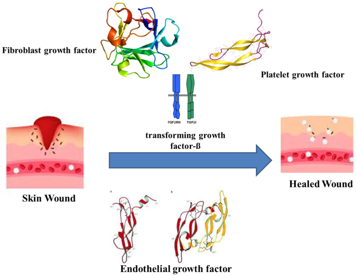
The healing of wounds combines the indulgence of various intricate factors and cell types, connective tissue, cytokines and the vascular system, along with growth and coagulation factors. As wound healing recasts the skin, it undergoes orderly meticulous phases. The major phases are hemostasis, inflammatory, proliferative and maturation [24]. Haemostasis occurs immediately after an injury by forming a blood clot to restrict bleeding. The clotting is assisted by thrombin activation, which in turn activates intravascular platelets [25]. The platelets eventually release growth factors, cytokines and vasoactive substances. The releasates, such as the fibroblast growth factor (FGF), platelet-derived growth factor (PDGF), platelet-derived angiogenesis factor, transforming growth factor-ß (TGF-ß), endothelial growth factor (EGF), bradykinin, thromboxane A2, platelet factor IV, platelet-activating factor, histamine, serotonin and prostaglandins, initiate the early wound-healing processes (Figure 1) [26][27].

3. Impact of Biofilms on Wounds

A prolonged open wound creates an inviting environment for opportunistic pathogens to adhere, proliferate and develop into mature biofilms capable of dodging immune systems or antimicrobials. The presence of necrotic debris, absconding blood circulation and a hypoxic environment offer a suitable surface for adhesion and growth media for proliferation to the microbes [21]. The repercussions of biofilm infections can range from mild discomfort to amputations, especially in the case of diabetic cutaneous wounds. According to an estimate, 80% of lower-limb amputations are succeeded by biofilm-infected foot ulcers [28]. Biofilms are noted to claim at least half of all chronic wounds [29][30] and an in vivo meta-analysis study reported the presence of biofilms among a minimum of 78% of all chronic wounds [31]. Similar evidences arise from reports on animal models, suggesting a persistent discomfort, as biofilms create a constant low inflammatory response and retard epithelialization and granulation of tissue formation [32]. Hence, wound-remodeling strategies must be able to identify and respond to the local infection without restricting tissue formation.
There is a correlation between the nonhealing of wounds and the bacterial existence of four or more species [33][34]. Such occurrence can transform into a multispecies biofilm, hampering cutaneous healing. Polymicrobial biofilm significantly delays wound closure by 12 more days than single-species biofilms, as observed in a murine model with four species infestations [35]. The non-healing wounds have a weakened molecular pathobiological mechanism, triggering abnormal cellular infiltration, hyperproliferation and infections arising from the colonization of polymicrobial biofilms. Such wounds hamper the ECM remodeling, fibroblast senescence and repress stem cell activation [36]. Typically, a wound infection marks the site of wound by the presence of replicating pathogenic organisms, capable of inducing host injury by dispersing virulence for their survival [37][38]. The virulence factors expressed by biofilms create hindrance in the recovery process. The biofilm-forming Staphylococci expresses a fibronectin receptor. This receptor is capable of blocking the migration of keratinocytes to the wound site, thereby disrupting the re-epithelialization of tissues [39].
Another advantage of biofilms is that they remain unaffected by the host immune phagocytes by promoting the production of leucocyte-inactivating substances, frustrating the phagocytosis that triggers inflammation [40]. Similarly, macrophage (M1) types are associated with the production of pro-inflammatory cytokines to confer appropriate host defences against pathogens.

4. Nanoparticles as Antimicrobials

Nanoparticles (NP) are observed to be efficient employees for escaping AMR [41], in addition to delivering antimicrobial agents [42] and growth promoters [43]. The generation of cost-effective biocompatible and biodegradable NPs and the route of biogenic synthesis is being encouraged [44]. The only setback of NPs is the compromised efficiency caused by the pre-mature or unintended release of drugs and promoters. Hence, the cultivation of localized response-inclusive strategies is necessary to attend this challenge. Engineering NPs with an inherent capability to activate in relation to their microenvironment or on being triggered by an external stimulus has surfaced as a remedy. Other nanoscale establishments also supplement benefits to wound reformation and biofilm extirpation. The undertone of nanomaterial-assisted wound therapy usually entails one of the two routes, one that possesses intrinsic properties that aid in wound closure and the other being delivery vehicles for therapeutic agents [45]. As described earlier, wound closure is at the mercy of microbial incompetence at the site. Hence, overviewing the key strategies of nanotechnology to obstruct microbial growth and encourage wound healing is imperative.
Typically, NPs are <100 nm in size [46]. This size renders them novel properties due to a large surface area–mass ratio [47]. Researchers have reported the rapid diffusion of smaller NPs through the pores of P. fluorescens biofilm, further stating an exponential decrease in the relative self-diffusion coefficients with an increase in the square of the radius of the nanoparticle [48]. Better prevention of biofilms is achieved with shapes delivering a higher surface area-to-mass ratio. For example; rod-like NPs destruct biofilms more effectively than spherical NPs [49]. Researchers have compared gold NP with different shapes to check their antibiofilm activity against S. aureus. The gold nano-stars and nanoflowers had higher antimicrobial activity than gold nanospheres. The authors attributed the difference to increased surface area, which generated a greater probability of interactions with the bacterial cell and biofilm components [50].
The mere availability of an alternative antimicrobial is not enough to consider against available agents. Silver sulfadiazine is a standard antimicrobial for burn wounds. However, this topical agent can cause adverse effects, such as leucopenia, argyria, renal and hepatic toxicity, making this unfit for long-term applications [51][52]. The major advantage of NPs against classical treatments is the ability to generate superior antimicrobial as well as healing properties.
Another route adopted by nanoparticles is to trigger the production of reactive oxygen species (ROS), succeeded by the phospholipid oxidation damaging the cell wall integrity before finally collapsing the internal nucleic acids and/or proteins, thereby staging a bactericidal impact [53].
Considering the ease of cost-effectiveness, nanoparticles can also be synthesized via the eco-friendly route. The topical application of green synthesized copper oxide NPs revealed antibacterial activity against S. dysenteriae, K. pneumoniae, S. aureus, S. typhimurium and E. coli through accelerating wound healing among Wistar Albino rats [54], thereby illustrating the antimicrobial effectivity of metallic and non-metallic nano topical agents. Another mode of tackling injuries is by the usage of bio-films that keep the toxicity in check. Investigation of the titanium oxide NP seeded in the gallium gum as a wound film dressing projected a versatile antimicrobial and healing potency. The biopolymer matrix of Gallium gum enacts as a suitable structure for skin regeneration, as it enhances cell proliferation and viability [55][56]. Meanwhile, the nano-titanium dioxide has an affinity for DNA. This was indicated by molecular docking that led to the proposition of TiO2 NPs binding to the G–C bonds of the DNA, creating hindrance in the bacterial multiplication [57]. The bio-film dressing was effective against S. aureus and E. coli, with no cytotoxicity on mouse fibroblast cells [58]. The in vivo and in vitro evidence declares nanotechnology to have a diversity of antimicrobial weaponry against wound biofilm remediation.

5. Nanoparticles in Chronic Wound Treatment

Cutaneous wound management grants biofilm formation as a major ingredient contributing to the chronicity of wounds [29][59][60]. Initially, the interaction of nanoparticles with the biofilm surface occurs at a bulk phase. It interacts with the lipids, lipopolysaccharides (LPS) or cell membrane proteins of the bacteria. The maturity, surface composition and chemistry of the biofilm are primary quotients governing the penetration of the NP. Further, the entry within the biofilm matrix is also dependent upon the physical and chemical characteristics of the NP, such as size, shape, charge, concentration, hydrophobicity and surface chemistry [61].
Clinical isolates of S. aureus, P. aeruginosa, S. epidermidis and Enterococcus spp. are the most predominant skin wound infections [62]. Thus, nanotechnology has to eradicate multiple species of bacteria at the same time and space. This can be achieved by modulating the wound microenvironment by inducing biophysical and biochemical alterations that can aid the removal of biofilms. For instance, the use of usnic acid derived from lichens is a bioactive compound with numerous biological activities, such as antioxidant, anti-inflammatory, wound healing and antimicrobial properties [63]. Clinical evidence has demonstrated accelerated healing and higher collagen deposition in porcine burn wounds by the application of usnic acid/liposomes based on gelatin membrane compared to silver sulfadiazine ointment and duoDerme® dressings [64]. Apart from addressing wound recovery, usnic acid demonstrates both a antibiofilm nature and the biocompatibility of nanocomposites. The study used anionic polymer dressings of carboximethylcellulose (CMC) or sodium alginate (AlgNa) with usnic acid-loaded magnetic NPs (Fe3O4@UA) to eradicate S. aureus biofilms. However, the exact mechanism of action by usinc acid on the biofilm is not clear. Further, they demonstrated biocompatibility against human endothelial cell lines and foetal progenitor cells to suggest the tissue regeneration capacity of the dressing [65].

6. Nanoplatforms against Wound Biofilms

The diversity of nanoplatforms delivers antibiofilm activity, according to the physiological condition of the wound site. The nature, depth, state, exudates, comorbidities and healing pace of the wound will suggest the necessary nanoplatform (Figure 2) to be allocated for infection control.
Figure 2. Nanopartile associated wound healing.

6.1. Organic Nanoplatforms

6.1.1. Nanoemulsions

Lipid nano formulations are the go-to carriers for antibacterial drugs, as they offer a variety of modes to choose from, namely—nano emulsions, solid lipid NPs, liquid lipid NPs and liposomes [66]. Liposome NPs can mimic the bacterial cell membrane structure, accelerate cellular uptake and prolong drug circulation time, casting it as a competent drug delivery vehicle [67][68][69]. Daptomycin-encapsulated nanoliposomes were found to be better at inhibiting S. aureus biofilm growth compared to the intravenous administration of daptomycin for treatment of subcutaneous infection in the mouse model [70].

6.1.2. Biopolymer NPs

The viability of polymeric nanomaterials is due to the privileges they encase, as both wound dressings and as enhancement conveyors that deliver antibacterial and reepithelialization benefits [45]. A carbohydrate polymer, chitosan has been used in several occasions of wound therapy, due to its enumerated biocompatibility. Chitosan has an inherent antibiofilm property owing to its polycationic nature that facilitates the disruption of the bacterial membrane [71]. The concoction of beneficial materials at a nanoscale improves the chances of restricting biofilm synthesis and helps in stimulating endothelial proliferation.

6.1.3. Synthetic Polymer NPs

The synthetic polymers recruited for skin tissue regeneration are: poly (lactic-co-glycolic acid) PLGA, PCL, poly (3-hydroxybutyrate-co-3-hydroxy valerate), poly (glycerol sebacate) and poly (etherurethane urea) [72][73]. PLGA is biocompatible [74], hence, it is vastly used as a delivery vector against infections, as microbial invasion is a determinant of wound repair. In 2019, Hasan et al. used cationic clindamycin-loaded PLGA-PEI (polyethyleneimine) NPs to reduce the bacterial burden in MRSA-infected wounds. The formulation was effective in demonstrating a sustained drug release >2 days, accelerated re-epithelization in a mouse wound model and was non-toxic to fibroblast cells. The wound model demonstrated higher bactericidal efficiency on being subjected to cationic NP compared to its anionic form, as the former bound more readily to MRSA surface [75].

6.2. Inorganic Nanoplatforms

6.2.1. Metal NPs

The most extensively studied metallic NPs are silver, gold and zinc [42]; among them, silver NPs act as a dual-edged sword. They are a potent antibacterial agent [76] and can modulate rapid wound closure [45] without increasing scarring. They also decrease keratinocyte viability and cell differentiation in a dose-dependent manner [77]. In order to overcome this shortfall, combinatorial or sustained release of the NP reduces its internalization in non-target cells. For instance, Ag NPs embedded in hydrogel delivered non-toxicity due to the sustained release of the NP. After treating the S. aureus wound biofilms with the nano-hydrogel, live/dead staining was performed. The staining gave remarkably superior results of reduced cell viability of mature biofilms and incurred a dispersion of biofilms within in vivo mice wounds.

6.2.2. Non-Metallic

There are numerous inorganic non-metallic antibiofilm variants. Metallic quantum dots (QD) can create a hassle in terms of toxicity. A better biocompatible substitute is the use of carbon QDs [78], which are arranged carbon QDs in self-healing hydrogels with anti-inflammatory and wound-healing benefits. They were studied as an injectable to hinder S. aureus and E. coli biofilms and enhance full-thickness wound healing. Additionally, they were relatively better at inhibiting mature S. aureus biofilms than gentamicin. The inference suggests the mechanism to be due to the cationic activity of the QD and its low drug resistance [79]. Other forms of carbon, such as fullerene, are also potent wound infection eliminators. Researchers have designed a fullerene NP that is functionalized with sulfur to terminate the MDR P. aeruginosa biofilm isolated from clinical chronic wounds, in order to explore the ascendary of the NP over the expression of toxA gene, which is encoding for exotoxin A, an important virulence factor of the strain. It reduced the gene expression [80].

7. Nanotechnology in Regeneration

7.1. Intrinsic Regenerative Properties

There are endless designs derived from nanotechnology. Nanoscale architecture helps in remodeling its microenvironment and liberating healing benefits to an injury. Interestingly, scholars have shed light on purposing nanotopography as a cell-aligning agent at the wound site [81]. The system was integrated with a microfluidic setup to which the nano-engraved patterns were aligned in parallel and perpendicular directions. This was used as a biomimicking profile of collagen fibers and fibroblasts to cue the swift migration of NIH-3T3 fibroblast cells to the wound site. The nanopattern that lay perpendicular to the microchannel of the setup displayed a speedy recovery due to collective migration guided by the nanotopography. Thus, the intrinsic capability of nanoengineered materials offer limitless sophisticated approaches to the remodeling of an injury.

7.2. Transdermal Nanocarriers

The use of transdermal drug deliveries is one of the recent and attractive methods that have a very convenient application, fewer systemic side effects and a less-pass effect. Due to its non-invasive properties, the transdermal carriers demonstrate the highest clinical potential, with very high drug delivery efficiency [82].
For an ideal shipment system, it is crucial to have a nontoxic vehicle that protects therapeutic integrity and increases its access to the injured site [83]. For instance, thrombin is an important tissue repair essential [84]. Conjugating magnetite (γ-Fe2O3) NP with thrombin is a means to expand its bioavailability without compromising its therapeutic value. In addition, the in vivo wound response on treatment with the conjugated NP resulted in heightened tensile strength. That, in turn, is essential for reducing wound dehiscence compared to treatment with free thrombin [85].

7.3. Nano Scaffold Tissue Engineering

Molding the topography at a nanoscale paves the way to directional remodeling of an injured site. The directional movement was put to test using uniformly spaced nanoscaffolds. The spaces were defined as dense (300–400 nm), intermediate (500–600 nm) and sparse (700–800 nm) based upon an increasing width range [86]. In an in vitro wound model, the healing trend displayed a direct correlation of fibroblast migration with dense nanotopography.
Bioengineered alternatives in wound care offer a plethora of substitutes, processes and conjugates. This makes them an enormous economic healthcare resource. Stem cell-based therapy facilitates the re-epithelialization of cutaneous wounds and boosts angiogenesis [87]. Delivery of mesenchymal stem cells at the site of the wound can result in its fastened cell death. Biomimetic delivery vehicles can overcome this challenge [88]. The engineered nanofiber-stem cell serves as an efficient mode for promoting wound healing. The bio nanocomposite was framed with mesenchymal stem cells derived from bone marrow that are functionalized over nanofiber scaffolds, demonstrating an escalated epidermal differentiation of burn wounds [89].

7.4. Nanotopography in Prevention of Biofilm

The topography of the surface greatly influences the development of biofilm by the sessile microbial colonies [90]. Various studies have demonstrated that the adhesion of the bacterial cells to a surface varies greatly with the change in the topography. Effective contact angle and surface hydrophilicity or hydrophobicity are key factors in the mechanism of the development of biofilm. The nanostructured materials prevent the adherence of the cells by setting off physicochemical changes, resulting in the development of cell deformation. The topographic changes brought about by nanostructures also develop gradient changes that prevent the adherence of the cells [90]. Studies have revealed that both 3D and 2D nanostructures have played an important role in the mechanism of inhibiting the development of biofilm. The 2D nanomaterials help in inhibiting the effective surface area of contact and air entrapment, thereby preventing the biofilm development on the biotic and abiotic surfaces. TiO2 NP and nanopillars help in the prevention of the development of biofilm [91].

8. Intelligent Nanotechnology

8.1. Wound Healing

8.1.1. Physically Responsive Nanomaterials

The use of thermo-responsive and photo-responsive nano therapy may lead to an uneven targeting of Gram-positive and Gram-negative bacteria. Coupling the two has proven to enact faster wound closure.

8.1.2. Chemically Responsive Nanomaterials

One of the environmental cues come from the fact that bacterial wound infestations create alkaline ambience. The pH of the wound bed needs to be considered while framing remedial dressings for chronic wound infections [92]. This pH is also indicative of bacterial proliferation [93]. Pathological conditions, such as diabetes and venous leg ulcers, can impair angiogenesis [94][95].

8.1.3. Bio-Responsive Nanomaterial

The usage of wound site facilities improvises the treatment processes. The generation of ROS at wounds orchestrates the healing cascade and may also result from an infection at the plot [96]. The development of ROS-reactive nanomaterials ensures the targeted release of wound-healing stimulants. One such example is that of the ROS-responsive nanomaterial poly-(1,4-phenyleneacetone dimethylene thioketal) loaded with the stromal cell-derived factor-1α (SDF-1α). Subsequently, the release of SDF-1a from the NPs stimulates a chemotaxis of bone marrow MSCs at the wound site. This recruitment was demonstrated among full-thickness skin wounds of mice and resulted in wound vascularization and accelerated healing [97].

8.2. Anti-Microbial Wound Care

The mechanism of bacterial colonizations can act as breeding grounds for intelligent wound care. The best proposal is to deliberately utilize the by-products of a pathogenic bacterial invasion, thereby framing “smart” wound dressings that act upon interaction with microbial prospects.
α-toxin, secreted by S. aureus, drills pores to impair cellular membranes. This virulence is incorporated in a liposome-based nanoreactor to ward off MDR bacteria. The components of the nanoreactor were calcium peroxide and rifampicin, which are then coated with lecithin and DSPE-PEG3400. Upon their interaction with a pathogenic environment, the a-toxins pierce through the nanoreactors. The pores then drown the nanoreactor with water, releasing hydrogen peroxide upon reaction with calcium peroxide. The decomposition of the H2O2 buds off oxygen, which liberates rifampicin. This smart nano system staged an anti-MRSA impact on the in vitro mode, alongside a significantly higher wound closure rate in the in vivo mode [98][99]. Apart from utilizing virulence, the enzyme-responsive bactericidal nano agents are also potent ammunition. This is based upon the secretion of hyaluronidase from pathogens as instigators for the nano agent [100]. On that note, for the development of a chemo-photothermal nano system to annihilate MDR bacteria, hyaluronic acid (HA) was preliminarily coated on ascorbic acid (AA), then the drug formulation was loaded in a mesoporous ruthenium NP. The nanocarrier was then mounted by catalyst—the ciprofloxacin-coated molybdenum disulphide (MoS2). On reaching the infection site, bacterial hyaluronidase decomposed the HA and subsequently liberated the AA that generated the in situ hydroxyl radicles by the MoS2 catalysis. This antibacterial strategy was increased by being accompanied with the photothermal nature of Ru NPs. Further, the AA@Ru@HA-MoS2 nanosystem was investigated against a skin-infected model. The results not only showcased the accelerated healing of the wound but also inhibited the formation, growth and multiplication of biofilms [101].

9. Advanced Nanotechnology

9.1. Wireless Monitoring

The upliftment of point-of-care wound monitoring requires hassle-free access to the injury-dependent factors. This is devised by either monitoring the microbial intensity or the wound in situ changes. This foresight in the wound care set-up can improvise the prompt healing of skin injuries. The detection accuracy is modified with the assistance of intelligent NPs to wirelessly indicate the physiology at wound site. Such a wireless-mediated outlook mostly employs a color-changing apparatus. For instance, antimicrobial agents with lipid vesicles were implanted in UV-photocrosslinkable methacrylated gelatin. The vesicles contained self-quenching fluorescent dyes resulting in a calorimetric indication upon interplay with infection. The toxins/enzymes secreted by P. aeruginosa and S. aureus disrupts the membrane bilayer of lipid vesicles, resulting in the expulsion of antimicrobials with a visual color alteration in the microenvironment [102]. Similar strategies were taken against the detection of wound biofilms with the help of intelligent WDs [103]. Researchers have designed a pH-responsive fluorescence scheme in a rabbit S. aureus wound model and terminated the biofilm presence with simultaneous imaging of the same. Such detection is critical, as it governs the success of the nanosystem in wound healing by an accompanying monitoring strategy [104].
The remote supervision of the state of healing dispatches easy and rapid diagnosis for the medical staff and the wounded victim. Real-time monitoring by wireless communication technology revolutionizes WD to a “smart” WDs [105]. The substrate-integrated circuit is the key to flexible surveillance of an injury. It has been reported that the combination of a biomimetic nanofiber membrane, a microenvironment sensor and a gelatine methacryloyl (GelMA)- β-cyclodextrin (β- cd) UV-crosslinked hydrogel is an integrated smart dressing. The dressing promotes angiogenesis and wound healing with instantaneous monitoring. While the GelMA -β- cd UV-crosslinked hydrogel boosts reformation [106][107][108], an integrated-chip supervises the wound microenvironment via transmission of data to a Bluetooth low-energy (BLE) 4.0 antennae on a smartphone mediated customized app [109]. Wound telemonitoring may encompass a wireless magnetic sensor using calor as a biomarker to develop smart WD technology [110].
The expansion of mobile surveillance of wound is infinite given the countless combinations of nanoscale operations from which to choose. One of them are the peroxide-sensing single-walled carbon nanotubes (SWCNTs) fabricated as wearable textiles [111]. The nanotubes were encapsulated within a polymer shell and soluble in organic solvent, enhancing its biocompatibility. This nanocomposite demonstrated a shift in NIR fluorescence in detecting physiologically relevant levels of peroxide in wounds. A differential response to hydrogen peroxide results from different bandgap energies of SWCNTs. The spectral attribute arises from their variable DNA sequence and chirality, as a response to the local environment [112][113].

9.2. Artificial Intelligence

Artificial intelligence (AI) can also be integrated into the functioning of nanoparticle-based dressings [114][115]. AI avails of health benefits by automated learning and proctoring myriads of clinical records for manging wound care decision making with predictive data [116]. Deep learning algorithms, such as the convolutional neural network (CNN), require low pre-processing time for differentiating input images [117]. This creates an advantageous mechanism for the automated detection of biofilms. In a rhinology diagnostic support study, a CNN-based biofilm scanning system results in an accuracy of ~98%, granting swift identification of biofilm in the images [118]. Existing machine learning also partakes in detection of biofilms. Machine learning and quantitative structure-activity relationship (QSAR) models predict the performance of different quorum-sensing compounds. This system accesses the trajectory of the range of effectiveness of the compounds across a biological setup, thus encouraging the intuitive design of countering biofilms [119]. Under similar contexts, AI can be replicated across platforms for monitoring cutaneous injuries. For example, it can be used as a diagnostic tool to aid the experts in evaluating the condition of the wound. Although there can be errors in the system, they can be escaped with the progression of the tool [120]. Combining AI with nanotechnology is a leap forward towards better health care.
There are immeasurable resources waiting to be discovered in the form of AI-nanoconjugate synergy. Volatile organic compounds (VOCs) are potential biomarkers for the diagnosis and surveillance of diabetic wounds [121]. Researchers have devised an ultra-selective detection of volatile organic compounds (VOCs) via an AI-nanomaterial sensor system. The system uses a modified Si nanowire field effect transistor (SiNW FETs) that is functionalized with varied saline molecules for the classification of VOCs. The model enables the selective detection of gas-phased chemical components in single component as well as a multicomponent environment. When conjugating an artificial neural network (ANN) model, the sensors were capable of recognizing 11 VOCs and retained its efficiency upon physical/chemical interferences. This enacts a beaming prospect to detect VOCs in wounds [122].

This entry is adapted from the peer-reviewed paper 10.3390/nano12050778

References

  1. Balas, M.; Popescu Din, I.M.; Hermenean, A.; Cinteza, L.O.; Dinischiotu, A. Exposure to Iron Oxide Nanoparticles Coated with Phospholipid-Based Polymeric Micelles Induces Renal Transitory Biochemical and Histopathological Changes in Mice. Materials 2021, 14, 2605.
  2. Hudita, A.; Radu, I.C.; Galateanu, B.; Ginghina, O.; Herman, H.; Balta, C.; Rosu, M.; Zaharia, C.; Costache, M.; Tanasa, E.; et al. Bioinspired silk fibroin nano-delivery systems protect against 5-FU induced gastrointestinal mucositis in a mouse model and display antitumor effects on HT-29 colorectal cancer cells in vitro. Nanotoxicology 2021, 15, 973–994.
  3. Vicas, S.I.; Laslo, V.; Timar, A.V.; Balta, C.; Herman, H.; Ciceu, A.; Gharbia, S.; Rosu, M.; Mladin, B.; Fritea, L.; et al. Functional Food Product Based on Nanoselenium-Enriched Lactobacillus casei against Cadmium Kidney Toxicity. Appl. Sci. 2021, 11, 4220.
  4. Gurdasani, K.; Li, L.; Rafter, M.A.; Daglish, G.J.; Walter, G.H. Nanoparticles as potential external markers for mark–release–recapture studies on Tribolium castaneum. Entomol. Exp. Appl. 2021, 169, 575–581.
  5. Cao, Z.; Li, B.; Sun, L.; Li, L.; Xu, Z.P.; Gu, Z. 2D Layered Double Hydroxide Nanoparticles: Recent Progress toward Preclinical/Clinical Nanomedicine. Small Methods 2020, 4, 1900343.
  6. Malekkhaiat Häffner, S.; Nyström, L.; Strömstedt, A.A.; Li, L.; van der Plas, M.J.A.; Malmsten, M. Nanoclay-induced bacterial flocculation for infection confinement. J. Colloid Interface Sci. 2020, 562, 71–80.
  7. Bharadwaj, K.K.; Rabha, B.; Pati, S.; Choudhury, B.K.; Sarkar, T.; Gogoi, S.K.; Kakati, N.; Baishya, D.; Kari, Z.A.; Edinur, H.A. Green Synthesis of Silver Nanoparticles Using Diospyros malabarica Fruit Extract and Assessments of Their Antimicrobial, Anticancer and Catalytic Reduction of 4-Nitrophenol (4-NP). Nanomater. 2021, 11, 1999.
  8. Sood, A.; Granick, M.S.; Tomaselli, N.L. Wound Dressings and Comparative Effectiveness Data. Adv. Wound Care 2014, 3, 511–529.
  9. Proksch, E.; Brandner, J.M.; Jensen, J.-M. The skin: An indispensable barrier. Exp. Dermatol. 2008, 17, 1063–1072.
  10. Madison, K.C. Barrier function of the skin: “la raison d’être” of the epidermis. J. Investig. Dermatol. 2003, 121, 231–241.
  11. Nethi, S.K.; Das, S.; Patra, C.R.; Mukherjee, S. Recent advances in inorganic nanomaterials for wound-healing applications. Biomater. Sci. 2019, 7, 2652–2674.
  12. Crovetti, G.; Martinelli, G.; Issi, M.; Barone, M.; Guizzardi, M.; Campanati, B.; Moroni, M.; Carabelli, A. Platelet gel for healing cutaneous chronic wounds. Transfus. Apher. Sci. Off. J. World Apher. Assoc. Off. J. Eur. Soc. Haemapheresis 2004, 30, 145–151.
  13. Lazarus, G.S.; Cooper, D.M.; Knighton, D.R.; Margolis, D.J.; Pecoraro, R.E.; Rodeheaver, G.; Robson, M.C. Definitions and guidelines for assessment of wounds and evaluation of healing. Arch. Dermatol. 1994, 130, 489–493.
  14. Sen, C.K. Human Wounds and Its Burden: An Updated Compendium of Estimates. Adv. wound care 2019, 8, 39–48.
  15. Khorasani, M.T.; Joorabloo, A.; Moghaddam, A.; Shamsi, H.; MansooriMoghadam, Z. Incorporation of ZnO nanoparticles into heparinised polyvinyl alcohol/chitosan hydrogels for wound dressing application. Int. J. Biol. Macromol. 2018, 114, 1203–1215.
  16. Das, U.; Behera, S.S.; Singh, S.; Rizvi, S.I.; Singh, A.K. Progress in the Development and Applicability of Potential Medicinal Plant Extract-Conjugated Polymeric Constructs for Wound Healing and Tissue Regeneration. Phytother. Res. 2016, 30, 1895–1904.
  17. Attinger, C.; Wolcott, R. Clinically Addressing Biofilm in Chronic Wounds. Adv. Wound Care 2012, 1, 127–132.
  18. Pastar, I.; Stojadinovic, O.; Yin, N.C.; Ramirez, H.; Nusbaum, A.G.; Sawaya, A.; Patel, S.B.; Khalid, L.; Isseroff, R.R.; Tomic-Canic, M. Epithelialization in Wound Healing: A Comprehensive Review. Adv. Wound Care 2014, 3, 445–464.
  19. Kirker, K.R.; Secor, P.R.; James, G.A.; Fleckman, P.; Olerud, J.E.; Stewart, P.S. Loss of viability and induction of apoptosis in human keratinocytes exposed to Staphylococcus aureus biofilms in vitro. Wound Repair Regen. 2009, 17, 690–699.
  20. Leung, K.P.; D’Arpa, P.; Seth, A.K.; Geringer, M.R.; Jett, M.; Xu, W.; Hong, S.J.; Galiano, R.D.; Chen, T.; Mustoe, T.A. Dermal wound transcriptomic responses to Infection with Pseudomonas aeruginosa versus Klebsiella pneumoniae in a rabbit ear wound model. BMC Clin. Pathol. 2014, 14, 20.
  21. Kirker, K.R.; James, G.A. In vitro studies evaluating the effects of biofilms on wound-healing cells: A review. APMIS 2017, 125, 344–352.
  22. Fazli, M.; Bjarnsholt, T.; Kirketerp-Møller, K.; Jørgensen, B.; Andersen, A.S.; Krogfelt, K.A.; Givskov, M.; Tolker-Nielsen, T. Nonrandom distribution of Pseudomonas aeruginosa and Staphylococcus aureus in chronic wounds. J. Clin. Microbiol. 2009, 47, 4084–4089.
  23. Kalashnikova, I.; Das, S.; Seal, S. Nanomaterials for wound healing: Scope and advancement. Nanomedicine 2015, 10, 2593–2612.
  24. Patil, S.; Chandrasekaran, R. Biogenic nanoparticles: A comprehensive perspective in synthesis, characterization, application and its challenges. J. Genet. Eng. Biotechnol. 2020, 18, 67.
  25. Martin, P. Wound healing--aiming for perfect skin regeneration. Science 1997, 276, 75–81.
  26. Stadelmann, W.K.; Digenis, A.G.; Tobin, G.R. Physiology and healing dynamics of chronic cutaneous wounds. Am. J. Surg. 1998, 176, 26S–38S.
  27. Greenhalgh, D.G. The Role of Growth Factors in Wound Healing. J. Trauma Acute Care Surg. 1996, 41, 159–167.
  28. Adler, A.I.; Boyko, E.J.; Ahroni, J.H.; Smith, D.G. Lower-extremity amputation in diabetes. The independent effects of peripheral vascular disease, sensory neuropathy, and foot ulcers. Diabetes Care 1999, 22, 1029–1035.
  29. James, G.A.; Swogger, E.; Wolcott, R.; deLancey Pulcini, E.; Secor, P.; Sestrich, J.; Costerton, J.W.; Stewart, P.S. Biofilms in chronic wounds. Wound Repair Regen. 2008, 16, 37–44.
  30. Kirketerp-Møller, K.; Jensen, P.Ø.; Fazli, M.; Madsen, K.G.; Pedersen, J.; Moser, C.; Tolker-Nielsen, T.; Høiby, N.; Givskov, M.; Bjarnsholt, T. Distribution, organization, and ecology of bacteria in chronic wounds. J. Clin. Microbiol. 2008, 46, 2717–2722.
  31. Malone, M.; Bjarnsholt, T.; McBain, A.J.; James, G.A.; Stoodley, P.; Leaper, D.; Tachi, M.; Schultz, G.; Swanson, T.; Wolcott, R.D. The prevalence of biofilms in chronic wounds: A systematic review and meta-analysis of published data. J. Wound Care 2017, 26, 20–25.
  32. Gurjala, A.N.; Geringer, M.R.; Seth, A.K.; Hong, S.J.; Smeltzer, M.S.; Galiano, R.D.; Leung, K.P.; Mustoe, T.A. Development of a novel, highly quantitative in vivo model for the study of biofilm-impaired cutaneous wound healing. Wound Repair Regen. 2011, 19, 400–410.
  33. Trengove, N.J.; Stacey, M.C.; McGechie, D.F.; Mata, S. Qualitative bacteriology and leg ulcer healing. J. Wound Care 1996, 5, 277–280.
  34. Davies, C.E.; Hill, K.E.; Newcombe, R.G.; Stephens, P.; Wilson, M.J.; Harding, K.G.; Thomas, D.W. A prospective study of the microbiology of chronic venous leg ulcers to reevaluate the clinical predictive value of tissue biopsies and swabs. Wound Repair Regen. 2007, 15, 17–22.
  35. Dalton, T.; Dowd, S.E.; Wolcott, R.D.; Sun, Y.; Watters, C.; Griswold, J.A.; Rumbaugh, K.P. An in vivo polymicrobial biofilm wound infection model to study interspecies interactions. PLoS ONE 2011, 6, e27317.
  36. Woo, K.Y.; Sibbald, R.G. The improvement of wound-associated pain and healing trajectory with a comprehensive foot and leg ulcer care model. J. Wound Ostomy Cont. Nurs. Off. Publ. Wound Ostomy Cont. Nurses Soc. 2009, 36, 183–184.
  37. Dow, G.; Browne, A.; Sibbald, R.G. Infection in chronic wounds: Controversies in diagnosis and treatment. Ostomy. Wound. Manage. 1999, 45, 29–40.
  38. Sharma, A.K.; Dhasmana, N.; Dubey, N.; Kumar, N.; Gangwal, A.; Gupta, M.; Singh, Y. Bacterial Virulence Factors: Secreted for Survival. Indian J. Microbiol. 2017, 57, 1–10.
  39. Kintarak, S.; Nair, S.P.; Speight, P.M.; Whawell, S.A. A recombinant fragment of the fibronectin-binding protein of Staphylococcus aureus inhibits keratinocyte migration. Arch. Dermatol. Res. 2004, 296, 250–257.
  40. Bjarnsholt, T.; Jensen, P.Ø.; Burmølle, M.; Hentzer, M.; Haagensen, J.A.J.; Hougen, H.P.; Calum, H.; Madsen, K.G.; Moser, C.; Molin, S.; et al. Pseudomonas aeruginosa tolerance to tobramycin, hydrogen peroxide and polymorphonuclear leukocytes is quorum-sensing dependent. Microbiology 2005, 151, 373–383.
  41. Lee, N.-Y.; Ko, W.-C.; Hsueh, P.-R. Nanoparticles in the Treatment of Infections Caused by Multidrug-Resistant Organisms. Front. Pharmacol. 2019, 10, 1153.
  42. Mihai, M.M.; Dima, M.B.; Dima, B.; Holban, A.M. Nanomaterials for Wound Healing and Infection Control. Materials 2019, 12, 2176.
  43. Losi, P.; Briganti, E.; Magera, A.; Spiller, D.; Ristori, C.; Battolla, B.; Balderi, M.; Kull, S.; Balbarini, A.; Di Stefano, R.; et al. Tissue response to poly(ether)urethane-polydimethylsiloxane-fibrin composite scaffolds for controlled delivery of pro-angiogenic growth factors. Biomaterials 2010, 31, 5336–5344.
  44. Bartolucci, C.; Antonacci, A.; Arduini, F.; Moscone, D.; Fraceto, L.; Campos, E.; Attaallah, R.; Amine, A.; Zanardi, C.; Cubillana-Aguilera, L.M.; et al. Green nanomaterials fostering agrifood sustainability. TrAC Trends Anal. Chem. 2020, 125, 115840.
  45. Hamdan, S.; Pastar, I.; Drakulich, S.; Dikici, E.; Tomic-Canic, M.; Deo, S.; Daunert, S. Nanotechnology-Driven Therapeutic Interventions in Wound Healing: Potential Uses and Applications. ACS Cent. Sci. 2017, 3, 163–175.
  46. Khan, I.; Saeed, K.; Khan, I. Nanoparticles: Properties, applications and toxicities. Arab. J. Chem. 2019, 12, 908–931.
  47. Huh, A.J.; Kwon, Y.J. “Nanoantibiotics”: A new paradigm for treating infectious diseases using nanomaterials in the antibiotics resistant era. J. Control. Release 2011, 156, 128–145.
  48. Peulen, T.-O.; Wilkinson, K.J. Diffusion of Nanoparticles in a Biofilm. Environ. Sci. Technol. 2011, 45, 3367–3373.
  49. Slomberg, D.L.; Lu, Y.; Broadnax, A.D.; Hunter, R.A.; Carpenter, A.W.; Schoenfisch, M.H. Role of Size and Shape on Biofilm Eradication for Nitric Oxide-Releasing Silica Nanoparticles. ACS Appl. Mater. Interfaces 2013, 5, 9322–9329.
  50. Penders, J.; Stolzoff, M.; Hickey, D.J.; Andersson, M.; Webster, T.J. Shape-dependent antibacterial effects of non-cytotoxic gold nanoparticles. Int. J. Nanomedicine 2017, 12, 2457–2468.
  51. Fraser, J.F.; Cuttle, L.; Kempf, M.; Kimble, R.M. Cytotoxicity of topical antimicrobial agents used in burn wounds in Australasia. ANZ J. Surg. 2004, 74, 139–142.
  52. Chaby, G.; Viseux, V.; Poulain, J.-F.; De Cagny, B.; Denoeux, J.-P.; Lok, C. Topical silver sulfadiazine-induced acute renal failure. Ann. Dermatol. Venereol. 2005, 132, 891–893.
  53. Karakoti, A.S.; Hench, L.L.; Seal, S. The potential toxicity of nanomaterials—The role of surfaces. JOM 2006, 58, 77–82.
  54. Sankar, R.; Baskaran, A.; Shivashangari, K.S.; Ravikumar, V. Inhibition of pathogenic bacterial growth on excision wound by green synthesized copper oxide nanoparticles leads to accelerated wound healing activity in Wistar Albino rats. J. Mater. Sci. Mater. Med. 2015, 26, 214.
  55. Ismail, N.A.; Razali, M.H.; Amin, K.A.M. Mechanical and physicochemical properties study on gellan gum thin film prepared using film casting method. AIP Conf. Proc. 2017, 1885, 20045.
  56. Oliveira, J.T.; Martins, L.; Picciochi, R.; Malafaya, P.B.; Sousa, R.A.; Neves, N.M.; Mano, J.F.; Reis, R.L. Gellan gum: A new biomaterial for cartilage tissue engineering applications. J. Biomed. Mater. Res. A 2010, 93, 852–863.
  57. Iram, N.E.; Khan, M.S.; Jolly, R.; Arshad, M.; Alam, M.; Alam, P.; Khan, R.H.; Firdaus, F. Interaction mode of polycarbazole-titanium dioxide nanocomposite with DNA: Molecular docking simulation and in-vitro antimicrobial study. J. Photochem. Photobiol. B 2015, 153, 20–32.
  58. Ismail, N.A.; Amin, K.A.M.; Majid, F.A.A.; Razali, M.H. Gellan gum incorporating titanium dioxide nanoparticles biofilm as wound dressing: Physicochemical, mechanical, antibacterial properties and wound healing studies. Mater. Sci. Eng. C 2019, 103, 109770.
  59. Dowd, S.E.; Wolcott, R.D.; Sun, Y.; McKeehan, T.; Smith, E.; Rhoads, D. Polymicrobial nature of chronic diabetic foot ulcer biofilm infections determined using bacterial tag encoded FLX amplicon pyrosequencing (bTEFAP). PLoS ONE 2008, 3, e3326.
  60. Bjarnsholt, T.; Kirketerp-Møller, K.; Jensen, P.Ø.; Madsen, K.G.; Phipps, R.; Krogfelt, K.; Høiby, N.; Givskov, M. Why chronic wounds will not heal: A novel hypothesis. Wound Repair Regen. 2008, 16, 2–10.
  61. Ikuma, K.; Decho, A.W.; Lau, B.L.T. When nanoparticles meet biofilms-interactions guiding the environmental fate and accumulation of nanoparticles. Front. Microbiol. 2015, 6, 591.
  62. Dryden, M.S. Complicated skin and soft tissue infection. J. Antimicrob. Chemother. 2010, 65 (Suppl. 3), iii35–iii44.
  63. Galanty, A.; Paśko, P.; Podolak, I. Enantioselective activity of usnic acid: A comprehensive review and future perspectives. Phytochem. Rev. 2019, 18, 527–548.
  64. Nunes, P.S.; Rabelo, A.S.; Souza, J.C.; Santana, B.V.; da Silva, T.M.; Serafini, M.R.; Dos, P.M.P.; Dos Santos, L.B.; Cardoso, J.C.; Alves, J.C.; et al. Gelatin-based membrane containing usnic acid-loaded liposome improves dermal burn healing in a porcine model. Int. J. Pharm. 2016, 513, 473–482.
  65. Grumezescu, A.M.; Holban, A.M.; Andronescu, E.; Mogoşanu, G.D.; Vasile, B.S.; Chifiriuc, M.C.; Lazar, V.; Andrei, E.; Constantinescu, A.; Maniu, H. Anionic polymers and 10 nm Fe3O4@UA wound dressings support human faetal stem cells normal development and exhibit great antimicrobial properties. Int. J. Pharm. 2014, 463, 146–154.
  66. Furneri, P.M.; Fuochi, V.; Pignatello, R. Lipid-based Nanosized Delivery Systems for Fluoroquinolones: A Review. Curr. Pharm. Des. 2017, 23, 6696–6704.
  67. Allen, T.M.; Cullis, P.R. Liposomal drug delivery systems: From concept to clinical applications. Adv. Drug Deliv. Rev. 2013, 65, 36–48.
  68. Fenske, D.B.; Cullis, P.R. Liposomal nanomedicines. Expert Opin. Drug Deliv. 2008, 5, 25–44.
  69. Rommasi, F.; Esfandiari, N. Liposomal Nanomedicine: Applications for Drug Delivery in Cancer Therapy. Nanoscale Res. Lett. 2021, 16, 95.
  70. Li, C.; Zhang, X.; Huang, X.; Wang, X.; Liao, G.; Chen, Z. Preparation and characterization of flexible nanoliposomes loaded with daptomycin, a novel antibiotic, for topical skin therapy. Int. J. Nanomed. 2013, 8, 1285.
  71. Khan, F.; Pham, D.T.N.; Oloketuyi, S.F.; Manivasagan, P.; Oh, J.; Kim, Y.M. Chitosan and their derivatives: Antibiofilm drugs against pathogenic bacteria. Colloids Surfaces B Biointerfaces 2020, 185, 110627.
  72. Kamoun, E.A.; Kenawy, E.R.S.; Chen, X. A review on polymeric hydrogel membranes for wound dressing applications: PVA-based hydrogel dressings. J. Adv. Res. 2017, 8, 217–233.
  73. Miguel, S.P.; Figueira, D.R.; Simões, D.; Ribeiro, M.P.; Coutinho, P.; Ferreira, P.; Correia, I.J. Electrospun polymeric nanofibres as wound dressings: A review. Colloids Surfaces B Biointerfaces 2018, 169, 60–71.
  74. Liu, H.; Slamovich, E.B.; Webster, T.J. Less harmful acidic degradation of poly(lactic-co-glycolic acid) bone tissue engineering scaffolds through titania nanoparticle addition. Int. J. Nanomed. 2006, 1, 541.
  75. Hasan, N.; Cao, J.; Lee, J.; Hlaing, S.P.; Oshi, M.A.; Naeem, M.; Ki, M.H.; Lee, B.L.; Jung, Y.; Yoo, J.W. Bacteria-Targeted Clindamycin Loaded Polymeric Nanoparticles: Effect of Surface Charge on Nanoparticle Adhesion to MRSA, Antibacterial Activity, and Wound Healing. Pharmaceutics 2019, 11, 236.
  76. Vijayakumar, V.; Samal, S.K.; Mohanty, S.; Nayak, S.K. Recent advancements in biopolymer and metal nanoparticle-based materials in diabetic wound healing management. Int. J. Biol. Macromol. 2019, 122, 137–148.
  77. Jura, J.; Szmyd, R.; Goralczyk, A.G.; Skalniak, L.; Cierniak, A.; Lipert, B.; Filon, F.L.; Crosera, M.; Borowczyk, J.; Laczna, E.; et al. Effect of silver nanoparticles on human primary keratinocytes. Biol. Chem. 2013, 394, 113–123.
  78. Zhu, C.; Chen, Z.; Gao, S.; Goh, B.L.; Samsudin, I.B.; Lwe, K.W.; Wu, Y.; Wu, C.; Su, X. Recent advances in non-toxic quantum dots and their biomedical applications. Prog. Nat. Sci. Mater. Int. 2019, 29, 628–640.
  79. Li, P.; Liu, S.; Yang, X.; Du, S.; Tang, W.; Cao, W.; Zhou, J.; Gong, X.; Xing, X. Low-drug resistance carbon quantum dots decorated injectable self-healing hydrogel with potent antibiofilm property and cutaneous wound healing. Chem. Eng. J. 2021, 403, 126387.
  80. Darabpour, E.; Doroodmand, M.M.; Halabian, R.; Imani Fooladi, A.A. Sulfur-Functionalized Fullerene Nanoparticle as an Inhibitor and Eliminator Agent on Pseudomonas aeruginosa Biofilm and Expression of toxA Gene. Microb. Drug Resist. 2019, 25, 594–602.
  81. Lee, I.; Kim, D.; Park, G.-L.; Jeon, T.-J.; Kim, S.M. Investigation of wound healing process guided by nano-scale topographic patterns integrated within a microfluidic system. PLoS ONE 2018, 13, e0201418.
  82. Yu, Z.; Meng, X.; Zhang, S.; Chen, Y.; Zhang, Z.; Zhang, Y. Recent Progress in Transdermal Nanocarriers and Their Surface Modifications. Molecules 2021, 26, 3093.
  83. Whittam, A.J.; Maan, Z.N.; Duscher, D.; Wong, V.W.; Barrera, J.A.; Januszyk, M.; Gurtner, G.C. Challenges and Opportunities in Drug Delivery for Wound Healing. Adv. Wound Care 2014, 5, 79–88.
  84. Carney, D.H.; Mann, R.; Redin, W.R.; Pernia, S.D.; Berry, D.; Heggers, J.P.; Hayward, P.G.; Robson, M.C.; Christie, J.; Annable, C.; et al. Enhancement of incisional wound healing and neovascularization in normal rats by thrombin and synthetic thrombin receptor-activating peptides. J. Clin. Investig. 1992, 89, 1469–1477.
  85. Glenn, K.C.; Frost, G.H.; Bergmann, J.S.; Carney, D.H. Synthetic peptides bind to high-affinity thrombin receptors and modulate thrombin mitogenesis. Pept. Res. 1988, 1, 65–73.
  86. Kim, J.; Bae, W.-G.; Kim, Y.J.; Seonwoo, H.; Choung, H.-W.; Jang, K.-J.; Park, S.; Kim, B.H.; Kim, H.-N.; Choi, K.S.; et al. Directional Matrix Nanotopography with Varied Sizes for Engineering Wound Healing. Adv. Healthc. Mater. 2017, 6, 1700297.
  87. Ojeh, N.; Pastar, I.; Tomic-Canic, M.; Stojadinovic, O. Stem Cells in Skin Regeneration, Wound Healing, and Their Clinical Applications. Int. J. Mol. Sci. 2015, 16, 25476–25501.
  88. Li, M.; Zhao, Y.; Hao, H.; Han, W.; Fu, X. Mesenchymal stem cell-based therapy for nonhealing wounds: Today and tomorrow. Wound Repair Regen. 2015, 23, 465–482.
  89. Ma, K.; Liao, S.; He, L.; Lu, J.; Ramakrishna, S.; Chan, C.K. Effects of nanofiber/stem cell composite on wound healing in acute full-thickness skin wounds. Tissue Eng. Part A 2011, 17, 1413–1424.
  90. Cheng, Y.; Feng, G.; Moraru, C.I. Micro- and Nanotopography Sensitive Bacterial Attachment Mechanisms: A Review. Front. Microbiol. 2019, 10, 191.
  91. Beltrán-Partida, E.; Valdez-Salas, B.; Curiel-Álvarez, M.; Castillo-Uribe, S.; Escamilla, A.; Nedev, N. Enhanced antifungal activity by disinfected titanium dioxide nanotubes via reduced nano-adhesion bonds. Mater. Sci. Eng. C Mater. Biol. Appl. 2017, 76, 59–65.
  92. Percival, S.L.; McCarty, S.; Hunt, J.A.; Woods, E.J. The effects of pH on wound healing, biofilms, and antimicrobial efficacy. Wound Repair Regen. 2014, 22, 174–186.
  93. Jones, E.M.; Cochrane, C.A.; Percival, S.L. The Effect of pH on the Extracellular Matrix and Biofilms. Adv. Wound Care 2015, 4, 431–439.
  94. Galiano, R.D.; Tepper, O.M.; Pelo, C.R.; Bhatt, K.A.; Callaghan, M.; Bastidas, N.; Bunting, S.; Steinmetz, H.G.; Gurtner, G.C. Topical vascular endothelial growth factor accelerates diabetic wound healing through increased angiogenesis and by mobilizing and recruiting bone marrow-derived cells. Am. J. Pathol. 2004, 164, 1935–1947.
  95. Drinkwater, S.L.; Burnand, K.G.; Ding, R.; Smith, A. Increased but ineffectual angiogenic drive in nonhealing venous leg ulcers. J. Vasc. Surg. 2003, 38, 1106–1112.
  96. Auf dem Keller, U.; Kümin, A.; Braun, S.; Werner, S. Reactive Oxygen Species and Their Detoxification in Healing Skin Wounds. J. Investig. Dermatol. Symp. Proc. 2006, 11, 106–111.
  97. Tang, T.; Jiang, H.; Yu, Y.; He, F.; Ji, S.; Liu, Y.; Wang, Z.; Xiao, S.; Tang, C.; Wang, G.-Y.; et al. A new method of wound treatment: Targeted therapy of skin wounds with reactive oxygen species-responsive nanoparticles containing SDF-1α. Int. J. Nanomed. 2015, 10, 6571–6585.
  98. Bhakdi, S.; Tranum-Jensen, J. Alpha-toxin of Staphylococcus aureus. Microbiol. Rev. 1991, 55, 733–751.
  99. Wu, Y.; Song, Z.; Wang, H.; Han, H. Endogenous stimulus-powered antibiotic release from nanoreactors for a combination therapy of bacterial infections. Nat. Commun. 2019, 10, 4464.
  100. Tyner, H.; Patel, R. Hyaluronidase in Clinical Isolates of Propionibacterium acnes. Int. J. Bacteriol. 2015, 2015, 218918.
  101. Liu, Y.; Lin, A.; Liu, J.; Chen, X.; Zhu, X.; Gong, Y.; Yuan, G.; Chen, L.; Liu, J. Enzyme-Responsive Mesoporous Ruthenium for Combined Chemo-Photothermal Therapy of Drug-Resistant Bacteria. ACS Appl. Mater. Interfaces 2019, 11, 26590–26606.
  102. Zhou, J.; Yao, D.; Qian, Z.; Hou, S.; Li, L.; Jenkins, A.T.A.; Fan, Y. Bacteria-responsive intelligent wound dressing: Simultaneous In situ detection and inhibition of bacterial infection for accelerated wound healing. Biomaterials 2018, 161, 11–23.
  103. Thet, N.T.; Alves, D.R.; Bean, J.E.; Booth, S.; Nzakizwanayo, J.; Young, A.E.R.; Jones, B.V.; Jenkins, A.T.A. Prototype Development of the Intelligent Hydrogel Wound Dressing and Its Efficacy in the Detection of Model Pathogenic Wound Biofilms. ACS Appl. Mater. Interfaces 2016, 8, 14909–14919.
  104. Sun, L.; Jiang, W.; Zhang, H.; Guo, Y.; Chen, W.; Jin, Y.; Chen, H.; Du, K.; Dai, H.; Ji, J.; et al. Photosensitizer-Loaded Multifunctional Chitosan Nanoparticles for Simultaneous in Situ Imaging, Highly Efficient Bacterial Biofilm Eradication, and Tumor Ablation. ACS Appl. Mater. Interfaces 2019, 11, 2302–2316.
  105. Zarrintaj, P.; Moghaddam, A.S.; Manouchehri, S.; Atoufi, Z.; Amiri, A.; Amirkhani, M.A.; Nilforoushzadeh, M.A.; Saeb, M.R.; Hamblin, M.R.; Mozafari, M. Can regenerative medicine and nanotechnology combine to heal wounds? The search for the ideal wound dressing. Nanomedicine 2017, 12, 2403–2422.
  106. Yue, K.; Trujillo-de Santiago, G.; Alvarez, M.M.; Tamayol, A.; Annabi, N.; Khademhosseini, A. Synthesis, properties, and biomedical applications of gelatin methacryloyl (GelMA) hydrogels. Biomaterials 2015, 73, 254–271.
  107. Niu, X.; Mo, Z.; Yang, X.; Sun, M.; Zhao, P.; Li, Z.; Ouyang, M.; Liu, Z.; Gao, H.; Guo, R.; et al. Advances in the use of functional composites of β-cyclodextrin in electrochemical sensors. Microchim. Acta 2018, 185, 328.
  108. Bezerra, F.M.; Lis, M.J.; Firmino, H.B.; Dias da Silva, J.G.; Curto Valle, R.d.C.S.; Borges Valle, J.A.; Scacchetti, F.A.P.; Tessaro, A.L. The Role of β-Cyclodextrin in the Textile Industry-Review. Molecules 2020, 25, 3624.
  109. Zhang, Y.; Li, T.; Zhao, C.; Li, J.; Huang, R.; Zhang, Q.; Li, Y.; Li, X. An Integrated Smart Sensor Dressing for Real-Time Wound Microenvironment Monitoring and Promoting Angiogenesis and Wound Healing. Front. Cell Dev. Biol. 2021, 9, 2172.
  110. Williams, S.; Okolie, C.L.; Deshmukh, J.; Hawco, L.; McNeil, J.; Nganou Assonkeng, A.C.; Bennett, C.; Mkandawire, M. Magnetizing Cellulose Fibers with CoFe2O4 Nanoparticles for Smart Wound Dressing for Healing Monitoring Capability. ACS Appl. Bio Mater. 2019, 2, 5653–5662.
  111. Safaee, M.M.; Gravely, M.; Roxbury, D. A Wearable Optical Microfibrous Biomaterial with Encapsulated Nanosensors Enables Wireless Monitoring of Oxidative Stress. Adv. Funct. Mater. 2021, 31, 2006254.
  112. Gravely, M.; Safaee, M.M.; Roxbury, D. Biomolecular Functionalization of a Nanomaterial to Control Stability and Retention within Live Cells. Nano Lett. 2019, 19, 6203–6212.
  113. O’ Connell, M.J.; Eibergen, E.E.; Doorn, S.K. Chiral selectivity in the charge-transfer bleaching of single-walled carbon-nanotube spectra. Nat. Mater. 2005, 4, 412–418.
  114. Ho, D.; Wang, P.; Kee, T. Artificial intelligence in nanomedicine. Nanoscale Horizons 2019, 4, 365–377.
  115. Ho, D.; Teo, G. Digital Medicine–The New Frontier for AI in Healthcare. Adv. Ther. 2020, 3, 2000015.
  116. Secinaro, S.; Calandra, D.; Secinaro, A.; Muthurangu, V.; Biancone, P. The role of artificial intelligence in healthcare: A structured literature review. BMC Med. Inform. Decis. Mak. 2021, 21, 125.
  117. Tharsanee, R.M.; Soundariya, R.S.; Kumar, A.S.; Karthiga, M.; Sountharrajan, S. Deep convolutional neural network–based image classification for COVID-19 diagnosis. In Data science for COVID-19; Kose, U., Gupta, D., de Albuquerque, V.H.C., Khanna, A., Eds.; Academic Press: Cambridge, MA, USA, 2021; pp. 117–145. ISBN 978-0-12-824536-1.
  118. Dimauro, G.; Deperte, F.; Maglietta, R.; Bove, M.; La Gioia, F.; Renò, V.; Simone, L.; Gelardi, M. A Novel Approach for Biofilm Detection Based on a Convolutional Neural Network. Electronics 2020, 9, 881.
  119. Stone, B.; Sapper, E. Machine Learning for the Design and Development of 2 Biofilm Regulators 3. 2018. Available online: https://doi.org/10.20944/preprints201803.0118.v1 (accessed on 6 December 2021).
  120. Howell, R.S.; Liu, H.H.; Khan, A.A.; Woods, J.S.; Lin, L.J.; Saxena, M.; Saxena, H.; Castellano, M.; Petrone, P.; Slone, E.; et al. Development of a Method for Clinical Evaluation of Artificial Intelligence-Based Digital Wound Assessment Tools. JAMA Netw. Open 2021, 4, e217234.
  121. Daneshkhah, A.; Siegel, A.P.; Agarwal, M. Volatile organic compounds: Potential biomarkers for improved diagnosis and monitoring of diabetic wounds. In Wound healing, tissue repairing, and regeneration in diabetes; Bagchi, D., Das, A., Roy, S., Eds.; Academic Press: Cambridge, MA, USA, 2020; pp. 491–512. ISBN 978-0-12-816413-6.
  122. Wang, B.; Cancilla, J.C.; Torrecilla, J.S.; Haick, H. Artificial sensing intelligence with silicon nanowires for ultraselective detection in the gas phase. Nano Lett. 2014, 14, 933–938.
More
This entry is offline, you can click here to edit this entry!
Video Production Service