Transience of the Retinal Output and Circuit Elements: History
Please note this is an old version of this entry, which may differ significantly from the current revision.
Subjects: Biology
Contributor:

Retinal ganglion cells (RGCs) encrypt stimulus features of the visual scene in action potentials and convey them toward higher visual centers in the brain. Although there are many visual features to encode, our recent understanding is that the ~46 different functional subtypes of RGCs in the retina share this task. In this scheme, each RGC subtype establishes a separate, parallel signaling route for a specific visual feature (e.g., contrast, the direction of motion, luminosity), through which information is conveyed. The efficiency of encoding depends on several factors, including signal strength, adaptational levels, and the actual efficacy of the underlying retinal microcircuits. Upon collecting inputs across their respective receptive field, RGCs perform further analysis (e.g., summation, subtraction, weighting) before they generate the final output spike train, which itself is characterized by multiple different features, such as the number of spikes, the inter-spike intervals, response delay, and the rundown time (transience) of the response. These specific kinetic features are essential for target postsynaptic neurons in the brain in order to effectively decode and interpret signals, thereby forming visual perception.

  • photoreceptor
  • bipolar cell
  • amacrine cell
  • ganglion cell
  • outer plexiform layer
  • inner plexiform layer
  • ganglion cell layer
  • retina
  • parallel signaling

1. The Wiring of the Mammalian Retina

The mammalian retina is a valuable and common model in neuroscience, due to its well-defined layered structure and its excellent potential for conducting in vitro electrophysiological studies. This has allowed scientists to map many of the neuronal connections and build a wiring blueprint (
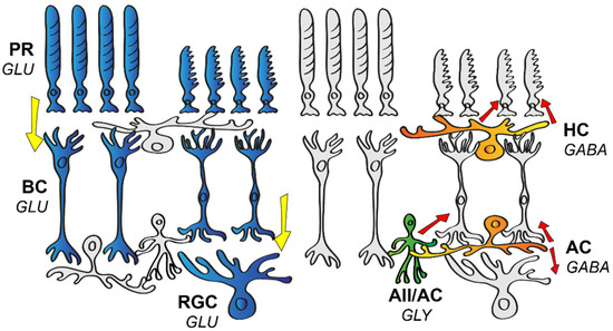
Figure 1) that brings functional and morphological connections in the retina together in one schematic [1]. Elements of this blueprint are generally found in all mammalian species, including both primate and non-primate models and humans as well. The researchers' current knowledge regarding neurons in the mouse retina points to at least 130 neuronal cell types, with 2 distinct photoreceptor types (PRs) [2], 1 type of horizontal cell (HC) [3], 15 different bipolar cell types (BCs) [4], roughly 63 diverse amacrine cell types (ACs) [5], and an estimated number of around 46 retinal ganglion cell types (RGCs) [6]. Just like in any other species, visual perception in mice begins at the level of PRs, where rods and cones create two separate pathways for daytime (high intensity or photopic) or nighttime (low intensity or scotopic) illumination, respectively. Rods are sensitive enough to detect a single photon [7], and thus they are most efficiently utilized in low-light environments. Cones, on the other hand, express photopigments that are less sensitive to the intensity of the stimulating light, but the variety in the expressed chromophores allows them to specialize to a certain wavelength, which has proven to be the first step in establishing color vision. The human retina contains 3 cone subtypes specifically adapted to peak sensitivity at short (S, ~430 nm), medium (M, ~530 nm), and long (L, ~560) wavelengths [8], while the mouse retina only possesses S and M sensitive cones [2].
Figure 1. Schematic drawing of the basic neuronal wiring of the mammalian retina. The vertical information stream encompasses the light-sensitive photoreceptors (PR), bipolar cells (BC), and retinal ganglion cells (RGC). These cells express glutamate (GLU), thus providing an excitatory (highlighted in blue) stream of signals (direction of information flow is indicated by yellow arrows). Horizontal cells (HC) and amacrine cells (AC) serve various forms of inhibition (red arrows) in the outer or the inner retina, respectively. HCs express GABA (highlighted in orange in the outer retina), whereas ACs release either glycine (GLY, highlighted in green) or GABA (highlighted in orange in the inner retina) as a neurotransmitter.
PRs form synapses with 15 different types of BCs, with one BC collecting inputs from about 5–10 presynaptic PRs [9]. BCs lay the foundation for a number of parallel signaling pathways at their level [10] and the segregation of information is maintained throughout the downstream circuitry to higher visual brain centers. Roughly half of these parallel pathways inform the brain about an increase in light intensity (ON pathways), whereas the other half signals decrements in light intensity (OFF pathways). This ON and OFF polarity distinction appears to be so fundamental for vision that ON and OFF signals are established as early as the first retinal synapse (the PR-BC synapse). BCs that express ionotropic glutamate receptors (BCs: 1(ab), 2, 3(a,b), 4) are responsible for forming the OFF signaling routes [11,12], whereas those that utilize metabotropic glutamate receptors (5(a–d), 6, 7, 8/9) are responsible for conveying ON signals [11]. The first synaptic layer of the retina is also equipped with HCs that express inhibitory GABA as a transmitter and signal laterally, thereby providing feedback and feedforward inhibition to PRs and BCs, respectively [13]. This inhibition serves mainly as a spatial filter and thus forms the basis for an inhibitory surround receptive field of downstream retinal neurons and thus contrast detection for vision [14].
ON and OFF BCs both release glutamate and synapse with ON, OFF, or ON–OFF RGCs and a large variety of ACs. In turn, BCs receive AC inhibitory inputs at their axon terminals through reciprocal feedback and/or feedforward mechanisms [15]. ACs are commonly classified into glycinergic narrow or GABAergic wide-field populations [16]. ACs in general exhibit great diversity in terms of morphology and function, and according to a recent study, their molecular makeup indicates as many as 63 separate subtypes [5]. While narrow-field ACs modulate local glutamate input of BCs, wide-field ACs generally provide a non-linear surround to RGCs [17]. Through gap junction (GJ) coupling, ACs can also relay excitatory input to neighboring ACs or even to neurons of other cell types. One of the best-known examples of ACs with GJ coupling are the narrow-field AII ACs that, upon receiving excitatory glutamate input from rod BCs [18], use gap junctions to excite ON cone BCs, and glycinergic chemical synapses to inhibit OFF BCs at the same time [19]. ACs might also provide a sign-inverting feedforward mechanism, such as in the case of S-M (short wavelength–medium wavelength) color opponency in the murine retina, where an S-ON/M-OFF RGC will receive direct excitatory input from S-ON BCs and inhibition through a sign-inverting AC connection from an M-ON BC [20]. Interestingly, there is at least one AC type (VGlut3 expressing AC) that utilizes a glutamatergic excitatory signaling mechanism to provide a feedforward (ON to ON, OFF to OFF) and crossover (ON to OFF, OFF to ON) glutamatergic input in the case of direction-selective ganglion cells (DSGCs) [21]. Once visual signals have passed through all the above-mentioned circuit elements, they are integrated by RGCs to forge spike trains that inform the brain about the change in only one specific visual aspect [22].

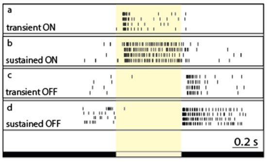
2. RGC Response Transience and Possible Visual Functions

Light-evoked RGC responses have been characterized by their polarity (ON, OFF, and ON–OFF), sensitivity to various stimuli, and their kinetics. In general, the two main response parameters that are considered and studied when it comes to RGC response kinetics are the response delay and the response decay. The response delay reflects the speed by which a certain neuron reacts to an incoming stimulus, and thus neurons including RGCs are generally considered either brisk or sluggish. On the other hand, response decay reveals the ability of a neuron to maintain its activity without inactivation in response to continuous stimulation. Based on different manifestations of the latter phenomenon, neurons are known to display either maintained spiking activity (sustained response) or present a brisk spike burst (transient response) in response to continuous stimulation (Figure 2). Both aspects (response delay and decay) likely contribute to signal efficiency on postsynaptic neuronal targets in higher visual centers [23,24,25]. The transient/sustained dichotomy of RGC response decay has been documented in a variety of vertebrate species, including cold-blooded animals, primates, and non-primate mammals as well [26,27,28,29,30,31,32,33,34,35,36], indicating that this kinetic feature has been conserved throughout vertebrate evolution and thus further attesting the importance of the role that RGC response kinetics play in visual coding.
Figure 2. The diversity of response transience across the retinal ganglion cell population. Peri-event rasters of representative RGCs (ad). Light-evoked spiking responses upon full-field illumination are rather similar across trials for each RGC, but they display a great variety in terms of their response length (or decay—expressed as the PSTHτ value in this work) for both the ON (cells 1 and 2) and OFF (cells 3 and 4) subpopulations. The white bar below the recordings represents the timing of the on- and off-set of the stimulus.

3. Circuit Elements That Contribute to Response Transience

3.1. The Photoreceptor to Bipolar Cell Synapses in the Outer Retina

Since all photoreceptors generate sustained responses upon illumination [37], a sustained-to-transient response transformation must occur along the vertical axis of the retinal circuitry. At the first synaptic site in the outer plexiform layer (OPL) of the retina, rods contact only a single subtype of BCs, known as the rod BC, whereas cones distribute information to 14 cone BC subtypes that can be distinguished using both morphological and physiological measures [4,9,10,38,39]. In the outer retina, the above-mentioned sustained-to-transient response transformation could potentially be performed by quick desensitization of postsynaptic BC glutamate receptors or through an inhibitory mechanism localized to the OPL.

3.2. Bipolar Cell Characteristics

Beyond subtype-specific differences in the synaptic interactions at the first synaptic layer, the differential passive and active membrane properties of various BCs may also account for some of the observed kinetic variations in BC responses. Given that BCs are, apart from input specificity in the OPL and selective stratification in the inner plexiform layer (IPL), relatively similar in shape, one does not normally expect considerable differences in their passive membrane features. Since the biological membrane acts as a low-pass filter, dendritic excitatory postsynaptic currents (EPSCs) are expected to become more sustained as they travel from the dendrites to the axonal ending, regardless of the initial kinetics of the response. In fact, it has been shown that axotomized BCs display relatively sustained responses compared to those recorded from intact counterparts of the same BC subtype [49]. This suggests that EPSCs go through low-pass filtering [10] and they all reach the inner retina as relatively sustained signals, meaning that transient BC outputs are recreated in the axons of certain BC subtypes. Nonetheless, it is expected that differential active membrane properties and/or synaptic interactions (e.g., inhibitory feedback) of the various BC subtypes play a significant role in shaping BC response kinetics.

3.3. Inner Retinal Contribution

Responses of axotomized BCs often differ from those of intact cells in slice preparation [49], indicating a heavy influence of inner retinal mechanisms on BC response kinetics. This finding has been supported more recently by demonstrating that outputs of a single BC can produce wildly different and type-specific RGC response kinetics, and that this diversity is mediated by ACs [97,98]. In fact, several inhibitory mechanisms in the inner retina have been suggested to shape BC responses prior to the information passing to RGCs [27,78,99,100,101,102,103,104,105,106].

3.4. Summation of Signals in the Inner Retina

If BC subtypes that feed RGCs were dominant in determining and shaping RGC response kinetics, then one would expect that all RGCs maintain comparable response transience when the same single BC type dominates their inputs. In contrast to photopic signals delivered by 5–6 bipolar cells to ON RGCs, low scotopic signals reach RGC targets mostly via the primary rod pathway that operates with the single rod BC type. Therefore, the same inputs are delivered to all RGC subtypes under scotopic conditions. In contrast, the unpublished data show that the scotopic RGC responses are just as diverse in terms of their kinetics as their photopic counterparts (Figure 3). This suggests that the observed kinetic variety of RGC responses is neither the result of the differential kinetics of the phototransduction cascade of rods and cones or the postsynaptic glutamate receptor on BC dendrites. In addition, different experiments demonstrated that outputs of a single BC can produce widely different RGC response patterns (including differences in kinetics), depending on the RGC type, and that this diversity is mediated by retinal ACs [97,98]. This suggests that irrespective of the kinetic nature of BC inputs, RGC response kinetics are likely reestablished in the inner retina both by passive and active membrane properties of RGCs. While circuit elements (mostly inhibitory) have been described in the previous section, there are a few more aspects to cover in this regard. These include the potential role of desensitization of postsynaptic glutamate receptors on RGC dendrites that may play a role in the generation of transient RGC responses [144], and also other voltage-gated currents that shape RGC responses [145].
Figure 3. Signal interference through parallel retinal pathways. (a) Representative ON retinal ganglion cell (RGC) light-evoked rasters (left column) and peristimulus time histogram (PSTH) cohorts (right column) were obtained to light stimuli of various strength (intensity is reflected in the right top corner). RGC PSTHτ values (for more information, see [146]) changed non-monotonously during this experiment while the stimulus intensity was gradually increased (see also panel (c)). PSTHs clearly show a very sensitive, relatively delayed response component (red arrow) and a less sensitive but fast response component (light blue arrow). These two components differ in their delays but appear similar in response decay, and therefore PSTHτ values are shifted towards the sustained range when the two signals are summated (mesopic conditions—2nd, 3rd, and 4th panels), whereas they appear transient when only one signal is present (scotopic condition—1st panel) or dominates over the other component (photopic conditions—5th, 6th, and 7th panels; see also panel (c)). (b) A similar experiment was performed for this representative OFF RGC. PSTHτ values of this RGC clearly changed during this experiment when the stimulus intensity was gradually increased. This OFF RGC also showed a very sensitive and delayed response (red arrow) as well as a faster but less sensitive (light blue arrow) response component. The two signal components differed in their delays and sensitivities and a slight alteration in PSTHτ values occurred as a result of the summation of components (mostly in mesopic conditions—middle panels). While the distinction of response components can clearly be differentiated for the ON RGC in (a), this OFF cell (and most examined RGCs) showed a less obvious and less separable summation of incoming signals. (c,d) Diagrams show that similar to cells shown in panels (a,b) (values of these cells appear in blue and red in the diagrams), most recorded RGCs displayed stimulus strength-driven changes of PSTHτ values (grey curves). (e) The diagram shows minimum/maximum PSTHτ value pairs for the recorded ON (blue) and OFF (red) cells during the course of the stimulus intensity recording paradigm. The examined RGCs showed ~18–73% PSTHτ changes during the course of this experiment.
Apart from RGC membrane properties, there is another crucial but often neglected factor that determines RGC response transience, namely the summation of all incoming inputs to the same RGC. One aspect of this issue, the feedforward inhibition from ACs to RGC, has already been discussed (see above in Section 3.3.2). However, RGCs may also integrate excitatory signals from more than one BC. Figure 3 provides one example, where an examined RGC clearly displays relative transient responses to both scotopic and photopic stimuli (small PSTHτ values, for the calculation of PSTHτ, refer to [146]), but the full-field light response of the same cell appeared more sustained (larger PSTHτ values) in the mesopic/low photopic range when both scotopic and photopic signaling streams were active. This is obviously the result of the summation of signals carried via a separate cohort of BCs serving at least two different signaling pathways. Under scotopic and photopic conditions, both the PSTH peak and τ values were influenced by only one signal component (the slow and sensitive component in scotopic conditions and the fast but less sensitive component in photopic conditions), whereas mid-range illumination brought about an intermingling of the fast signal component peak and a shoulder of the slow response component, thereby resulting in an increased PSTHτ value. The summation of these signals evidently caused the spiking pattern of this otherwise transient cell to appear more sustained. A similar signal summation should occur as well when two or more photopic signals are conveyed parallel to each other to the same RGC targets. In this scheme, a certain RGC subtype receives inputs from two or more signaling streams (BCs). This, in fact, is the case for ON alpha RGCs of the mouse retina that receive a mixture of inputs from type 6, 7, and 8 bipolar cells [147]. In addition, sustained OFF alpha cells are postsynaptic to type 2 bipolar and GluMI cells, whereas transient OFF alpha cells are targeted by type 3a and type 4 bipolar cells [148,149]. Therefore, the mixing of inputs originating from several parallel signaling streams seems to be a general feature for most RGCs in the mammalian retina. Furthermore, it appears that the summation of inputs with slightly different kinetics (delay and decay) and sensitivity plays a crucial role in determining the ultimate kinetics of RGC responses. Again, these results are not necessarily in conflict with other reports regarding mechanisms that determine BC response kinetics, however BC response characteristics are not simply inherited by postsynaptic RGCs but greatly altered via signal summation. The results of such signal summation can be exemplified for some RGC subtypes that have been studied more deeply, including the sustained and the transient OFF alpha cell populations. Sustained OFF alpha RGC responses are generated by the summation of excitatory inputs from the transient type 2 bipolar cells and GluMI cells [43,148,150], whereas the synaptic partners transient OFF alpha cells are the transient type 3a and sustained type 4 bipolar cells [149,150].
Clearly, rather than simply inheriting response kinetics of presynaptic BCs, these two RGC subtypes performed a transformation of incoming signals. One issue remains: while the summation of excitatory signals can provide an example of how transient inputs are transformed into intermediary or sustained signals by RGCs, the reverse (sustained-to-transient transformation) cannot be explained by the same mechanism. It is known that in addition to excitatory BC inputs to RGCs, some signaling routes also provide inhibitory signals via intermediary ACs. In fact, the sections above detailed how GABAergic inhibition may alter RGC response transience. In this scheme, the early onset of inhibition truncates the response of the RGC target, thus making it more sustained. GABAergic AC input alters RGC response kinetics and functionality, as a fast inhibition is known to be capable of truncating excitatory signals [33,151], whereas delayed inhibitory inputs can shift the signal towards the more transient domain of the spectrum (Figure 4) [49,121,152].
Figure 4. Summary of potential signal summation mechanisms affecting RGC response transience. (a) Two bipolar cells (BCs) of different subtypes provide transient inputs to the same retinal ganglion cell (RGC; light blue EPSC curves). These two inputs have dissimilar delays (due to differential BC signaling and/or a different location of synapses over the RGC dendritic arbor), and therefore the summation of the responses results in an intermediate or sustained RGC spiking response. (b) This RGC receives excitatory inputs from two sources: from a transient BC (light blue EPSC) and from a gap junction-coupled amacrine cell (AC; purple depolarization). If the dynamics of these two inputs differ, their summation will induce intermediate and/or sustained RGC spiking. (c) This RGC receives excitation from a BC (light blue EPSC) and delayed inhibition (red IPSC) from an AC, resulting in a transient RGC response. (d) An RGC that receives excitation from a BC (light blue EPSC) and inhibition (red IPSC) from an AC. In this scenario, the two inputs have about the same delays, and therefore the excitation will be truncated and the RGC output is an intermediate/sustained spiking.

This entry is adapted from the peer-reviewed paper 10.3390/cells11050810

This entry is offline, you can click here to edit this entry!
ScholarVision Creations