Resveratrol-Induced Resensitization of Acquired Drug-Resistant Cancer Cells: History
Please note this is an old version of this entry, which may differ significantly from the current revision.
Subjects: Cell Biology
Contributor:

Multidrug resistance (MDR) refers to a phenomenon wherein tumors exhibit cross-resistance to an array of drugs with different structures or action mechanisms once they become resistant to one anticancer drug. MDR to anticancer drugs remains a serious obstacle to the success of cancer chemotherapy. Resveratrol, a polyphenol, present in natural products exerts anticancer activity and acts as a potential MDR inhibitor in various drug-resistant cancer cells.

  • resveratrol
  • cancer
  • chemotherapy
  • drug-resistance

1. Introduction

Many patients often experience recurrence of cancer after chemotherapy due to the development of multidrug resistance (MDR), which is one of the most crucial hurdles in cancer treatment [1]. MDR refers to a phenomenon wherein tumors exhibit cross-resistance to an array of drugs with different structures or action mechanisms once they become resistant to one anticancer drug [2].
Acquired drug resistance may result due to the modification of various cellular and molecular mechanisms, including (1) removal of drug by increased efflux transporters; (2) inactivation of drug due to metabolization; (3) promotion of DNA damage repair; (4) modification of drug targets; (5) regulation of cell cycle progression; (6) inhibition of programmed cell death pathways; and (7) induction of epithelial to mesenchymal transition (EMT) and cancer stem cells (CSCs) [3,4,5,6].
Recently, various plant-derived compounds with anticancer properties have emerged as attractive drug candidates due to their advantages such as low toxicity and immediate availability. Among them, polyphenols, which are natural compounds present in fruits and vegetables, have been proven to have multiple benefits in the treatment of cancer as well as in several chronic diseases.
Resveratrol (3,5,4′-trans-trihydroxystilbene, RES) is abundantly produced in a wide variety of plants, such as grapes, berries, and peanuts [8], and is a major polyphenol possessing anti-inflammatory, cardiovascular protective, as well as cancer chemopreventive activities [9,10]. In particular, the prominence of RES is increasing owing to its chemosensitizing and radiosensitizing effects [11,12,13].

2. In Vitro and In Vivo Activity of RES in Different Tumor Models
Chemotherapeutic agents, such as vinca alkaloids (vincristine (VCR) and vinblastine), anthracyclines (doxorubicin (DOX), daunorubicin, and epirubicin), anthracenediones (mitoxantrone (MX)), antimetabolites (5-fluorouracil (5-FU), methotrexate, and gemcitabine), taxanes (paclitaxel (PTX) and docetaxel (DTX)), and platinum salts (cisplatin (CIS), carboplatin, and oxaliplatin (OXT)), are most frequently associated with drug resistance [19,20].
Due to multiple resistance responses to classical MDR drugs, the paradigm is gradually shifting toward the development of targeted anticancer drugs, which block cancer-specific pathways. The selected targeted anticancer drugs are tyrosine kinase inhibitors, including imatinib (IM), which interferes with BCR-ABL; vemurafenib (VEM), which inhibits BRAF; and cetuximab (CET) and gefitinib (GEF), which block EGFR. However, the emergence of drug resistance still seems to be an unavoidable issue [21,22]. Moreover, tamoxifen (TAM), a selective estrogen receptor (ER) modulator that has been used for all stages of ER-positive breast cancers, is also associated with acquired resistance in breast cancer cells, after long-term treatment that initially responded to antiestrogen therapy [23].
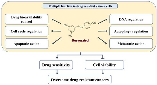
Cancer cells may eventually develop resistance to almost all types of drugs, and strategies to deal with the resistance response need to be considered during chemotherapy. However, RES has been shown to suppress acquired drug resistance caused by multiple types of anticancer drugs, which occurs in a variety of cancer tissues, including bladder, breast, colon, stomach, white blood cells, lungs, and prostate cancer [7,24,25,26] (Figure 1 and Table 1).
Figure 1. A schematic model of RES function in drug-resistant cancer cells.
Table 1. RES-induced regulation of cellular signaling molecules and gene regulatory factors in various drug-resistant cancer cells.

Target

Regulatory Molecules

Cellular Effect

↑ Upregulation

↓ Downregulation

↑ Upregulation

↓ Downregulation

Drug transporters and drug-metabolizing enzymes

AMPK

ABCG2, GST, LRP1, MDR1, MRP1, Nrf2, p-AKT, p-CREB, p-NF-κB, PI3K

Cellular accumulation

ABC transporters

ATPase activity

Detoxification

DNA damage, repair, and replication

APC, Topo-II, γ-H2AX

DDB2, FEN-1, POLH,

POL-β, Rad51

DNA damage

DNA repair

DNA replication

Cell cycle regulation

miR-122-5p, p21, p53, PTEN

CDC2, CDK2, CDK4, CDK6,

Cyclin D1, ERα, IRS1

Cell cycle arrest

-

Pro-apoptotic and anti-apoptotic action

AIF, AMPK, Apaf-1, Bad, Bax,

Caspae-3, Caspase-7, Caspase-8, Caspase-9, CHK2, CK1, Endo G,

miR-122-5p, p53, p-p53(S20), PTEN, PUMA, TSC1, TSC2

Bcl-2, Bcl-xL, Clusterin, Integrin β1,

p-AKT, p-Bad(s136), p-EGFR,

p-ERK1/2, p-FAK, PI3K, p-IkBα,

p-Jak, p-mTOR, p-NF-κB,

p-p53(S15, S46), p-Src, p-Stat1,

Survivin, uPAR

Apoptosis

Cell death

Senescence

Sub-G1 arrest

Cell proliferation

Tumor volume

Autophagy regulation

Atg3, Atg5, Atg7, Atg14, Atg12, Atg16L1, Beclin-1, LC3-Ⅱ, p62,

p-AMPKα, p-JNK

p-AKT, p-mTOR, Rubicon

Autophagy

-

Migration, invasion, metastasis, EMT, and CSC

E-cadherin, SIRT1, γ-catenin

ALDH1, CD133, CD44, CXCR4, Fibronectin, MMP-2, MMP-9,

N-Cadherin, p-ERK, p-NF-κB, p-p38,

p-Smad2, p-Smad3, Slug, Snail, TGF-β, Vimentin, β-Catenin

Intracellular junction

Cell migration, invasion, and metastasis

Colony formation

CSC

EMT

3. Biological Effects and Mechanisms of RES in Acquired Drug-Resistant Cancer Cells

3.1. Inhibition of Drug Transporters and Drug-Metabolizing Enzymes

The overexpression of various ABC efflux transporters such as P-glycoprotein (P-gp/MDR1/ABCBl), multidrug-resistance-associated protein 1 (MRP1/ABCC1), breast cancer resistance protein (BCRP/ABCG2), and lung resistance protein (LRP) in cancer cells can significantly eliminate anticancer drugs from the cell, thus causing persistent resistance in cancer chemotherapy [27]. One way to overcome MDR is to prevent the expression or activity of ABC transporters, allowing chemotherapeutic drugs to remain in cancer cells [28]. Additionally, drug resistance can be suppressed by inhibiting the expression and activity of drug-metabolizing enzymes, including cytochrome P450s (CYPs) and glutathione-S-transferases (GSTs), resulting in altered metabolic control in cancerous cells [29].

3.2. Promotion of DNA Damage and Inhibition of DNA Repair and Replication

DNA is a critical target for numerous chemotherapeutic drugs [39]. However, elevated DNA repair and tolerance to DNA damage may induce resistance to DNA-targeting drugs [40]. Inhibition of the DNA repair system may be a beneficial strategy to restore drug sensitivity in resistant cells. Notably, RES has been shown to enhance the DNA-damaging effect of anticancer drugs in several drug-resistant cancer cells.
DNA topoisomerases are critical enzymes that maintain DNA structure during DNA replication and transcription [41]. The expression levels of DNA topoisomerase-II (Topo-II) in cancers is a predictive factor of responsiveness to therapy. However, a typical feature of MDR, induced by Topo-II-interacting drugs, is reduced Topo-II amount or activity [42,43]. The anthracycline class of drugs, such as DOX, interferes with DNA replication and induces DNA strand breaks by forming drug–Topo-II–DNA complexes in cancer cells. Although Topo-II levels were reduced in pumc-91/ADM cells, a significant increase in Topo-II was detected in the RES-treated group compared to that in the RES-untreated group [24]. Hence, RES appears to promote drug-induced DNA damage in DOX-resistant cancer cells through upregulation of Topo-II expression. In addition, RES has been shown to enhance the DNA damaging effect in 5-FU-R cells, established from 5-FU-sensitive HCT116 cells to acquire resistance to 5-FU. 5-FU-R cells highly expressed the 5-FU-resistance protein, thymidylate synthase, and anti-apoptotic proteins, such as FLICE-like inhibitory protein, DNA polymerase eta (POL-H), DNA polymerase beta (POL-β), DNA damage-binding protein 2 (DDB2), and flap endonuclease 1 (FEN1), in comparison to parental HCT116 cells. As expected, upon exposure to a synthetic DNA damaging agent, 1,3-bis(2-chloroethyl)-1-nitrosourea (BCNU), 5-FU-R cells were highly resistant. However, a combination of RES and BCNU significantly increased the sensitivity and DNA damage of 5-FU-R cells; inhibited DNA repair proteins, POL-H, POL-β, FEN1, and DDB2; and increased the expression of the DNA damage response protein, adenomatous polyposis coli (APC). DNA damage and apoptosis were elevated with increasing concentrations of RES and a constant BCNU concentration, suggesting the critical role of RES in the sensitization of 5-FU-R cells [44].
The inhibitory effects of RES in the repair of DNA double-strand breaks have also been reported in CIS-resistant cancer cells. Platinum drugs, such as CIS, induce DNA damage by forming platinum–DNA adducts that interfere with DNA replication and transcription. CIS treatment increased the levels of Rad51 protein, which is essential for the homologous recombination repair of DNA double-strand breaks in human breast cancer MCF-7 cells and the CIS-resistant subline, MCF-7R cells. However, RES decreased the relative levels of Rad51 and the transcript levels of homologous recombination initiation complex components (Nbs-1, Mre-11, and Rad-50) and increased H2AX (p-serine139) levels, which are used as a marker for DNA damage.

3.3. Cell Cycle Regulation

Cell cycle arrest may be ambivalent in determining the cancer cell’s fate in response to chemical drugs. Inhibition of the cell cycle may result in a relative insensitivity to drugs and act as a defense mechanism since cells become less responsive to toxic stimuli in their resting phases [45]. Conversely, deregulation of the cell cycle also acts to enhance the sensitivity of resistant cells to chemotherapy since blockage of cell cycle progression often escapes alternative cell death. In the most studies, cell cycle arrest induced by RES in drug-resistant cancer cells was observed along with the activation of apoptosis, which is proved by an increase in apoptotic signal molecules, including PTEN, p53, and active caspases-3, -7, -8, and -9, and a decrease in anti-apoptotic regulators, such as p-AKT and EGFR [16,37,44,46,47].
Therefore, RES-induced cell cycle arrest may proceed toward apoptosis induction and overcoming drug resistance rather than acting as a defense mechanism. Progression of the cell cycle is regulated by cyclin-dependent kinases (CDKs), cyclins, and Cdk inhibitors (CDKIs). CDKs are upregulated by cyclins (A, B, D, and E) and downregulated by CDKIs [48].
RES has been shown to inhibit cell cycle progression in drug-resistant cancer cells as well as in parental cancer cells, including colon, breast, and prostate cancer cells [10,16,49,50]. Indeed, RES arrested the cell cycle in the G0–G1 phases in SPC-A-1/CDDP cells, which are generated from SPC-A-1 lung cancer cells to acquire resistance to CIS [16]. In addition, RES significantly induced G1 arrest in TAM-resistant breast cancer cells, MCF-7 TR1, as well as in parental MCF-7 cells by increasing p53-dependent p21 expression. In a study using MCF-7 TR1 cells, the sustained activation of p38MAPK by RES was suggested to be a critical mechanism in the modulation of p53 and ERα expression. Moreover, the expression of cyclin D1 and the estrogen-regulated gene, IRS1, was significantly decreased by RES treatment [51].
Furthermore, RES has been shown to act as a potential chemosensitizer in MCF-7-ADR cells, which are DOX-resistant MCF-7 breast cancer cells. During this process, RES increased the expression of miR-122-5p, and both RES and miR-122-5p mimic significantly downregulated CDKs (CDK2, CDK4, and CDK6) and induced G1 arrest in MCF-7-ADR cells. However, inhibitors of miR-122-5p significantly reversed the effects of RES. Thus, it has been suggested that miR-122-5p is involved in RES-mediated cell cycle arrest in a CDK-dependent manner [46].
Certain drugs, such as CIS, have also been reported to induce cell cycle arrest through p21 induction in multiple cancer cells. In parental MCF-7 and CIS-resistant MCF-7R cells, treatment with CIS with or without RES as well as treatment with RES alone upregulated p21 gene expression levels. Notably, combined treatment with CIS and RES resulted in further increase in p21 expression and sensitized CIS-induced resistance in MCF-7R cells [47]. The synergistic effects of RES and CIS were consistently observed in HCT116 colorectal cancer cells. Simultaneous treatment with CIS and RES resulted in a significant increase in the percentage of cells in the G0 phase in parental and CIS-resistant HCT-116 cells [30]. RES was also reported to affect cell cycle regulation in 5-FU-R, which are 5-FU-resistant HCT-116 cells. In this cell line, combined treatment with BCNU and RES significantly intensified the increase in cell cycle inhibitory protein p21 and phosphatase and tensin homolog (PTEN) and the decrease in cyclin-dependent kinase-1 (CDC2) as the concentration of RES increased [44].

3.4. Pro-Apoptotic and Antisurvival Actions

Induction of cell death and suppression of cell survival are fundamental principles of chemotherapy [53]. Chemotherapeutic drugs can initiate apoptosis, the major type of programmed cell death in cancer cells. The extrinsic and intrinsic pathways are well-known apoptotic processes, which ultimately activate the cysteine proteases (caspases), which are critical apoptotic executioners [54]. During the apoptotic process, the efficiency generally depends on prosurvival signaling factors, such as Akt and ERK1/2, and apoptosis regulatory proteins, such as Bcl-2 family members [55]. RES has been reported to affect multiple pro-apoptotic and antisurvival regulators in drug-resistant cancer cells.

3.5. Autophagy Regulation

Autophagy is an evolutionarily conserved adaptive mechanism that enables cells to maintain homeostasis and survive stressful environments by facilitating the degradation and recycling of cytoplasmic constituents and organelles [66]. The autophagic process is initiated by phagophore assembly, autophagosome formation, and fusion of the autophagosomes with lysosomes, leading to lysosomal degradation of autophagosomal contents by lysosomal acid hydrolases [67,68]. These processes are tightly regulated by distinct signaling pathways that control autophagy-related (ATG) proteins [69]. The role of autophagy in the treatment of cancer MDR can be both beneficial and harmful. It contributes to the development of MDR and protects cancer cells from cytotoxic drugs but also kills drug-resistant cells in which apoptotic pathways are disabled [3]. In the latter case, autophagy acts as a death executioner to trigger a form of type II programmed cell death, which utilizes a signal pathway distinct from type I programmed cell death, apoptosis [53].
RES has been reported to induce autophagic cell death through the modulation of AMPK and Akt signaling in CIS-resistant CAR cells. Exposure to RES increased the protein levels of AMPKα and phosphorylated AMPKα at Thr172 but decreased the phosphorylation of Akt on Ser473 and of mTOR on Ser2448. It has also been shown to increase the protein levels of key autophagy markers such as Atg5, Atg7, Atg12, Atg14, Atg16L1, Beclin-1, and microtubule-associated protein 1 light chain 3 (LC3)-II and to decrease the protein levels of Rubicon, a negative regulator of autophagy [63].
RES-induced LC3-II accumulation has also been observed in several other cancer cells, including IM-sensitive and IM-resistant chronic myelogenous leukemia (CML) cells [70]; human ovarian A2780 and CIS-resistant subline, A2780CP cells [71]; and PC9/G cells [37]. RES treatment resulted in the loss of cell viability and antileukemic effects via the induction of apoptosis and autophagic death in IM-sensitive and IM-resistant CML cells. However, in these cells, loss of cell viability is only partly affected in the presence of the pan-caspase inhibitor, z-VAD-fmk, and mainly due to autophagic death. RES-triggered autophagy is controlled by increased expression of ATG3, JNK-dependent accumulation of p62, and inhibition of the mTOR pathway [70].
Therefore, RES may regulate autophagy-related molecules (ATGs, beclin, LC3, and Rubicon) and trigger autophagic cell death by interfering with the PI3K/Akt/mTOR pathway, AMPK activation, and JNK-mediated p62 expression in drug-resistant cancer cells.

3.6. Inhibition of EMT and CSCs

EMT plays an essential role not only in cancer cell invasion and metastasis but also in drug resistance. Numerous EMT-related signaling pathways are associated with drug resistance in cancer cells [72]. The critical hallmarks of EMT are loss of E-cadherin expression and upregulation of vimentin, snail, and slug proteins [73]. CSCs, a subset of cancer cells possessing self-renewal capacity, are also responsible for the development of resistance to anticancer drugs [74]. CD44, CD133, epithelial-specific antigen, and aldehyde dehydrogenase 1 (ALDH1) are markers of CSCs [75]. EMT and CSCs remain under control at the gene level by multiple signaling pathways, such as the MEK/ERK, TGF-β/SMAD, JAK/STAT, PI3K/Akt/NF-κB, and the WNT/β-catenin pathway [76]. Moreover, some populations of CSCs share properties with EMT-like cells [77]. Targeting EMT and CSCs has been recognized as a potential therapeutic strategy to overcome chemoresistance, and RES has been shown to reverse EMT and CSC features in several drug-resistant cancer cells.
In MCF7/DOX cells possessing DOX resistance, combinatorial treatment with RES and DOX inhibited DOX-induced cell migration, invasion, and metastasis [58]. RES reversed EMT properties by upregulating SIRT1 and downregulating vimentin, N-cadherin, and β-catenin in MCF7/ADR cells [78]. Similarly, in SGC7901/DOX cells, RES antagonized DOX-induced EMT by downregulating vimentin and β-catenin and upregulating E-cadherin, thereby preventing cell migration [26]. RES also affects TGF-β-related signaling during the acquisition of EMT- and CSC-like features. For instance, TAM-resistant MCF-7/TR cells undergo EMT driven by enhanced endogenous TGF-β/Smad signaling. However, RES restored the expression of epithelial markers such as E-cadherin and γ-catenin and downregulated the expression of mesenchymal markers, including fibronectin, vimentin, and N-cadherin. Furthermore, RES suppressed TGF-β production and the phosphorylation of Smad2 and Smad3 in these cells [56].
On the other hand, the invasive ability of HCT116 colorectal cancer cells cultured in 3D alginate matrix was increased in the presence of TNF-α or TNF-β. However, addition of RES dramatically suppressed TNF-α- or TNF-β-induced survival and invasion of parental HCT116 and 5-FU-resistant HCT116 (HCT116R) cells. In addition, RES sensitized TNF-β-enhanced chemoresistance of HCT116R cells to 5-FU. This study showed that RES decreased TNF-β-induced expression of CSC markers (CD133, CD44, and ALDH1) and the activation of tumor-promoting factors (NF-κB(p65), MMP-9, and CXCR4) in both parental HCT116 and HCT116R cells. RES also reduced vimentin and slug levels and elevated E-cadherin expression in both cell lines [25]. Thus, RES may contribute to the inhibition of CSCs induced by TNF-α and TNF-β, as well as the anticancer drug, 5-FU. Another study showed that RES promoted the transition of 5-FU-induced formation of microvilli to a planar cell surface, which occurred concurrently with the upregulation of desmosomes, gap and tight junctions, and E-cadherin expression, and attenuated drug resistance by preventing EMT factors including vimentin, slug, and MMP-9 in HCT116 and 5-FU-resistant HCT116R cells [57]. Prevention of EMT-like characteristics by RES has also been observed in CIS-resistant cancer cells. RES reduced the migration and invasive capacity of CIS-resistant CAR cells by inhibiting the phosphorylation of ERK and p-38MAPK as well as suppressing MMP2 and MMP9 expression [79]. Likewise, RES reversed CIS-induced snail expression by blocking the ERK pathway, thereby inhibiting the morphological changes and cell migration in ovarian cancer A2780 and A2780CP (resistant to CIS) cells [71].

This entry is adapted from the peer-reviewed paper 10.3390/nu14030699

This entry is offline, you can click here to edit this entry!
Video Production Service