Glycosyltransferases (GTs) catalyze the glycosylation reaction between activated sugar and acceptor substrate to synthesize a wide variety of glycans. GTs are involved in metabolic processes, signal pathways, cell wall polysaccharide biosynthesis, cell development, and growth. Glycosylation mainly takes place in the endoplasmic reticulum (ER) and Golgi, where GTs and glycosidases involved in this process are distributed to different locations of these compartments and sequentially add or cleave various sugars to synthesize the final products of glycosylation. Therefore, delivery of these enzymes to the proper locations in the cell is essential and involves numerous secretory pathway components.
- glycosyltransferases
- ER-Golgi trafficking
- mechanism of protein sorting
- COPI and COPII complexes
- sequences and motifs involved in trafficking
1. Introduction
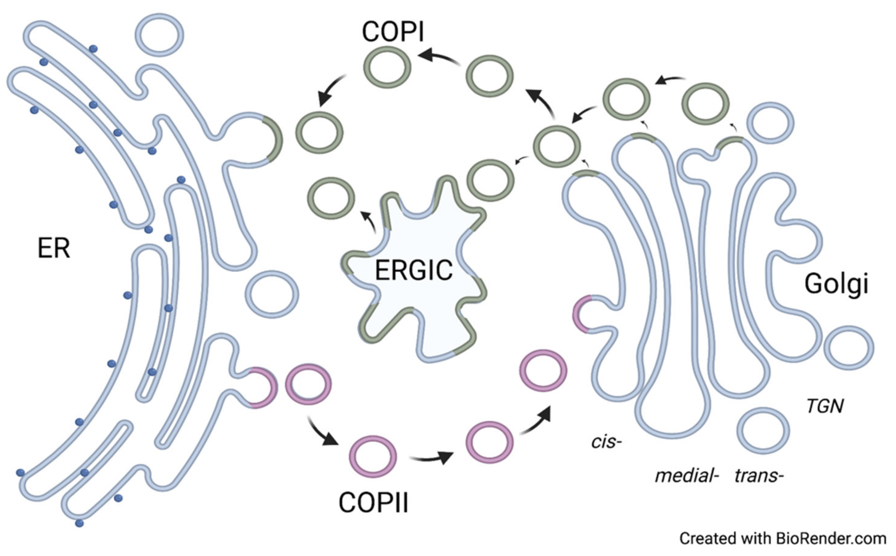
2. Main Components of the Secretory Pathway
3. Specific Sequence Motifs Involved in GTs and Glycosidases Sorting and Trafficking
4. Other Protein Domains Essential for the Trafficking of Enzymes Involved in Glycosylation
5. Recycling of Glycosyltransferase and Glycosidases Involved in Glycosylation.
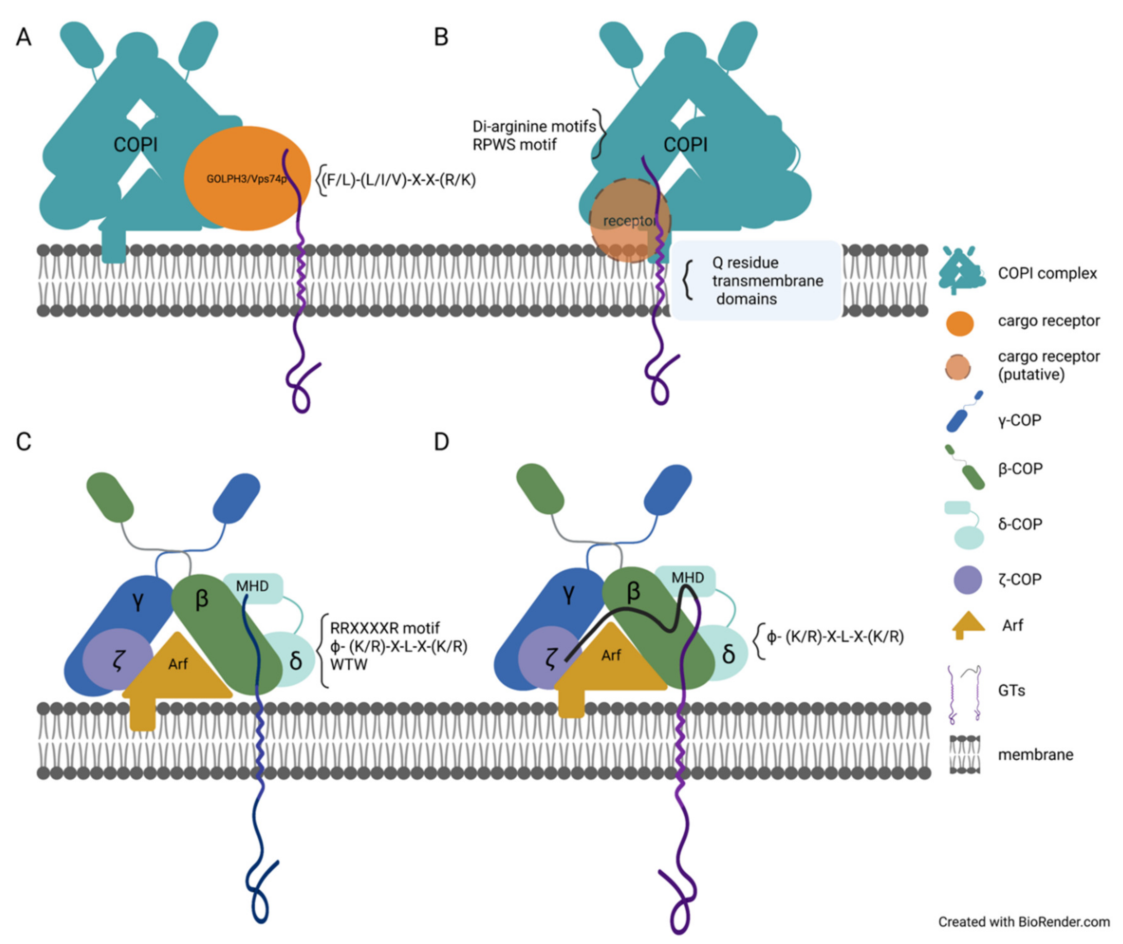
The transport cargo carriers (i.e., the COPI and COPII complexes) are critical for GTs recycling. It was shown in plants when the N-terminal domain of GnTI and Sar1p were co-expressed in N. benthamiana leaf epidermal cells, both proteins were co-localized in the punctate structure at ER-exit sites (ERES) [59]. However, a mutant version of GnTI, where basic amino acids within its cytoplasmic tail were mutated, was not able to recruit Sar1 to ERES, indicating that COPII proteins are involved in GnTI transport. Even though the studies about the transport of the plant GTs via the COPI and COPII complexes are limited, the results indicate that the mechanism of membrane proteins transport in plant and mammalian cells are similar. For example, the LxxLE motif functions as the ER-export signal in animals and plants [67][68][69]. Hence, the advanced knowledge about trafficking of GTs via COPI and COPII-coated cargo carriers in animal cells might offer some clues to the GTs transport via COPI and COPII complexes in plants. For example, the silencing of the coatomer subunits δCOP or εCOP results in the mislocalization of the Golgi-resident A.thaliana MNS3-GFP protein [55]. During the formation of the COPI and COPII-coated cargo carriers harboring GTs as cargo, indirect or direct interactions between GTs and COPI/COPII complex proteins were observed. For example, the Vps74p protein was detected as the intermediate protein in interaction with the COPI complex in yeast, and the knockout of Vps74p impacted the localization of Kre2p, Mnn2, Mnn9, and Ktr6 [54]. Vps74p was shown to bind to Sec26p (β-COP) and Ret2p (δ-COP) in in vitro experiments (Figure 2A) [54].
In animal cells, the Vps74p ortholog protein GOLPH3 functions similarly to Vps74p in yeast. GOLPH3 binds to C2GnT and SiaTI in vitro, and all three proteins, GOLPH3, C2GnT, and SiaTI, were detected in COPI vesicles [70]. Meanwhile, the knockout of GOLPH3 triggered the mislocalization of C2GnT and SiaTI from ER/Golgi to Golgi only. At the same time, the content of C2GnT and SiaTI in COPI vesicles was significantly decreased [70]. In recent studies, GOLPH3 was proved to interact with not only the LxxR motif but the positively charged amino acids upstream of the LxxR motif, [71], which further confirmed the function of GOLPH3/ Vps74p in retaining the cargo protein in the Golgi cisternae and preventing cargo from leaving to the TGN [71][72]. The protein was transported to lysosomes when it escaped the GOLPH3-mediated cisternal inter-conversion mechanism. This indicated that GOLPH3/ Vps74p controls the lysosomal degradation of the protein [71][72]. The ER-target signal, R11R12XXXXR, in the cytosolic tail of M1-SAT-I has been proven to interact with β-COP or δ-COP (Figure 2C), while the mutation, M1-SAT-I-R11/12S, interrupted this interaction [58]. This indicates that the RR residues may directly bind to β-COP or δ-COP (Figure 2C). The Golgi protein GlcNAc-1-phosphotransferase (Ptase) synthesizes the mannose 6-phosphate recognition marker. The utilization of the recently developed BioID2 assay revealed the interactions among the Ptase, δ-, and ζ-COP subunit proteins [60]. The direct interaction between δ-/ζ-COP and Ptase was confirmed by pull-down assay, which also detected traces of β-COP and γ-COP (Figure 2D) [60]. It has been shown that Ptase directly binds to the highly reserved sequence, VRFSTE, in the MHD domain of δ-COP [60]. The mutations of K4 to Q, R8 to G, and S15 to Y in the cytosolic tail of Ptase impaired and weakened its interaction with δ-/ζ-COP [60]. The ϕ- (K/R)-X-L-X-(K/R) sequence is also found in the cytosolic tail of other GTs, such as C2GNT1, GALNT3, GALNT6, and GALNT8 [60]. C2GNT1, GALNT3, and GALNT8 directly bind to β-COP, ζ-COP, and the MHD domain of δ-COP (Figure 2D); GALNT6 interacts with β-COP and the MHD domain of δ-COP (Figure 2C) [60]. The arginine residues in the R3TLLR7R8R9 sequence in the cytosolic tail of C2GNT1 are essential for recruiting C2GNT1 to the Golgi. The mutation of arginine residues impaired the interaction between C2GNT1 and ζ-COP protein, and the interaction between C2GNT1 and the MHD domain of δ-COP (Figure 2D) [60]. In previous studies on CHO-K1 cells, C2GNT1 was shown to interact with GOLPH3 and later with COPI subunits [70]. However, in HeLa cells, the knockout of GOLPH3 did not affect the localization of C2GNT1 [60]. UBIAD1 is localized in the Golgi in L02 cells, but UBIAD1 is localized in the ER and the Golgi in both HEK293 and T24 cells [66], which indicates that the mechanism of trafficking of GTs might vary for different cell types. Although there is no ϕ- (K/R)-X-L-X-(K/R) sequence in the cytosolic tail of GALNT4, the WTW motif was found to be responsible for its interaction with the MHD domain of δ-COP and β-COP (Figure 2C) [60]. In addition, the Sar1 protein has been proven to interact with GTs directly. Synthetic cytosolic tails with RR motifs of GalNAcT and GalT2 interacted with Sar1 in vitro. The mutation of RR to AA impaired the interaction between Sar1 and GalT2 or GalNAcT (Figure 3) [61]. The cytosolic tails of GalNAcT and GalT2 bond to Sec23p in vitro, and the presence of active Sar1 increased interaction between GalNAcT or GalT2 with Sec23p [61].
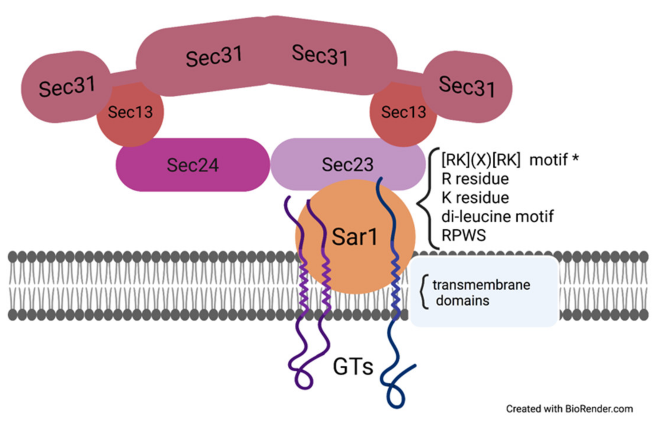
Figure 3. The protein–protein interaction between cargo sorting signal motifs in GTs and COPII coatomers.
In addition to Vps74p and GOLPH3 being shown to affect GT-localization via direct interaction with the COPI complex, other proteins are involved in determining the localization of GTs. The Golgi-localized STELLO1 and STELLO2 proteins (STL1 and STL2) from A.thaliana, which contain the glycosyltransferase-like domain, were shown to alter the CesA distribution and assembly via direct interaction with the latter [73]. The genes encoding the STL1 and STL2 proteins were co-expressed together with CesA genes in the A.thaliana stl1stl2 mutant recovering the cellulose content that was reduced in the stl1stl2 mutant [73]. In animal cells, the GlcNAcT-I inhibitory protein (GnT1IP) shares a similar protein sequence with GlcNAcT-IV glycosyltransferases and inhibits GlcNAcT-I activity [74]. Two GnT1IP transcripts were named GnT1IP-L and GnT1IP-S, and the GnT1IP-L protein was shown to be the type II membrane protein [74]. GnT1IP-L can interact directly with GlcNAcT-I, causing its mislocalization from the medial-Golgi to the ER, ERGIC, and cis-Golgi [74]. Golgi-resident GRASP55 regulated the subcellular localization of glycosylation protein involved in glycosphingolipid biosynthesis by direct interaction [75]. The L95LGV98 sequence in the GRASP domain of GRASP55 interacted with the cytosolic tail of GlcCer synthase (GCS), which catalyzes the critical step in glycosphingolipid biosynthesis [75]. The direct binding with GRASP55 promoted the correct subcellular localization of GCS by preventing GCS from entering in the retrograde transportation [75]. The GTP exchange factor GBF1 facilitated the phosphorylation of Arf1-GDP, and the Src tyrosine kinase (Src) played an essential role in the ARF GTP formation [76]. Src phosphorylated the Y876 and Y898 in the GEF domain C-terminus of GBF1, further increasing the binding between GBF1 and Arf1 and the GALNT relocation [76].
This entry is adapted from the peer-reviewed paper 10.3390/plants11030428
References
- Cantarel, B.L.; Coutinho, P.M.; Rancurel, C.; Bernard, T.; Lombard, V.; Henrissat, B. The Carbohydrate-Active EnZymes database (CAZy): An expert resource for Glycogenomics. Nucleic Acids Res. 2009, 37, D233–D238.
- Lairson, L.L.; Henrissat, B.; Davies, G.J.; Withers, S.G. Glycosyltransferases: Structures, functions, and mechanisms. Annu. Rev. Biochem. 2008, 77, 521–555.
- Zabotina, O.A.; Zhang, N.; Weerts, R. Polysaccharide Biosynthesis: Glycosyltransferases and Their Complexes. . Front. Plant Sci. 2021, 12, 625307.
- Yuan, Y.; Barrett, D.; Zhang, Y.; Kahne, D.; Sliz, P.; Walker, S. Crystal structure of a peptidoglycan glycosyltransferase suggests a model for processive glycan chain synthesis. Proc. Natl. Acad. Sci. USA 2007, 104, 5348–5353.
- Nagashima, Y.; von Schaewen, A.; Koiwa, H. Function of N-glycosylation in plants. Plant Sci. 2018, 274, 70–79.
- Varki, A.; Cummings, R.D.; Esko, J.D.; Stanley, P.; Hart, G.W.; Aebi, M.; Darvill, A.G.; Kinoshita, T.; Packer, N.H.; Prestegard, H.H. (Eds.) Essentials of Glycobiology, 3rd ed.; Cold Spring Harbor Laboratory Press: Cold Spring Harbor, NY, USA, 2015.
- Aebi, M. N-linked protein glycosylation in the ER. Biochim. Biophys. Acta 2013, 1833, 2430–2437.
- Li, Y.; Liu, Y.; Zhu, H.; Chen, X.; Tian, M.; Wei, Y.; Gong, Y.; Jiang, J. N-acetylglucosaminyltransferase I promotes glioma cell proliferation and migration through increasing the stability of the glucose transporter GLUT1. FEBS Lett. 2020, 594, 358–366.
- Vanier, G.; Lucas, P.L.; Loutelier-Bourhis, C.; Vanier, J.; Plasson, C.; Walet-Balieu, M.-L.; Tchi-Song, P.C.; Remy-Jouet, I.; Richard, V.; Bernard, S.; et al. Heterologous expression of the N-acetylglucosaminyltransferase I dictates a reinvestigation of the N-glycosylation pathway in Chlamydomonas reinhardtii. Sci. Rep. 2017, 7, 10156.
- Yoo, J.Y.; Ko, K.S.; Vu, B.N.; Lee, Y.E.; Yoon, S.H.; Pham, T.T.; Kim, J.-T.; Lim, J.-M.; Kang, Y.J.; Hong, J.C.; et al. N-acetylglucosaminyltransferase II Is Involved in Plant Growth and Development under Stress Conditions. Front. Plant Sci. 2021, 12, 761064.
- Kadirvelraj, R.; Yang, J.-Y.; Sanders, J.H.; Liu, L.; Ramiah, A.; Prabhakar, P.K.; Boons, G.-J.; Wood, Z.A.; Moremen, K.W. Human N-acetylglucosaminyltransferase II substrate recognition uses a modular architecture that includes a convergent exosite. Proc. Natl. Acad. Sci. USA 2018, 115, 4637–4642.
- Wong, L.-J.; Sheu, K.-F.R.; Lee, S.-L.; Frey, P.A. Galactose-1-phosphate uridylyltransferase: Isolation and properties of a uridylyl-enzyme intermediate. Biochemistry 1977, 16, 1010–1016.
- Garnham, R.; Scott, E.; Livermore, K.E.; Munkley, J. ST6GAL1: A key player in cancer (Review). Oncol. Lett. 2019, 18, 983–989.
- Khoder-Agha, F.; Harrus, D.; Brysbaert, G.; Lensink, M.F.; Harduin-Lepers, A.; Glumoff, T.; Kellokumpu, S. Assembly of B4GALT1/ST6GAL1 heteromers in the Golgi membranes involves lateral interactions via highly charged surface domains. J. Biol. Chem. 2019, 294, 14383–14393.
- Huang, G.; Li, Z.; Li, Y.; Liu, G.; Sun, S.; Gu, J.; Kameyama, A.; Li, W.; Dong, W. Loss of core fucosylation in both ST6GAL1 and its substrate enhances glycoprotein sialylation in mice. Biochem. J. 2020, 477, 1179–1201.
- Yu, M.; Wang, H.; Liu, J.; Qin, H.; Liu, S.; Yan, Q. The sialyltransferase ST3Gal3 facilitates the receptivity of the uterine endometrium in vitro and in vivo. FEBS Lett. 2018, 592, 3696–3707.
- Zhang, X.; Yang, X.; Chen, M.; Zheng, S.; Li, J.; Lin, S.; Wang, X. ST3Gal3 confers paclitaxel-mediated chemoresistance in ovarian cancer cells by attenuating caspase-8/3 signaling. Mol. Med. Rep. 2019, 20, 4499–4506.
- Khoder-Agha, F.; Sosicka, P.; Escriva Conde, M.; Hassinen, A.; Glumoff, T.; Olczak, M.; Kellokumpu, S. N-acetylglucosaminyltransferases and nucleotide sugar transporters form multi-enzyme-multi-transporter assemblies in golgi membranes in vivo. Cell. Mol. Life Sci. 2019, 76, 1821–1832.
- Kellokumpu, S.; Hassinen, A.; Glumoff, T. Glycosyltransferase complexes in eukaryotes: Long-known, prevalent but still unrecognized. Cell. Mol. Life Sci. 2016, 73, 305–325.
- Wang, J.; Chen, J.; Enns, C.A.; Mayinger, P. The First Transmembrane Domain of Lipid Phosphatase SAC1 Promotes Golgi Localization. PLoS ONE 2013, 8, e71112.
- Béthune, J.; Wieland, F.T. Assembly of COPI and COPII Vesicular Coat Proteins on Membranes. Annu. Rev. Biophys. 2018, 47, 63–83.
- Arakel, E.C.; Schwappach, B. Formation of COPI-coated vesicles at a glance. . 37073 Göttingen, Germany. J. Cell Sci. 2018, 131, jcs209890.
- Luo, P.M.; Boyce, M. Directing Traffic: Regulation of COPI Transport by Post-translational Modifications. Front. Cell Dev. Biol. 2019, 7, 190.
- Sager, G.; Szul, T.; Lee, E.; Kawai, R.; Presley, J.F.; Sztul, E. Modeling the dynamic behaviors of the COPI vesicle formation regulators, the small GTPase Arf1 and its activating Sec7 guanine nucleotide exchange factor GBF1 on Golgi membranes. Mol. Biol. Cell 2021, 32, 446–459.
- Bui, Q.T.; Golinelli-Cohen, M.P.; Jackson, C.L. Large Arf1 guanine nucleotide exchange factors: Evolution, domain structure, and roles in membrane trafficking and human disease. Mol. Genet. Genom. 2009, 282, 329–350.
- Beck, R.; Adolf, F.; Weimer, C.; Bruegger, B.; Wieland, F.T. ArfGAP1 activity and COPI vesicle biogenesis. Traffic 2009, 10, 307–315.
- Shiba, Y.; Luo, R.; Hinshaw, J.E.; Szul, T.; Hayashi, R.; Sztul, E.; Nagashima, K.; Baxa, U.; Randazzo, P.A. ArfGAP1 promotes COPI vesicle formation by facilitating coatomer polymerization. Cell. Logist. 2011, 1, 139–154.
- Stagg, S.M.; LaPointe, P.; Razvi, A.; Gurkan, C.; Potter, C.S.; Carragher, B.; Balch, W.E. Structural Basis for Cargo Regulation of COPII Coat Assembly. Cell 2008, 134, 474–484.
- Peotter, J.; Kasberg, W.; Pustova, I.; Audhya, A. COPII-mediated trafficking at the ER/ERGIC interface. Traffic 2019, 20, 491–503.
- Fath, S.; Mancias, J.D.; Bi, X.; Goldberg, J. Structure and Organization of Coat Proteins in the COPII Cage. Cell 2007, 129, 1325–1336.
- McCaughey, J.; Stephens, D.J. COPII-dependent ER export in animal cells: Adaptation and control for diverse cargo. Histochem. Cell Biol. 2018, 150, 119–131.
- Melville, D.B.; Studer, S.; Schekman, R. Small sequence variations between two mammalian paralogs of the small GTPase SAR1 underlie functional differences in coat protein complex II assembly. J. Biol. Chem. 2020, 295, 8401–8412.
- Sánchez-Simarro, J.; Bernat-Silvestre, C.; Gimeno-Ferrer, F.; Selvi-Martínez, P.; Montero-Pau, J.; Aniento, F.; Marcote, M.J. Loss of Arabidopsis β-COP Function Affects Golgi Structure, Plant Growth and Tolerance to Salt Stress. Front. Plant Sci. 2020, 11, 430.
- Bernat-Silvestre, C.; De Sousa Vieira, V.; Sanchez-Simarro, J.; Pastor-Cantizano, N.; Hawes, C.; Marcote, M.J.; Aniento, F. p24 Family Proteins Are Involved in Transport to the Plasma Membrane of GPI-Anchored Proteins in Plants. Plant Physiol. 2020, 184, 1333–1347.
- Sánchez-Simarro, J.; Bernat-Silvestre, C.; Aniento, F.; Marcote, M.J. ß-COP mutants show specific high sensitivity to chloride ions. Plant Signal. Behav. 2021, 16, 1858629.
- Gimeno-Ferrer, F.; Pastor-Cantizano, N.; Bernat-Silvestre, C.; Selvi-Martínez, P.; Vera-Sirera, F.; Gao, C.; Perez-Amador, M.A.; Jiang, L.; Aniento, F.; Marcote, M.J. α2-COP is involved in early secretory traffic in Arabidopsis and is required for plant growth. J. Exp. Bot. 2017, 68, 391–401.
- Pastor-Cantizano, N.; Bernat-Silvestre, C.; Marcote, M.J.; Aniento, F. Loss of Arabidopsis p24 function affects ERD2 trafficking and Golgi structure, and activates the unfolded protein response. J. Cell Sci. 2018, 131, jcs203802.
- Cabada Gomez, D.A.; Chavez, M.I.; Cobos, A.N.; Gross, R.J.; Yescas, J.A.; Balogh, M.A.; Indriolo, E. COPI complex isoforms are required for the early acceptance of compatible pollen grains in Arabidopsis thaliana. Plant Reprod. 2020, 33, 97–110.
- Ahn, H.K.; Kang, Y.W.; Lim, H.M.; Hwang, I.; Pai, H.S. Physiological Functions of the COPI Complex in Higher Plants. Mol. Cells 2015, 38, 866–875.
- Min, M.K.; Jang, M.; Lee, M.; Lee, J.; Song, K.; Lee, Y.; Choi, K.Y.; Robinson, D.G.; Hwang, I. Recruitment of Arf1-GDP to Golgi by Glo3p-type ArfGAPs is crucial for golgi maintenance and plant growth. Plant Physiol. 2013, 161, 676–691.
- Pastor-Cantizano, N.; García-Murria, M.J.; Bernat-Silvestre, C.; Marcote, M.J.; Mingarro, I.; Aniento, F. N-Linked Glycosylation of the p24 Family Protein p24δ5 Modulates Retrograde Golgi-to-ER Transport of K/HDEL Ligands in Arabidopsis. Mol. Plant 2017, 10, 1095–1106.
- Montesinos, J.C.; Pastor-Cantizano, N.; Robinson, D.G.; Marcote, M.J.; Aniento, F. Arabidopsis p24δ5 and p24δ9 facilitate Coat Protein I-dependent transport of the K/HDEL receptor ERD2 from the Golgi to the endoplasmic reticulum. Plant J. 2014, 80, 1014–1030.
- Chung, K.P.; Zeng, Y.; Jiang, L. COPII Paralogs in Plants: Functional Redundancy or Diversity? Trends Plant Sci. 2016, 21, 758–769.
- Chang, M.; Wu, S.Z.; Ryken, S.E.; O’Sullivan, J.E.; Bezanilla, M. COPII Sec23 proteins form isoform-specific endoplasmic reticulum exit sites with differential effects on polarized growth. Plant Cell 2022, 34, 333–350.
- Liu, X.; Tong, M.; Zhang, A.; Liu, M.; Zhao, B.; Liu, Z.; Li, Z.; Zhu, X.; Guo, Y.; Li, R. COPII genes SEC31A/B are essential for gametogenesis and interchangeable in pollen development in Arabidopsis. Plant J. 2021, 105, 1600–1614.
- Liang, X.; Li, S.W.; Gong, L.M.; Li, S.; Zhang, Y. COPII Components Sar1b and Sar1c Play Distinct yet Interchangeable Roles in Pollen Development. Plant Physiol. 2020, 183, 974–985.
- Zeng, Y.; Chung, K.P.; Li, B.; Lai, C.M.; Lam, S.K.; Wang, X.; Cui, Y.; Gao, C.; Luo, M.; Wong, K.-B.; et al. Unique COPII component AtSar1a/AtSec23a pair is required for the distinct function of protein ER export in Arabidopsis thaliana. Proc. Natl. Acad. Sci. USA 2015, 112, 14360–14365.
- Aboulela, M.; Nakagawa, T.; Oshima, A.; Nishimura, K.; Tanaka, Y. The Arabidopsis COPII components, AtSEC23A and AtSEC23D, are essential for pollen wall development and exine patterning. J. Exp. Bot. 2018, 69, 1615–1633.
- Zhao, B.; Shi, H.; Wang, W.; Liu, X.; Gao, H.; Wang, X.; Zhang, Y.; Yang, M.; Li, R.; Guo, Y. Secretory COPII Protein SEC31B Is Required for Pollen Wall Development. Plant Physiol. 2016, 172, 1625–1642.
- Li, B.; Zeng, Y.; Cao, W.; Zhang, W.; Cheng, L.; Yin, H.; Wu, Q.; Wang, X.; Huang, Y.; Lau, W.C.Y.; et al. A distinct giant coat protein complex II vesicle population in Arabidopsis thaliana. Nat. Plants 2021, 7, 1335–1346.
- Robinson, D.G.; Brandizzi, F.; Hawes, C.; Nakano, A. Vesicles versus Tubes: Is Endoplasmic Reticulum-Golgi Transport in Plants Fundamentally Different from Other Eukaryotes? Plant Physiol. 2015, 168, 393–406.
- Mironov, A.A. ER-Golgi transport could occur in the absence of COPII vesicles. Nat. Rev. Mol. Cell Biol. 2014, 15, 1.
- Ito, Y.; Uemura, T.; Nakano, A. The Golgi entry core compartment functions as a COPII-independent scaffold for ER-to-Golgi transport in plant cells. J. Cell Sci. 2018, 131, jcs203893.
- Tu, L.; Tai, W.C.; Chen, L.; Banfield, D.K. Signal-mediated dynamic retention of glycosyltransferases in the Golgi. Science 2008, 321, 404–407.
- Schoberer, J.; König, J.; Veit, C.; Vavra, U.; Liebminger, E.; Botchway, S.W.; Altmann, F.; Kriechbaumer, V.; Hawes, C.; Strasser, R. A signal motif retains Arabidopsis ER-α-mannosidase I in the cis-Golgi and prevents enhanced glycoprotein ERAD. Nat. Commun. 2019, 10, 3701.
- Boulaflous, A.; Saint-Jore-Dupas, C.; Herranz-Gordo, M.-C.; Pagny-Salehabadi, S.; Plasson, C.; Garidou, F.; Kiefer-Meyer, M.-C.; Ritzenthaler, C.; Faye, L.; Gomord, V. Cytosolic N-terminal arginine-based signals together with a luminal signal target a type II membrane protein to the plant ER. BMC Plant Biol. 2009, 9, 144.
- Saint-Jore-Dupas, C.; Nebenführ, A.; Boulaflous, A.; Follet-Gueye, M.-L.; Plasson, C.; Hawes, C.; Driouich, A.; Faye, L.; Gomord, V. PlantN-Glycan Processing Enzymes Employ Different Targeting Mechanisms for Their Spatial Arrangement along the Secretory Pathway. Plant Cell 2006, 18, 3182–3200.
- Uemura, S.; Yoshida, S.; Shishido, F.; Inokuchi, J. The cytoplasmic tail of GM3 synthase defines its subcellular localization, stability, and in vivo activity. Mol. Biol. Cell 2009, 20, 3088–3100.
- Schoberer, J.; Vavra, U.; Stadlmann, J.; Hawes, C.; Mach, L.; Steinkellner, H.; Strasser, R. Arginine/lysine residues in the cytoplasmic tail promote ER export of plant glycosylation enzymes. Traffic 2009, 10, 101–115.
- Liu, L.; Doray, B.; Kornfeld, S. Recycling of Golgi glycosyltransferases requires direct binding to coatomer. Proc. Natl. Acad. Sci. USA 2018, 115, 8984–8989.
- Giraudo, C.G.; Maccioni, H.J. Endoplasmic reticulum export of glycosyltransferases depends on interaction of a cytoplasmic dibasic motif with Sar1. Mol. Biol. Cell 2003, 14, 3753–3766.
- Schoberer, J.; Liebminger, E.; Vavra, U.; Veit, C.; Castilho, A.; Dicker, M.; Maresch, D.; Altmann, F.; Hawes, C.; Botchway, S.W.; et al. The transmembrane domain of N -acetylglucosaminyltransferase I is the key determinant for its Golgi subcompartmentation. Plant J. 2014, 80, 809–822.
- Schoberer, J.; Liebminger, E.; Vavra, U.; Veit, C.; Grünwald-Gruber, C.; Altmann, F.; Botchway, S.W.; Strasser, R. The Golgi Localization of GnTI Requires a Polar Amino Acid Residue within Its Transmembrane Domain. Plant Physiol. 2019, 180, 859–873.
- Becker, J.L.; Tran, D.T.; Tabak, L.A. Members of the GalNAc-T family of enzymes utilize distinct Golgi localization mechanisms. Glycobiology 2018, 28, 841–848.
- Franke, M.; Braulke, T.; Storch, S. Transport of the GlcNAc-1-phosphotransferase α/β-Subunit Precursor Protein to the Golgi Apparatus Requires a Combinatorial Sorting Motif. J. Biol. Chem. 2013, 288, 1238–1249.
- Wang, X.; Wang, D.; Jing, P.; Wu, Y.; Xia, Y.; Chen, M.; Hong, L. A Novel Golgi Retention Signal RPWS for Tumor Suppressor UBIAD1. PLoS ONE 2013, 8, e72015.
- Mancias, J.D.; Goldberg, J. Structural basis of cargo membrane protein discrimination by the human COPII coat machinery. EMBO J. 2008, 27, 2918–2928.
- Dancourt, J.; Barlowe, C. Protein sorting receptors in the early secretory pathway. Annu. Rev. Biochem. 2010, 79, 777–802.
- Chung, K.P.; Zeng, Y.; Li, Y.; Ji, C.; Xia, Y.; Jiang, L. Signal motif-dependent ER export of the Qc-SNARE BET12 interacts with MEMB12 and affects PR1 trafficking in Arabidopsis. J. Cell Sci. 2018, 131, jcs202838.
- Eckert, E.S.; Reckmann, I.; Hellwig, A.; Röhling, S.; El-Battari, A.; Wieland, F.T.; Popoff, V. Golgi Phosphoprotein 3 Triggers Signal-mediated Incorporation of Glycosyltransferases into Coatomer-coated (COPI) Vesicles. J. Biol. Chem. 2014, 289, 31319–31329.
- Rizzo, R.; Russo, D.; Kurokawa, K.; Sahu, P.; Lombardi, B.; Supino, D.; A Zhukovsky, M.; Vocat, A.; Pothukuchi, P.; Kunnathully, V.; et al. Golgi maturation-dependent glycoenzyme recycling controls glycosphingolipid biosynthesis and cell growth via GOLPH3. EMBO J. 2021, 40, e107238.
- Welch, L.G.; Peak-Chew, S.Y.; Begum, F.; Stevens, T.J.; Munro, S. GOLPH3 and GOLPH3L are broad-spectrum COPI adaptors for sorting into intra-Golgi transport vesicles. J. Cell Biol. 2021, 220, e202106115.
- Zhang, Y.; Nikolovski, N.; Sorieul, M.; Vellosillo, T.; McFarlane, H.E.; DuPree, R.; Kesten, C.; Schneider, R.; Driemeier, C.; Lathe, R.; et al. Golgi-localized STELLO proteins regulate the assembly and trafficking of cellulose synthase complexes in Arabidopsis. Nat. Commun. 2016, 7, 11656.
- Huang, H.H.; Stanley, P. A testis-specific regulator of complex and hybrid N-glycan synthesis. J. Cell Biol. 2010, 190, 893–910.
- Pothukuchi, P.; Agliarulo, I.; Pirozzi, M.; Rizzo, R.; Russo, D.; Turacchio, G.; Nüchel, J.; Yang, J.; Gehin, C.; Capolupo, L.; et al. GRASP55 regulates intra-Golgi localization of glycosylation enzymes to control glycosphingolipid biosynthesis. EMBO J. 2021, 40, e107766.
- Chia, J.; Wang, S.C.; Wee, S.; Gill, D.J.; Tay, F.; Kannan, S.; Verma, C.S.; Gunaratne, J.; Bard, F.A. Src activates retrograde membrane traffic through phosphorylation of GBF1. eLife 2021, 10, e68678.