Endotoxin Adsorption in the Treatment of Septic Shock: History
Please note this is an old version of this entry, which may differ significantly from the current revision.
Contributor:

Lipopolysaccharide, the main component of the outer membrane of Gram-negative bacteria is a highly potent endotoxin responsible for organ dysfunction in sepsis. It is present in the blood stream not only in Gram-negative infections, but also in Gram-positive and fungal infections, presumably due to sepsis-related disruption of the intestinal barrier. Various pathways, both extra- and intracellular, are involved in sensing endotoxin and non-canonical activation of caspase-mediated pyroptosis is considered to have a major role in sepsis pathophysiology. Endotoxin induces specific pathological alterations in several organs, which contributes to poor outcomes. The adverse consequences of endotoxin in the circulation support the use of anti-endotoxin therapies, yet more than 30 years of experience with endotoxin adsorption therapies have not provided clear evidence in favor of this treatment modality. The results of small studies support timely endotoxin removal guided by measuring the levels of endotoxin; unfortunately, this has not been proven in large, randomized studies. The presence of endotoxemia can be demonstrated in the majority of patients with COVID-19, yet only case reports and case series describing the effects of endotoxin removal in these patients have been published to date. The place of blood purification therapies in the treatment of septic shock has not yet been determined.

  • endotoxin
  • septic shock
  • blood purification
  • bacterial translocation

1. Introduction

Sepsis is recognized as a global health problem with an estimated nearly 50 million cases and 11 million deaths recorded worldwide in 2017, representing almost 20% of all global deaths [1]. Recent meta-analysis of epidemiological evidence, related to the burden of hospital-acquired sepsis, showed mortality between 30.1% and 64.6% among ICU-treated patients [2]. Septic shock is characterized by persistent hypotension requiring vasopressor support and a serum lactate level > 2 mmol/L, despite adequate fluid resuscitation. In the continuum of sepsis severity it carries the worst prognosis, with mortality reaching up to 92% in some studies [3].
In addition to standard therapy, which includes infection control (antibiotics, controlling the source), cardiovascular resuscitation (administering fluids, vasoactive agents), and organ support, modulation of the host response is assumed to improve outcome, with low-dose corticosteroids being most commonly advocated [4][5]. An alternative approach includes extracorporeal therapies aimed at removing molecules that are involved in the immune reaction to invading microorganisms. Endotoxin plays a prominent role in the pathogenesis of sepsis, and the idea to neutralize its detrimental capacities continues to attract the attention of researchers and clinicians.

2. Lipopolysaccharide Sensing Pathways

Lipopolysaccharide is sensed via extracellular and intracellular pathways that lead to the activation of the immune response.

2.1. Toll-like Receptor 4–Myeloid Differentiation Protein 2 (TLR4-MD-2) Pathway

The toll-like receptor 4 (TLR4) is the main sensing receptor for LPS, and it is one of the pattern recognition receptors responsible for the early detection of invading microbes by the innate immune system. TLR4 is expressed on the surface of macrophages, monocytes, neutrophils, dendritic, and epithelial cells, as well as within endosomes, forming the front line of the host’s defense against Gram-negative bacteria. LPS molecules in the bacterial cell wall and also soluble LPS-aggregates are dissociated and bound by LPS Binding Protein (LBP), carried to form a complex with either a soluble or membrane bound cluster of differentiation-14 (CD14), and subsequently transferred to the toll-like receptor 4/myeloid differentiation-2 (MD-2) complex, which promotes the TLR4/MD-2 dimerization necessary for activating intracellular MyD88-dependent and TRIF-dependent pathways. Both pathways lead to the production and release of pro-inflammatory cytokines and type I interferones (IFNs), respectively [6][7][8]. Immune hyperactivation from the inappropriate triggering by pathogens and the cytokine storm leads to organ damage, multi-organ failure, and death [9].
The progress in research on LPS recognition systems, witnessed in the last decade, led to important discoveries of TLR4-independent LPS-sensing pathways that may have a central role in the pathophysiology of sepsis and related mortality.

2.2. Transient Receptor Potential (TRP) Ion Channels

Transient receptor potential ion channels are membrane-bound channels that serve as cellular sensors of environmental and intracellular stimuli. LPS sensing by TRP channels has been demonstrated in neurons and airway epithelial cells [10][11]. The activation of TRPA1 channels in nociceptive neurons by the LPS of pathogenic bacteria generates pain during inflammation [12]. Activation of the TRPV4 channels in the airway epithelium boosts ciliary beat frequency and the production of bactericidal nitric oxide, which facilitates the pathogen clearance from the airways. LPS sensing by TRP channels provides an immediate response to invading pathogens, which is faster and independent of the canonical TLR4 immune pathway [11].

2.3. Intracellular LPS Sensing

The activation of caspases plays a crucial role in intracellular pathogen detection and defense. LPS can enter the cytosol as LPS/outer-membrane-vesicle (OMV)-high mobility-group-box-1 (HMGB1) complexes internalized through a receptor for advanced glycation (RAGE). LPS that enters the cytoplasm of macrophages, as well as endothelial and epithelial cells, is sensed by inflammatory caspases—caspase-11 in mice and caspase-4/5 in humans—and leads to the induction of pyroptosis, an inflammatory form of cell death. Activated caspases cleave gasdermin D, which causes pore formation in the cell membrane with subsequent cell lysis and the release of proinflammatory IL-1β and IL-18 [13]. Inflammasome activation and pyroptosis are important mechanisms of the innate immune defense against pathogens that are capable of invading the cytosol and play a major role in sepsis pathophysiology. Caspase-11 has been found to be responsible for bacterial clearance in Klebsiella pneumoniae and Acinetobacter baumannii, as well as Burkholderia lung infections [13]. It is speculated that caspases may be responsible for sensing penta-acylated LPS, which is not detected by TLR4 [14]. Caspase-mediated pyroptosis of endothelial cells has a fundamental role in the host’s defense and immune surveillance functions of the microvasculature [15]. Excessive activation of pyroptosis causes extensive cell death and immense inflammation leading to organ failure and septic shock [16].

3. Organ Damage Caused by Sensing Endotoxin
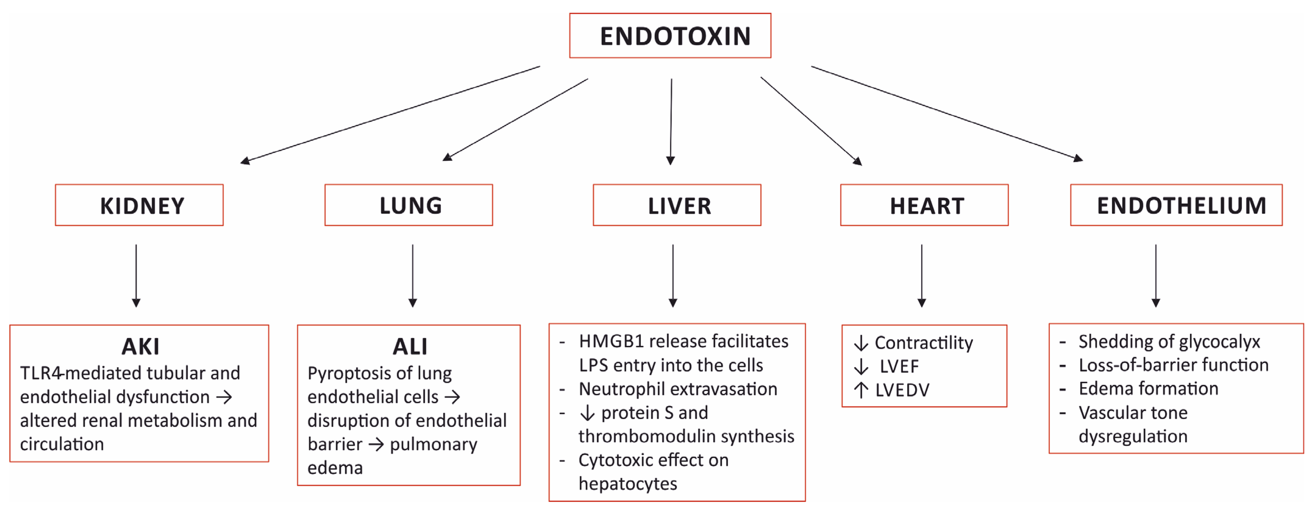
Endotoxin plays a very prominent role in the pathogenesis of sepsis. It is one of the most important pathogen-associated molecular patterns (PAMP), and a large burden of endotoxin triggers an excessive, uncontrolled systemic inflammatory response that leads to multi-organ failure and death. Moreover, endotoxin induces specific pathological alterations in several organs that contribute to the outcome (Figure 1).
/media/item_content/202202/6209bdd9203f8jcm-11-00619-g001.pngFigure 1. Selected organ damage induced by sensing endotoxin. AKI—acute kidney injury; ALI—acute lung injury; TLR4—toll-like receptor 4; HMGB1—high mobility group box-1; LPS—lipopolysaccharide; LVEF—left ventricular ejection fraction; LVEDV—left ventricular end diastolic volume.

3.1. The Kidney

Acute kidney injury (AKI) develops in at least 40–50% of patients with sepsis or septic shock and is associated with significantly higher mortality [17][18][19][20]. In addition to septic alterations, AKI presents with metabolic and fluid abnormalities, necessitating adjustments in volume therapy and pharmacotherapy, most notably limiting antimicrobial choice. The pathophysiology of septic AKI is complex and, in addition to hypoperfusion, interactions between vascular, tubular, and inflammatory factors are involved. Although the exact mechanism underlying renal dysfunction in sepsis remains unknown, there is strong experimental evidence supporting the prominent role of the toll-like receptor 4 (TLR-4), which is expressed in the kidney [21]. Its activation causes cytokine and chemokine release; leukocyte infiltration, which results in endothelial dysfunction; tubular dysfunction and altered renal metabolism and circulation [22]. TLR-4 receptors are located in the tubular epithelium and in the glomeruli and vascular endothelium. Endotoxin is filtered in renal glomeruli, internalized by S1 proximal tubules through TLR4 receptors, and interactions between endotoxin and S1 tubules result in severe oxidative stress and damage to the neighboring S2 segments [22][23]. TLR4 directly inhibits bicarbonate absorption in the medullary-thick ascending limb, downregulates renal sodium, chloride, and glucose transporters, causes luminal obstruction, and reduces tubular flow, among other effects [22]. Endothelial activation and alterations to glomerular glycocalyx and the deposit of NETs in kidney tissue secondary to endotoxic shock also contribute to kidney injury [24][25]. Direct renal damage by endotoxin can explain the occurrence of AKI in sepsis, even when hemodynamic parameters are well-controlled [23]. In fact, protocolized hemodynamic resuscitation did not influence either the development or the course of AKI in patients with septic shock [20]. As a result, the concept of equating sepsis-induced AKI to acute tubular necrosis, attributed to ischemia from hemodynamic changes, has been replaced by the theory of the interplay between inflammation and oxidative stress, microvascular dysfunction, and the adaptive response of the tubular epithelial cells to the septic insult [26].

3.2. The Lung

In mice subjected to LPS-induced sepsis, pronounced histological alterations in the lungs were found, with thickening of the septum, edema, congestion, and high leukocyte infiltration into the interstitium, which correlated with a significant increase in the serum concentrations of NETs and the extent of lung injury [25]. In another experimental study, lung injury was attributed to LPS-triggered pyroptosis of the endothelial cells in the lungs; LPS sensing in the endothelial cytoplasm via caspase-4/5/11-mediated pyroptosis led to disruption of the endothelial barrier resulting in pulmonary edema, the release of pro-inflammatory cytokines, fluid protein leakage, and a massive influx of leukocytes [15]. The pyroptotic response was augmented when the expression of caspase-4/5/11 was enhanced by concomitant priming with extracellular LPS via LPS binding to TLR4 [15].

3.3. The Heart

Toll-like receptors 4 are expressed in cardiomyocytes and their activation elicits an inflammatory response with the production of cytokines and chemokines with a negative effect on cardiac contractility [27]. In healthy volunteers, endotoxemia resulted in a reduction in the left ventricular ejection fraction and an increase in the left ventricular end diastolic volume [28]. In mice, LPS administration resulted in significant pathological changes in the myocardial bundles, congestion of the capillaries with the presence of leukocytes attached to the endothelium, and pathological changes in the cardiomyocytes seen upon histological examination [25]. The results of other studies indicated that sepsis-associated cardiac dysfunction was also mediated by mechanisms other than TLR4 [29].

3.4. The Liver

The liver is an important participant in the body’s reaction to endotoxemia. Murine studies demonstrated that endotoxin uses both TLR4 and caspase-11/gasdermin D (GsdmD) pathways to induce the release of HMGB1 from hepatocytes—the major source of circulating HMGB1 in sepsis [30]. Complexes of hepatocyte-released HMGB1 and LPS are delivered via RAGE into the cytosol of macrophages and endothelial cells, where LPS activates caspase-11 and induces pyroptosis and cell death [31]. The intracellular LPS-sensing pathway is considered to have a central role in the pathogenesis of sepsis [13].
In the liver, LPS affects the architecture of the sinusoidal endothelium and blood flow velocities, which leads to extravasation of neutrophils and neutrophil–hepatocyte interactions, decreases protein S and thrombomodulin synthesis, which contributes to a pro-coagulant state and has a direct cytotoxic effect on hepatocytes [24][32]. In mice subjected to LPS-induced endotoxemia, histological changes in the liver included enlarged sinusoids, an increased volume of endothelial cells with rounded nuclei, a high number of leukocytes in the lumen, Kupffer cell hypertrophy and hyperplasia, along with the presence of leukocytes close to periportal areas and congestion of the central vein with swollen hepatocytes [25].

3.5. The Vascular Endothelium

Endothelial cell dysfunction is thought to be the key factor in the progression from sepsis to organ failure [24]. The presence of endotoxin in the blood causes shedding of the glycocalyx lining of the vascular endothelium that leads to the loss-of-barrier function, the formation of edema, and the dysregulation of vascular tone, among other effects [24]. LPS triggered, caspase-dependent pyroptosis in endothelial cells results in disruption of the endothelial barrier, fluid leakage, and the development of ALI [15].

4. Investigating Aspects of Endotoxin Removal

4.1. Timing of the Initiation of Endotoxin Adsorption

Non-randomized studies that compared an early vs. late initiation of the PMX HP treatment in patients with septic shock found better survival or reduced catecholamine requirements in the early treatment group. The initiation of PMX hemoperfusion within 6, 8, or 9 h after the administration of catecholamine or the diagnosis of septic shock resulted in a more favorable outcome compared to a later initiation [33][34][35]. According to the results of these studies, PMX HP therapy should be performed as early as possible in patients with septic shock, and a delay in PMX HP therapy may contribute to increased mortality [34].

4.2. Extended Endotoxin Adsorption Treatment

The recommended period for PMX HP treatment is 2 h. In studies where the time of treatment ranged from 8 to 24 h, there were improved hemodynamics and improved pulmonary oxygenation, but no improved mortality was observed [36][37][38].

4.3. Endotoxin Removal Treatment Guided by Measuring the Endotoxin Level

In 11 patients diagnosed with postsurgical sepsis and who had a high EAA (≥0.6), when the PMX HP treatment was performed and repeated every 24 h until the EAA was low (<0.4), all patients survived until the 28-day follow-up [39]. These findings are similar to an observation from the post-hoc analysis of the EUPHRATES trial. A trend toward lower mortality and a significant increase in ventilation-free days was found in patients with septic shock and a greater than median reduction in EAA on day 3 after the PMX HP treatment. The same was true for patients who achieved an EAA of less than 0.65 on day 3 [40]. The authors of the study suggested that the dosing regimen of PMX therapy should be tailored according to measured endotoxin levels and/or patient’s clinical response, but this hypothesis needs to be validated in a prospective study [40].

This entry is adapted from the peer-reviewed paper 10.3390/jcm11030619

References

  1. Rudd, K.E.; Johnson, S.C.; Agesa, K.M.; Shackelford, K.A.; Tsoi, D.; Kievlan, D.R.; Colombara, D.V.; Ikuta, K.S.; Kissoon, N.; Finfer, S.; et al. Global, Regional, and National Sepsis Incidence and Mortality, 1990–2017: Analysis for the Global Burden of Disease Study. Lancet 2020, 395, 200–211.
  2. Markwart, R.; Saito, H.; Harder, T.; Tomczyk, S.; Cassini, A.; Fleischmann-Struzek, C.; Reichert, F.; Eckmanns, T.; Allegranzi, B. Epidemiology and Burden of Sepsis Acquired in Hospitals and Intensive Care Units: A Systematic Review and Meta-Analysis. Intensive Care Med. 2020, 46, 1536–1551.
  3. Putzu, A.; Schorer, R.; Lopez-Delgado, J.C.; Cassina, T.; Landoni, G. Blood Purification and Mortality in Sepsis and Septic Shock. Anesthesiology 2019, 131, 580–593.
  4. Vincent, J.-L. Septic Shock. In Textbook of Critical Care; Vincent, J.-L., Abraham, E., Moore, F.A., Kochanek, P.M., Fink, M.P., Eds.; Elsevier: Philadelphia, PA, USA, 2017; pp. 843–848. ISBN 978-0-323-37638-9.
  5. Evans, L.; Rhodes, A.; Alhazzani, W.; Antonelli, M.; Coopersmith, C.M.; French, C.; Machado, F.R.; Mcintyre, L.; Ostermann, M.; Prescott, H.C.; et al. Surviving Sepsis Campaign: International Guidelines for Management of Sepsis and Septic Shock 2021. Intensive Care Med. 2021, 47, 1181–1247.
  6. Palsson-McDermott, E.M.; O’Neill, L.A.J. Signal Transduction by the Lipopolysaccharide Receptor, Toll-like Receptor-4. Immunology 2004, 113, 153–162.
  7. Rosadini, C.V.; Kagan, J.C. Early Innate Immune Responses to Bacterial LPS. Curr. Opin. Immunol. 2017, 44, 14–19.
  8. Aluri, J.; Cooper, M.A.; Schuettpelz, L.G. Toll-Like Receptor Signaling in the Establishment and Function of the Immune System. Cells 2021, 10, 1374.
  9. Fajgenbaum, D.C.; June, C.H. Cytokine Storm. N. Engl. J. Med. 2020, 383, 2255–2273.
  10. Meseguer, V.; Alpizar, Y.A.; Luis, E.; Tajada, S.; Denlinger, B.; Fajardo, O.; Manenschijn, J.-A.; Fernández-Peña, C.; Talavera, A.; Kichko, T.; et al. TRPA1 Channels Mediate Acute Neurogenic Inflammation and Pain Produced by Bacterial Endotoxins. Nat. Commun. 2014, 5, 3125.
  11. Alpizar, Y.A.; Boonen, B.; Sanchez, A.; Jung, C.; López-Requena, A.; Naert, R.; Steelant, B.; Luyts, K.; Plata, C.; De Vooght, V.; et al. TRPV4 Activation Triggers Protective Responses to Bacterial Lipopolysaccharides in Airway Epithelial Cells. Nat. Commun. 2017, 8, 1059.
  12. Mazgaeen, L.; Gurung, P. Recent Advances in Lipopolysaccharide Recognition Systems. IJMS 2020, 21, 379.
  13. Rathinam, V.A.K.; Zhao, Y.; Shao, F. Innate Immunity to Intracellular LPS. Nat. Immunol. 2019, 20, 527–533.
  14. Zamyatina, A.; Heine, H. Lipopolysaccharide Recognition in the Crossroads of TLR4 and Caspase-4/11 Mediated Inflammatory Pathways. Front. Immunol. 2020, 11, 585146.
  15. Cheng, K.T.; Xiong, S.; Ye, Z.; Hong, Z.; Di, A.; Tsang, K.M.; Gao, X.; An, S.; Mittal, M.; Vogel, S.M.; et al. Caspase-11–Mediated Endothelial Pyroptosis Underlies Endotoxemia-Induced Lung Injury. J. Clin. Investig. 2017, 127, 4124–4135.
  16. Zhang, W.; Coopersmith, C.M. Dying as a Pathway to Death in Sepsis. Anesthesiology 2018, 129, 238–240.
  17. Hoste, E.A.J.; Bagshaw, S.M.; Bellomo, R.; Cely, C.M.; Colman, R.; Cruz, D.N.; Edipidis, K.; Forni, L.G.; Gomersall, C.D.; Govil, D.; et al. Epidemiology of Acute Kidney Injury in Critically Ill Patients: The Multinational AKI-EPI Study. Intensive Care Med. 2015, 41, 1411–1423.
  18. Uchino, S. Acute Renal Failure in Critically Ill PatientsA Multinational, Multicenter Study. JAMA 2005, 294, 813.
  19. Vincent, J.-L.; Sakr, Y.; Sprung, C.L.; Ranieri, V.M.; Reinhart, K.; Gerlach, H.; Moreno, R.; Carlet, J.; Le Gall, J.-R.; Payen, D. Sepsis in European Intensive Care Units: Results of the SOAP Study. Crit. Care Med. 2006, 34, 344–353.
  20. Kellum, J.A.; Chawla, L.S.; Keener, C.; Singbartl, K.; Palevsky, P.M.; Pike, F.L.; Yealy, D.M.; Huang, D.T.; Angus, D.C. The Effects of Alternative Resuscitation Strategies on Acute Kidney Injury in Patients with Septic Shock. Am. J. Respir. Crit. Care Med. 2016, 193, 281–287.
  21. Fenhammar, J.; Rundgren, M.; Forestier, J.; Kalman, S.; Eriksson, S.; Frithiof, R. Toll-Like Receptor 4 Inhibitor TAK-242 Attenuates Acute Kidney Injury in Endotoxemic Sheep. Anesthesiology 2011, 114, 1130–1137.
  22. Anderberg, S.B.; Luther, T.; Frithiof, R. Physiological Aspects of Toll-like Receptor 4 Activation in Sepsis-Induced Acute Kidney Injury. Acta Physiol. 2017, 219, 575–590.
  23. Kalakeche, R.; Hato, T.; Rhodes, G.; Dunn, K.W.; El-Achkar, T.M.; Plotkin, Z.; Sandoval, R.M.; Dagher, P.C. Endotoxin Uptake by S1 Proximal Tubular Segment Causes Oxidative Stress in the Downstream S2 Segment. J. Am. Soc. Nephrol. 2011, 22, 1505–1516.
  24. Ince, C.; Mayeux, P.R.; Nguyen, T.; Gomez, H.; Kellum, J.A.; Ospina-Tascón, G.A.; Hernandez, G.; Murray, P.; De Backer, D. The Endothelium in Sepsis. Shock 2016, 45, 259–270.
  25. Czaikoski, P.G.; Mota, J.M.S.C.; Nascimento, D.C.; Sônego, F.; Castanheira, F.V.e.S.; Melo, P.H.; Scortegagna, G.T.; Silva, R.L.; Barroso-Sousa, R.; Souto, F.O.; et al. Neutrophil Extracellular Traps Induce Organ Damage during Experimental and Clinical Sepsis. PLoS ONE 2016, 11, e0148142.
  26. Gomez, H.; Ince, C.; De Backer, D.; Pickkers, P.; Payen, D.; Hotchkiss, J.; Kellum, J.A. A Unified Theory of Sepsis-Induced Acute Kidney Injury: Inflammation, Microcirculatory Dysfunction, Bioenergetics, and the Tubular Cell Adaptation to Injury. Shock 2014, 41, 3–11.
  27. Boyd, J.; Mathur, S.; Wang, Y.; Bateman, R.; Walley, K. Toll-like Receptor Stimulation in Cardiomyoctes Decreases Contractility and Initiates an NF-ΚB Dependent Inflammatory Response. Cardiovasc. Res. 2006, 72, 384–393.
  28. Suffredini, A.F.; Fromm, R.E.; Parker, M.M.; Brenner, M.; Kovacs, J.A.; Wesley, R.A.; Parrillo, J.E. The Cardiovascular Response of Normal Humans to the Administration of Endotoxin. N. Engl. J. Med. 1989, 321, 280–287.
  29. Martin, L.; Derwall, M.; Al Zoubi, S.; Zechendorf, E.; Reuter, D.A.; Thiemermann, C.; Schuerholz, T. The Septic Heart. Chest 2019, 155, 427–437.
  30. Li, W.; Deng, M.; Loughran, P.A.; Yang, M.; Lin, M.; Yang, C.; Gao, W.; Jin, S.; Li, S.; Cai, J.; et al. LPS Induces Active HMGB1 Release From Hepatocytes Into Exosomes Through the Coordinated Activities of TLR4 and Caspase-11/GSDMD Signaling. Front. Immunol. 2020, 11, 229.
  31. Deng, M.; Tang, Y.; Li, W.; Wang, X.; Zhang, R.; Zhang, X.; Zhao, X.; Liu, J.; Tang, C.; Liu, Z.; et al. The Endotoxin Delivery Protein HMGB1 Mediates Caspase-11-Dependent Lethality in Sepsis. Immunity 2018, 49, 740–753.e7.
  32. Wang, Y.; Liu, Y. Neutrophil-Induced Liver Injury and Interactions Between Neutrophils and Liver Sinusoidal Endothelial Cells. Inflammation 2021, 44, 1246–1262.
  33. Takeyama, N.; Noguchi, H.; Hirakawa, A.; Kano, H.; Morino, K.; Obata, T.; Sakamoto, T.; Tamai, F.; Ishikura, H.; Kase, Y.; et al. Time to Initiation of Treatment with Polymyxin B Cartridge Hemoperfusion in Septic Shock Patients. Blood Purif. 2012, 33, 252–256.
  34. Chihara, S.; Masuda, Y.; Tatsumi, H.; Nakano, K.; Shimada, T.; Murohashi, T.; Yamakage, M. Early Induction of Direct Hemoperfusion with a Polymyxin-B Immobilized Column Is Associated with Amelioration of Hemodynamic Derangement and Mortality in Patients with Septic Shock. J. Artif. Organs 2017, 20, 71–75.
  35. Tanaka, T.; Tabata, T.; Fujino, K.; Tsujita, Y.; Eguchi, Y. Impact of Timing of Polymyxin B-immobilized Fiber Column Direct Hemoperfusion on Outcome in Patients with Septic Shock: A Single-center Observational Study. Acute Med. Surg. 2020, 7, e446.
  36. Yamashita, C.; Hara, Y.; Kuriyama, N.; Nakamura, T.; Nishida, O. Clinical Effects of a Longer Duration of Polymyxin B-Immobilized Fiber Column Direct Hemoperfusion Therapy for Severe Sepsis and Septic Shock: Longer Duration of PMX-DHP Therapy. Apher. Dial. 2015, 19, 316–323.
  37. Miyamoto, K.; Kawazoe, Y.; Kato, S. Prolonged Direct Hemoperfusion Using a Polymyxin B Immobilized Fiber Cartridge Provides Sustained Circulatory Stabilization in Patients with Septic Shock: A Retrospective Observational before-after Study. J. Intensive Care 2017, 5, 19.
  38. Mitaka, C.; Kusao, M.; Kawagoe, I.; Satoh, D.; Iba, T.; Ronco, C. Impact of Extended Duration of Polymyxin B-Immobilized Fiber Column Direct Hemoperfusion on Hemodynamics, Vasoactive Substance Requirement, and Pulmonary Oxygenation in Patients with Sepsis: An Observational Study. Blood Purif. 2021, 51, 62–69.
  39. Novelli, G.; Ferretti, G.; Ruberto, F.; Morabito, V.; Pugliese, F. Early Management of Endotoxemia Using the Endotoxin Activity Assay and Polymyxin B-Based Hemoperfusion. In Contributions to Nephrology; Ronco, C., Piccinni, P., Rosner, M.H., Eds.; KARGER: Basel, Switzerland, 2010; Volume 167, pp. 91–101. ISBN 978-3-8055-9484-4.
  40. Rachoin, J.-S.; Foster, D.; Giese, R.; Weisberg, L.S.; Klein, D.J. Importance of Endotoxin Clearance in Endotoxemic Septic Shock: An Analysis from the Evaluating Use of PolymyxinB Hemoperfusion in a Randomized Controlled Trial of Adults Treated for Endotoxemic Septic Shock (EUPHRATES) Trial. Crit. Care Explor. 2020, 2, e0083.
More
This entry is offline, you can click here to edit this entry!
ScholarVision Creations