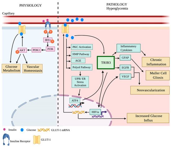
2.1.1. Neovascularization and Microvascular Changes in Diabetic Rodents
The most critical pathologic findings of PDR are neovascularization, hemorrhage, and fibro-vascular proliferation, leading to retinal traction and detachment and vitreous hemorrhage [
63]. Oxygen-induced retinopathy (OIR) in rodents is an accurate and reproducible model of vascular proliferative changes in the retina [
55]. Hypoxia-driven vascular proliferative changes seem to be similar to those seen in the retinopathy of prematurity, age-related macular degeneration, and diabetic retinopathy. OIR was developed in canine models for the first time in the early 1950s. In this model, Arnall Patz and colleagues investigated the effects of hyperoxia on retinal vessel development to study proliferative retinopathy [
56,
57]. To develop this model, one-day-old pups were exposed to hyperoxia for four consecutive days. In the early 1990s, this approach was introduced in rodents by Dr. Smith and her colleagues and has gained increasing popularity. In addition to OIR canines and rodents, aberrant angiogenesis has also been reported in zebrafish, rabbit, and monkey models.
The rodent OIR model is the most common approach to investigating the effect of hypoxia on the retina since it mimics the characteristics of human retinal proliferative changes [
55,
58,
64]. Because retinal vasculature develops in the first two weeks of birth in rodents, researchers can leverage this opportunity to analyze the aberrant vascular development triggered by hypoxia. In this model, hypoxia is induced at postnatal day (P) 7 after the regression of hyaloid vessels to avoid the development of mixed hyaloidopathy. The rodent pups were then exposed to hyperoxia (75% oxygen) for five consecutive days from P7 to P12 and then observed at room air from P13 to P17 [
55]. The peak changes of neovascularization are usually observed at P17, and these are resolved by P25. The C57BL/6 mice or the Sprague Dawley (SD) rats are the common strains employed in this model due to their neovascular susceptibility to hypoxia [
58,
64,
65]. The OIR mice developed irregular blood vessels and a reduction in the retinal inner and deep plexuses at P18, mimicking retinal proliferative events triggered by hypoxia in patients with diabetic complications [
66]. Downie and colleagues reported an increase in extraretinal neovascularization and impaired pericyte distribution in the OIR SD rat retinas as early as P18 [
67].
Genetically modified Akimba, Akita, and Kimba mice manifest vascular dysfunction. Akimba mice were specifically developed to study the microvascular changes of DR and showed these changes at the early age of eight weeks old [
47]. Thus, at eight months of age, these mice developed neovascularization, retinal edema, and detachment that progressed further through 25 weeks of age [
47]. In the Kimba mice, abnormal blood vessel development was seen as early as P28, while an increase in vascular permeability and adherent leukocytes was observed at six weeks of age. Additionally, loss of retinal capillaries, neovascularization, an increased avascular area, alteration in the vessel length, and pericyte loss were reported from nine weeks to the advanced age of 24 weeks [
46,
68]. Vascular dysfunction in Ins2
Akita mice presents as an increase in leukocytosis at eight weeks, compromised vascular permeability at 12 weeks, microaneurysms at six months, and neovascularization at nine months of hyperglycemia [
42,
69].
STZ mice also show microvascular changes earlier in the course of diabetes compared to STZ-induced hyperglycemic rats. For example, vascular permeability detected by imaging the distribution of fluorescein-conjugated dextran is compromised in these animals as early as eight days post-STZ injection [
70]. However, a decrease in arteriolar diameter and velocity were reported at four weeks and eight weeks post-STZ injection, respectively [
26]. Later in the course of diabetes (six to nine months), the STZ-induced hyperglycemic mice manifested pericyte loss and developed acellular capillaries [
71].
In albino Wistar–Kyoto rats, the blood retinal barrier (BRB) disruption occurs as early as two weeks post-STZ injection. Several studies reported early neovascular changes such as adherent leukocytosis and thickening of the basement membrane occurring at 8 and 12 weeks, respectively [
8,
72,
73]. Gong et al. noted that neovascularization in STZ-injected SD rats can be observed at three to four months after induction of hyperglycemia. An increase in VEGFR1 and VEGFR2 expression levels was associated with neovascularization in STZ-induced rats [
74]. Similar findings were observed in the Alloxan-induced diabetic rats; leukocytosis and neovascularization were reported at two and nine months after induction of hyperglycemia, respectively. At two months of sustained hyperglycemia, the authors observed pericyte loss, the formation of acellular capillaries, and basement membrane thickening [
75,
76]. In contrast, several other studies reported that BB rats with autoimmune T1D manifested these retinal changes as early as four months, while this model as well as genetic ZDF and obese OLETF rat models demonstrated BRB breakdown and pericyte loss at six to eight months [
48,
49,
51,
53,
77,
78]. Overall, these studies imply that the observed vascular dysfunction could vary in rat models of DR triggered by different insults. In addition to rats, hyperglycemia induced by a high-fat diet in db/db mice with T2D also leads to an increase in vascular permeability and basement thickness at 13–14 weeks of hyperglycemia [
79,
80]. Moreover, these mice also demonstrate pericyte loss, blood retinal barrier disruption, and vascular leakage at 12 months of age [
39].
Neuronal cell death and gliosis are observed in the diabetic retina of animals with diabetes. Thus, in hyperglycemic rats, GFAP activation has been reported. STZ injection results in an increase in GFAP immunoreactivity in the retina as early as six to seven weeks [
81] and as late as 8–16 weeks post-injection [
81,
82]. Retinal cell loss and functional changes have also been reported as early as two weeks and as late as 24 weeks post-STZ injection. Moreover, an increase in apoptotic cell death in the ONL, INL, and RGC layers resulting in a decrease in the total retinal thickness has been detected between 12 and 16 weeks post-STZ injections in rats [
82,
83]. In contrast, necrotic RGC death was reported at four weeks post-STZ treatment in rats [
83]. These rats also manifested severe loss of photoreceptors at 12 and 24 weeks, [
83] while in WBN/ Kob rat retinas, photoreceptor degeneration occurs earlier, at four weeks of age [
50]. Our recent study also confirmed RGC function loss and cell death in STZ-induced hyperglycemic mice at 32 weeks post-injection [
84] and tree shrews at 16 weeks post-injection [
31]. In addition to retinal neurons, RPE degeneration was reported in diabetic retinas. Thus, in four-month-old diabetic BB rats, hyperglycemia induces RPE degeneration through focal necrosis [
85]. In hyperglycemic OLETF rat retinas, the decrease in the thickness of the RPE layer along with a reduction in the INL and ONL thicknesses occurs later, at nine months after induction of hyperglycemia [
53]. Much later, at 50 weeks post-hyperglycemia induction, retinal detachment and fibrous proliferation occurs in Torii (SDT) rats with spontaneous diabetes [
54]. In other model of spontaneous diabetes, ZFD rats, extensive glial activation along with photoreceptor outer segment (POS) degeneration occurs in 32-week-old retinas [
86].The latter agrees with multiple studies demonstrating the thinning of the INL and IPL in OIR rat pups at P18 [
67,
84,
87,
88]. In addition, the thinning of the inner limiting membrane (ILM) is observed in STZ-induced SD retinas [
85].
In STZ-induced diabetic mice, RGC loss occurs between 6 and 12 weeks [
89]. RGC death occurs through apoptosis. The number of RGC apoptotic positive cells measured by TUNEL is 25% higher than that in control retinas [
90]. These data are similar to our observation of about a 30% RGC death with this model, [
84] although another study reported that the RGC density across the retina varies at 20 weeks post-STZ treatment [
91]. A few studies with Ins2
Akita mice detected early cone photoreceptor cell loss at three months. The authors observed a significant reduction in the IPL and INL thicknesses along with a diminishing number of RGCs at 22 weeks and 36 weeks of hyperglycemia [
42,
92]. Similarly, the OCT analysis of 16- and 28-week-old diabetic db/db mice retinas showed thinning in the NFL and RGC layer at a rate of 0.104 μm per week, resulting in a reduction of the total retinal thickness by 28 weeks [
91,
93]. The 28-week-old diabetic db/db mice also showed TUNEL positive photoreceptor cells and reduction in the ONL thickness. STZ-induced hyperglycemia in mice also leads to GFAP overexpression in retinal astrocytes at five weeks post-STZ treatment, while Müller cell gliosis are not seen even after 15 months of DM [
71,
94]. In contrast, the OIR mice demonstrated a reduction in the total retinal, INL, and IPL thicknesses, as well as distorted photoreceptor OS, neuronal loss, hyperactivity of Müller cells, and microglial activation at P18 [
66].

