Redox-Regulation of α-Globin in Vascular Physiology: History
Please note this is an old version of this entry, which may differ significantly from the current revision.
Subjects: Cell Biology
Contributor:

Interest in the structure, function, and evolutionary relations of circulating and intracellular globins dates back more than 60 years to the first determination of the three-dimensional structure of these proteins. Non-erythrocytic globins have been implicated in circulatory control through reactions that couple nitric oxide (NO) signaling with cellular oxygen availability and redox status. Small artery endothelial cells (ECs) express free α-globin, which causes vasoconstriction by degrading NO. This reaction converts reduced (Fe2+) α-globin to the oxidized (Fe3+) form, which is unstable, cytotoxic, and unable to degrade NO. Therefore, (Fe3+) α-globin must be stabilized and recycled to (Fe2+) α-globin to reinitiate the catalytic cycle. The molecular chaperone α-hemoglobin-stabilizing protein (AHSP) binds (Fe3+) α-globin to inhibit its degradation and facilitate its reduction. The mechanisms that reduce (Fe3+) α-globin in ECs are unknown, although endothelial nitric oxide synthase (eNOS) and cytochrome b5 reductase (CyB5R3) with cytochrome b5 type A (CyB5a) can reduce (Fe3+) α-globin in solution.

  • α-globin
  • endothelial nitric oxide synthase (eNOS)
  • cytochrome
  • redox system
  • arteries
  • blood pressure

1. Introduction

The globin protein superfamily shares an active motif conserved across eukaryotes, bacteria, and archaea [1][2][3]. These proteins present a conserved three-dimensional structure, the “globin fold” [4]. Globin-family proteins contain a central heme prosthetic group that mediates numerous biological functions related to the transport or metabolism of molecular oxygen (O2), carbon monoxide (CO), or nitric oxide (NO). Hemoglobin and myoglobin contain pentacoordinate heme and are involved primarily in O2 transport and storage [5]. In contrast, neuroglobin and cytoglobin contain hexacoordinate heme, in which the sixth coordinate position of the central iron atom is bound by a globin amino acid that competes for external ligand binding [6]. These globins, particularly cytoglobin, may serve as reversible ligand carriers and may also participate in redox reactions, but their biological functions are currently unclear [7]. Each member of the globin family of proteins has a distinct pattern of tissue expression, subcellular localization, and affinity for external ligands, including O2, carbon dioxide (CO2), and NO [8][9]. Globin proteins in the cells of the vascular wall can regulate vasodilatory NO signaling between endothelium and smooth muscle, inactivating and detoxifying NO by transforming it to nitrate (NO3) or reducing nitrite (NO2) to active NO under hypoxic conditions to enable autoregulation of tissue perfusion by smooth muscle [9].
Recently, α-globin and cytoglobin have been identified in arteriolar endothelial cells (ECs) and smooth muscle cells (SMCs), respectively, and have been noted to cause small artery vasoconstriction [10][11][12][13][14] by degrading the vasodilator NO in a redox reaction termed dioxygenation [NO + O2 (Fe2+) globin → NO3 + (Fe3+) globin] [13][15][16][17]. Moreover, studies of individuals with α-thalassemia demonstrated increased flow-mediated dilation [18]. The increased vasodilation may be explained by the loss of α-globin genes resulting in lower α-globin levels in the vasculature, as none of the patients presented with anemia or hemolysis. Nitric oxide generated in blood vessel ECs by endothelial nitric oxide synthase (eNOS) diffuses into adjacent vascular SMCs (VSMCs) to induce their relaxation by activating soluble guanylyl cyclase [19][20][21]. Two identical eNOS monomers can associate to form a dimeric active enzyme; each monomer includes a C-terminal reductase domain and an N-terminal oxygenase domain (the heme domain). The reductase domain contains two subdomains, each with its own flavin nucleotide prosthetic group: flavin mononucleotide (FMN) or flavin adenine dinucleotide (FAD). The reductase domain is connected to the oxygenase domain by a flexible linker containing a calmodulin (CaM) binding sequence. Calmodulin with associated Ca2+ regulates electron shuttling between the reductase and oxygenase domains. When eNOS is in the dimeric state, the heme group of one monomer receives electrons from the FMN domain of the other monomer. The oxygenase domain contains a tetrahydrobiopterin (BH4) prosthetic group that supports electron delivery to the heme group and enhances its affinity for the substrate l-arginine [22]. The catalytic activity mediated by the oxygenase domain uses l-arginine and O2 as substrates for generating NO, with nicotinamide adenine dinucleotide phosphate (NADPH) and CaM being essential cofactors [23].
Globin heme iron can bind ligands reversibly or participate in chemical reactions via electron transfer. Accordingly, the iron exists mainly in one of two interconvertible redox states—oxidized (Fe3+) or reduced (Fe2+)—depending on the direction of electron transfer. In reactions in which globin is used as an electron donor, including dioxygenation, heme iron is converted from Fe2+ to Fe3+ [13][16][17][24]. To participate in subsequent reactions, oxidized (Fe3+) globin must be recycled to the reduced (Fe2+) form. In erythrocytes, the major globin reductase system consists of soluble cytochrome b5 type A (CyB5a) and NADH–cytochrome b5 reductase 3 (CyB5R3) [25][26]. Specifically, CyB5R3 transfers electrons to CyB5a, which then donates electrons to oxidized hemoglobin. Previous studies have demonstrated that CyB5R3 participates in α-globin reduction in ECs [11], although whether this occurs through CyB5a or via other mechanisms is unknown.
Unlike erythrocytes, ECs express α-globin, but not β-globin [11][12]. Free α-globin rapidly autoxidizes and denatures. The molecular chaperone α-hemoglobin-stabilizing protein (AHSP) protects free α-globin by preferentially binding to the (Fe3+) form and rapidly inducing the formation of a stable hexacoordinate state that limits further chemical reactions with reactive oxygen and nitrogen oxide species [27]. α-Globin can bind either eNOS or AHSP, with the latter being favored when heme iron is oxidized [11][12][28]. In solution, (Fe3+) α-globin–AHSP can be reduced by CyB5R3 + CyB5a or by the reductase domain of eNOS. As free α-globin is unstable, we have proposed that most or all (Fe3+) α-globin in ECs is bound to AHSP, which facilitates its reduction by cellular enzymes, its release, and its transfer to eNOS to participate in dioxygenation [12]. According to this model, α-globin is stabilized by AHSP, and its availability to degrade NO is regulated by the cellular redox status. The present study investigated the model further by examining (Fe3+) α-globin reductase systems in ECs.

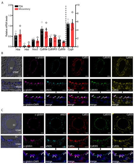
2. α-Globin, CyB5R3/CyB5a, and eNOS Are Expressed in Arterioles

To characterize the expression of α-globin, CyB5R3/CyB5a, eNOS, and cytoglobin in the thoracodorsal artery (TDA) (non-resistant) and mesenteric (resistant) arteries, we determined the mRNA steady-state levels in normal arteries by RT-qPCR analysis. The steady-state levels of α-globin (Hba1 and Hba2) and eNOS (Nos3) mRNA expression are equivalent in the TDA and mesenteric arterioles (Figure 1A) (Supplemental Table S2, could be found in https://www.mdpi.com/2076-3921/11/1/159#supplementary). Similarly, CyB5a and CyB5R3 are also expressed in equivalent steady-state levels in the TDA and mesenteric arterioles. In contrast, cytoglobin, characterized as being expressed in SMCs [13], is expressed at a level 10 times that of α-globin in both of these tissues (Figure 1A). As β-globin was previously not detected in arteriolar extracts [12][28], we assume that the Hbb mRNA detected came from erythroid cell contamination.
Figure 1. α-Globin and redox systems are co-expressed in small arteries. (A) mRNA levels in the thoracodorsal artery (TDA) and mesenteric arteries. The bar chart shows data from three or four 6-month-old mice. (B,C) Indirect immunofluorescence multiplexing staining for α-globin, endothelial nitric oxide synthase (eNOS), CyB5a, CyB5b, and CyB5R3 (white arrows) in TDA (B) and mesenteric artery (C) preparations. The dashed line across the internal elastic lamina demarcates the ECs and VSMCs. Merges represent the overlap of “α-globin+DAPI” with other staining based on the black arrow upstream. DAPI-stained nuclei are shown in blue, in combination with differential interference contrast. Scale bars are equivalent to 10 μm and 20 μm (for the higher magnification).
Immunofluorescence multiplex staining of TDA and mesenteric arteriole sections from 6-month-old mice detected α-globin and eNOS, which is consistent with our previous reports (Figure 1B,C, white arrows) [11][12]. In ECs, the distribution of CyB5a and CyB5R3 overlapped that of α-globin, consistent with the co-localization of the three proteins. α-Globin and eNOS signals were detected almost exclusively in arteriolar ECs, whereas CyB5a and CyB5R3 were detected in both ECs and VSMCs.
Another cytochrome, cytochrome b5 type B (CyB5b), has been suggested to be a donor of electrons to cytoglobin [29], so we investigated its expression and distribution and found that it is also expressed in arteriolar ECs and VSMCs. Therefore, CyB5b is indeed a potential electron donor, although the mitochondrial localization of this cytochrome [30] should limit its bimolecular interactions with α-globin and cytoglobin.

3. eNOS Reductase and Oxygenase Domain Activities

The discovery that eNOS is an α-globin-reducing agent was unanticipated [12]. To study this reduction reaction further, we tested its dependence on eNOS cofactors required for NO production. We first studied the effect of the cofactors BH4 and CaM/Ca2+ ± L-arginine on the reduction of the oxygenase domain of one monomer by the FMN domain of the other monomer in the functional eNOS dimer (Figure 2 and Table 1). We estimated the reduction rate by measuring the CO binding to the (Fe2+) eNOS oxygenase domain (Supplemental Figure S1). CO binds rapidly and with high affinity to the (Fe2+) heme domain; the limiting factor in this condition is the rate of reduction of the oxygenase domain by the reductase domain (electrons flow through the flavin domains until the final interdomain transfer toward the oxygenase domain). The reduction of the oxygenase domain was monophasic, and the presence of L-arginine had only a low influence on these rates (Table 1). The rate of reduction, 0.003 s−1, is consistent with what has been reported previously [31]. In the steady state, with the cofactors (BH4 and CaM/Ca2+ ± l-arginine) present, we consistently observed a partial (~60%) reduction of the oxygenase domain of eNOS, as compared to the complete reduction observed with sodium dithionite (Figure 2). In the presence of cofactors (BH4 and CaM/Ca2+), eNOS exhibited a slow rate of reduction of the oxygenase domain, probably to avoid, especially in the presence of O2, the catabolism of NO, its own catalysis product (Figure 2 and Table 1). At equilibrium under these conditions (with approximately 60% of the heme reduced), we assume that only a single oxygenase domain of the eNOS coupled dimeric protein is being reduced. We anticipate the involvement of an additional mechanism of negative cooperativity in the reduction of a second oxygenase domain. The kinetic features may be also explained, at least in part, by the electron repartition between the FAD reductase subdomain and both reduced conformations (semiquinone and hydroquinone) of the FMN subdomain.
Figure 2. eNOS reduction. (A) Spectra for full-length Fe3+ eNOS (black line), after the addition of NADPH (red line) or with sodium dithionite (cyan line), under anaerobic conditions. The red spectrum is simulated as a combination of 40% ferric and 60% ferrous eNOS. The insert shows the differential spectrum for the oxygenase domain redox transition of the full-length eNOS after addition of NADPH, which also involves flavin reduction in the reductase domain (485 nm, black arrow). (B) eNOS reduction kinetics in the presence of cofactors and L-arginine after mixing NADPH under anaerobic conditions. The dashed lines represent the exponential rise fit with the associated determination coefficient (R2).
Table 1. Reduction rates for α-globin–AHSP and eNOS.
  Enzymatic System Reduction Rate (s−1) ± SD Conditions
α-Globin–AHSP
(1 µM)
CyB5R3/CyB5a/NADH
(0.1 µM/1 µM/100 µM)
0.003 ± 0.00004 76 Torr CO
CyB5R3/NADH
(0.1 µM/100 µM)
0 76 Torr CO/10 Torr O2
eNOS/NADPH/BH4/CaM/Ca2+
(1 µM/50 µM/50 µM/3 µM/1.4 mM)
0.04 ± 0.0007 76 Torr CO
eNOS/NADPH/BH4/CaM/Ca2+/L-arginine
(1 µM/50 µM/50 µM/3 µM/1.4 mM/0.2 mM)
0.05 ± 0.0019 Anaerobic
eNOS
(1 µM)
NADPH/BH4/CaM/Ca2+
(50 µM/50 µM/3 µM/1.4 mM)
0.008± 0.00005 76 Torr CO
NADPH/BH4//CaM/Ca2+/L-arginine
(50 µM/50 µM/3 µM/1.4 mM/0.2 mM)
0.003 ± 0.00002 Anaerobic
Reduction of eNOS with its cofactors was initiated after addition of NADH. Reduction of α-globin–AHSP by CyB5R3 in the presence or absence of its partner protein CyB5a or by eNOS was initiated after addition of the globin to the enzymatic reaction components. All reaction were performed at 25 °C.

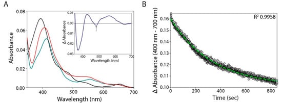
4. Reduction of α-Globin by eNOS

After establishing that full-length eNOS protein was dimeric and functionally active, we measured the reduction of (Fe3+) α-globin–AHSP by eNOS under anaerobic conditions in the presence of BH4 and CaM/Ca2+ and in the presence or absence of L-arginine and CO (Figure 3A and Table 1). We limited the use of L-arginine in our experiments to avoid NO production and associated redox side reactions in the presence of O2 (even if we used an O2 scavenging system). We compared the results to the eNOS oxygenase reduction rate to evaluate a potential interdomain electron transfer from reduced eNOS-deoxygenated (Fe2+) heme. Full reduction was completed within a few tens of seconds with 1 µM protein concentrations. When CO was added, we observed fully saturated α-globin (100% heme reduced) (Figure 3B: the dotted gray line compared to the solid black line at 420 nm) and approximately 60% saturation of the reduced eNOS oxygenase domain (Figure 3B: the dotted gray line compared to the solid black line at 445 nm). When we added NADPH after mixing eNOS and (Fe3+) α-globin–AHSP under CO to measure the competition for electron capture between the two heme domains, α-globin was reduced well before the eNOS oxygenase domain, in accordance with the reduction rates reported in Table 1 (data not shown). The reduction rates for the eNOS oxygenase domain were slower than those for the reduction of (Fe3+) α-globin–AHSP by at least an order of magnitude (Table 1), which indicates that α-globin reduction can compete in the range of nanomolar concentrations, even with BH4 and CaM/Ca2+ bound to the eNOS dimeric structure. Adding CO did not decrease the rate of electron transfer, probably because the intermolecular pathway for α-globin reduction passes through the eNOS reductase domain, with reduced FMN serving as the final electron donor. The kinetics of α-globin reduction by eNOS were monophasic (Figure 3A, Supplemental Figure S2, and Table 1).
Figure 3. α-Globin reduction by eNOS. (A) α-Globin–AHSP reduction kinetics after mixing with eNOS pre-incubated under anaerobic conditions in the presence of NADPH (as presented in Figure 2A). The chosen detection wavelength of 410 nm is close to the isosbestic point of the eNOS redox transition (as presented in Figure 2B). The dashed line represents the exponential decay fit with the associated determination coefficient (R2). The insert shows the transition spectrum of the oxidized (black line) and reduced (red line) α-globin. (B) The second derivative of the eNOS/NADPH and α-globin–AHSP mixture after the completion of globin reduction under anaerobic conditions with CO. The (Fe3+) α-globin–AHSP second derivative is centered at 414 nm, which is characteristic of bis-histidyl hexacoordination (black solid line). After reduction, the (Fe2+) α-globin–CO is revealed with the minima of its second derivative centered at 419 nm, whereas the (Fe2+) eNOS–CO is revealed with the minima centered at 445 nm (orange line). Upon the addition of dithionite, the amplitude of the (Fe2+) eNOS–CO second derivative increases almost two-fold (purple dashed line) by comparison with that of (Fe2+) α-globin–CO, which does not change (orange line). This underlines the fact that (Fe3+) α-globin–AHSP is fully reduced by eNOS after a few tens of seconds, whereas eNOS is only partially reduced by approximately 50%. All reaction were performed at 25 °C.

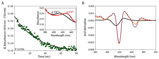
5. Reduction of α-Globin by the CyB5R3/CyB5a System

CyB5a and CyB5R3 are proteins with methemoglobin reductase activity. We determined the ability of CyB5R3 to reduce (Fe3+) α-globin–AHSP in the presence or absence of its partner protein CyB5a. Experiments conducted in the absence of CyB5a showed that CyB5R3 in the presence of NADH cannot reduce α-globin directly. The rate of CyB5R3 reduction (without CyB5a) was too low for this reduction to be involved in a turnover reaction for the NO dioxygenase activity of α-globin in ECs (Table 1). Therefore, CyB5R3 requires CYB5a as an intermediate electron acceptor that in turn reduces α-globin (Table 1). The major difference in redox potential between CyB5R3 (−265 mV) and CyB5a (+20 mV) favors electron transfer [32], but CyB5a and (Fe3+) α-globin are required to form a complex whose bonding is principally determined by complementary charge interactions between acidic groups of CyB5a and basic groups of α-globin [33]. α-Globin as a monomer and as incorporated in the hemoglobin tetramer exhibits the same reactivity with CyB5a [34]. Considering the slower consensus rate of 0.04 s−1, the eNOS reductase domain reduced (Fe3+) α-globin at a rate at least an order of magnitude faster than that observed with a CyB5R3/CyB5a system (Table 1).

This entry is adapted from the peer-reviewed paper 10.3390/antiox11010159

References

  1. Freitas, T.A.K.; Hou, S.; Dioum, E.M.; Saito, J.A.; Newhouse, J.; Gonzalez, G.; Gilles-Gonzalez, M.-A.; Alam, M. Ancestral hemoglobins in Archaea. Proc. Natl. Acad. Sci. USA 2004, 101, 6675–6680.
  2. Vinogradov, S.N.; Hoogewijs, D.; Bailly, X.; Arredondo-Peter, R.; Gough, J.; Dewilde, S.; Moens, L.; Vanfleteren, J.R. A phylogenomic profile of globins. BMC Evol. Biol. 2006, 6, 31.
  3. Vinogradov, S.N.; Tinajero-Trejo, M.; Poole, R.K.; Hoogewijs, D. Bacterial and archaeal globins—A revised perspective. Biochim. Biophys. Acta 2013, 1834, 1789–1800.
  4. Hoppe, F. Ueber das Verhalten des Blutfarbstoffes im Spectrum des Sonnenlichtes. Virchows Arch. 1862, 23, 446–449.
  5. Gell, D.A. Structure and function of haemoglobins. Blood Cells Mol. Dis. 2018, 70, 13–42.
  6. Kiger, L.; Tilleman, L.; Geuens, E.; Hoogewijs, D.; Lechauve, C.; Moens, L.; Dewilde, S.; Marden, M.C. Electron transfer function versus oxygen delivery: A comparative study for several hexacoordinated globins across the animal kingdom. PLoS ONE 2011, 6, e20478.
  7. Burmester, T.; Hankeln, T. Function and evolution of vertebrate globins. Acta Physiol. 2014, 211, 501–514.
  8. Saha, D.; Patgaonkar, M.; Shroff, A.; Ayyar, K.; Bashir, T.; Reddy, K.V.R. Hemoglobin expression in nonerythroid cells: Novel or ubiquitous? Int. J. Inflam. 2014, 2014, 803237.
  9. Keller, T.C.S.; Lechauve, C.; Keller, A.S.; Brooks, S.; Weiss, M.J.; Columbus, L.; Ackerman, H.C.; Cortese-Krott, M.M.; Isakson, B.E. The role of globins in cardiovascular physiology. Physiol. Rev. 2021.
  10. Halligan, K.E.; Jourd’heuil, F.L.; Jourd’heuil, D. Cytoglobin is expressed in the vasculature and regulates cell respiration and proliferation via nitric oxide dioxygenation. J. Biol. Chem. 2009, 284, 8539–8547.
  11. Straub, A.C.; Lohman, A.W.; Billaud, M.; Johnstone, S.R.; Dwyer, S.T.; Lee, M.Y.; Schoppee Bortz, P.; Best, A.K.; Columbus, L.; Gaston, B.; et al. Endothelial cell expression of haemoglobin α regulates nitric oxide signalling. Nature 2012, 491, 473–477.
  12. Lechauve, C.; Butcher, J.T.; Freiwan, A.; Biwer, L.A.; Keith, J.M.; Good, M.E.; Ackerman, H.; Tillman, H.S.; Kiger, L.; Isakson, B.E.; et al. Endothelial cell α-globin and its molecular chaperone α-hemoglobin–stabilizing protein regulate arteriolar contractility. J. Clin. Investig. 2018, 128, 5073–5082.
  13. Liu, X.; El-Mahdy, M.A.; Boslett, J.; Varadharaj, S.; Hemann, C.; Abdelghany, T.M.; Ismail, R.S.; Little, S.C.; Zhou, D.; Thuy, L.T.T.; et al. Cytoglobin regulates blood pressure and vascular tone through nitric oxide metabolism in the vascular wall. Nat. Commun. 2017, 8, 14807.
  14. Shu, X.H.; Ruddiman, C.A.; Keller, T.C.S., 4th; Keller, A.S.; Yang, Y.; Good, M.E.; Best, A.K.; Columbus, L.; Isakson, B.E. Heterocellular contact can dictate arterial function. Circ. Res. 2019, 124, 1473–1481.
  15. Gardner, A.M.; Cook, M.R.; Gardner, P.R. Nitric-oxide dioxygenase function of human cytoglobin with cellular reductants and in rat hepatocytes. J. Biol. Chem. 2010, 285, 23850–23857.
  16. Doyle, M.P.; Hoekstra, J.W. Oxidation of nitrogen oxides by bound dioxygen in hemoproteins. J. Inorg. Biochem. 1981, 14, 351–358.
  17. Gladwin, M.T.; Lancaster, J.R., Jr.; Freeman, B.A.; Schechter, A.N. Nitric oxide’s reactions with hemoglobin: A view through the SNO-storm. Nat. Med. 2003, 9, 496–500.
  18. Denton, C.C.; Shah, P.; Suriany, S.; Liu, H.; Thuptimdang, W.; Sunwoo, J.; Chalacheva, P.; Veluswamy, S.; Kato, R.; Wood, J.C.; et al. Loss of alpha-globin genes in human subjects is associated with improved nitric oxide-mediated vascular perfusion. Am. J. Hematol. 2021, 96, 277–281.
  19. Morgado, M.; Cairrão, E.; Santos-Silva, A.J.; Verde, I. Cyclic nucleotide-dependent relaxation pathways in vascular smooth muscle. Cell. Mol. Life Sci. 2012, 69, 247–266.
  20. Huang, P.L.; Huang, Z.; Mashimo, H.; Bloch, K.D.; Moskowitz, M.A.; Bevan, J.A.; Fishman, M.C. Hypertension in mice lacking the gene for endothelial nitric oxide synthase. Nature 1995, 377, 239–242.
  21. Scotland, R.S.; Madhani, M.; Chauhan, S.; Moncada, S.; Andresen, J.; Nilsson, H.; Hobbs, A.J.; Ahluwalia, A. Investigation of vascular responses in endothelial nitric oxide synthase/cyclooxygenase-1 double-knockout mice: Key role for endothelium-derived hyperpolarizing factor in the regulation of blood pressure in vivo. Circulation 2005, 111, 796–803.
  22. Tejero, J.; Shiva, S.; Gladwin, M.T. Sources of vascular nitric oxide and reactive oxygen species and their regulation. Physiol. Rev. 2019, 99, 311–379.
  23. Mas, M. A close look at the endothelium: Its role in the regulation of vasomotor tone. Eur. Urol. Suppl. 2009, 8, 48–57.
  24. Angelo, M.; Hausladen, A.; Singel, D.J.; Stamler, J.S. Interactions of NO with hemoglobin: From microbes to man. Methods Enzymol. 2008, 436, 131–168.
  25. Passon, P.G.; Hultquist, D.E. Soluble cytochrome b5 reductase from human erythrocytes. Biochim. Biophys. Acta 1972, 275, 62–73.
  26. Passon, P.G.; Reed, D.W.; Hultquist, D.E. Soluble cytochrome b5 from human erythrocytes. Biochim. Biophys. Acta 1972, 275, 51–61.
  27. Feng, L.; Zhou, S.; Gu, L.; Gell, D.A.; Mackay, J.P.; Weiss, M.J.; Gow, A.J.; Shi, Y. Structure of oxidized α-haemoglobin bound to AHSP reveals a protective mechanism for haem. Nature 2005, 435, 697–701.
  28. Straub, A.C.; Butcher, J.T.; Billaud, M.; Mutchler, S.M.; Artamonov, M.V.; Nguyen, A.T.; Johnson, T.; Best, A.K.; Miller, M.P.; Palmer, L.A.; et al. Hemoglobin α/eNOS coupling at myoendothelial junctions is required for nitric oxide scavenging during vasoconstriction. Arterioscler. Thromb. Vasc. Biol. 2014, 34, 2594–2600.
  29. Amdahl, M.B.; Sparacino-Watkins, C.E.; Corti, P.; Gladwin, M.T.; Tejero, J. Efficient reduction of vertebrate cytoglobins by the cytochrome b5/cytochrome b5 reductase/NADH system. Biochemistry 2017, 56, 3993–4004.
  30. James, R.; Searcy, J.L.; Le Bihan, T.; Martin, S.F.; Gliddon, C.M.; Povey, J.; Deighton, R.F.; Kerr, L.E.; McCulloch, J.; Horsburgh, K. Proteomic analysis of mitochondria in APOE transgenic mice and in response to an ischemic challenge. J. Cereb. Blood Flow Metab. 2012, 32, 164–176.
  31. Abu-Soud, H.M.; Ichimori, K.; Presta, A.; Stuehr, D.J. Electron transfer, oxygen binding, and nitric oxide feedback inhibition in endothelial nitric-oxide synthase. J. Biol. Chem. 2000, 275, 17349–17357.
  32. Henderson, C.J.; McLaughlin, L.A.; Wolf, C.R. Evidence that cytochrome b5 and cytochrome b5 reductase can act as sole electron donors to the hepatic cytochrome P450 system. Mol. Pharmacol. 2013, 83, 1209–1217.
  33. Gacon, G.; Lostanlen, D.; Labie, D.; Kaplan, J.C. Interaction between cytochrome b5 and hemoglobin: Involvement of β66 (E10) and β95 (FG2) lysyl residues of hemoglobin. Proc. Natl. Acad. Sci. USA 1980, 77, 1917–1921.
  34. Wheeler, K.E.; Nocek, J.M.; Cull, D.A.; Yatsunyk, L.A.; Rosenzweig, A.C.; Hoffman, B.M. Dynamic docking of cytochrome b5 with myoglobin and α-hemoglobin: Heme-neutralization “squares” and the binding of electron-transfer-reactive configurations. J. Am. Chem. Soc. 2007, 129, 3906–3917.
More
This entry is offline, you can click here to edit this entry!