Clotting Dysfunction in Sepsis: History
Please note this is an old version of this entry, which may differ significantly from the current revision.
Contributor:

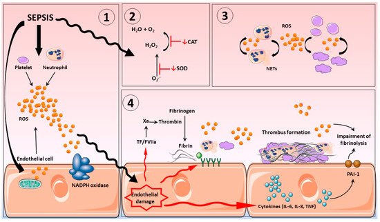
Sepsis is regarded as one of the main causes of death among the critically ill. Pathogen infection results in a host-mediated pro-inflammatory response to fight infection; as part of this response, significant endogenous reactive oxygen (ROS) and nitrogen species (RNS) production occurs, instigated by a variety of sources, including activated inflammatory cells, such as neutrophils, platelets, and cells from the vascular endothelium. Inflammation can become an inappropriate self-sustaining and expansive process, resulting in sepsis. Patients with sepsis often exhibit loss of aspects for normal vascular homeostatic control, resulting in abnormal coagulation events and development of disseminated intravascular coagulation. Diagnosis and treatment of sepsis remains a significant challenge for health care providers globally. Targeting the drivers of excessive oxidative/nitrosative stress using antioxidant treatments might be a therapeutic option.

  • sepsis
  • oxidative stress
  • nitric oxide
  • netosis
  • platelets
  • clotting dysfunction

1. Introduction

Sepsis is a life-threatening condition that affects 30 million people worldwide per year and is considered one of the main causes of death amongst critically ill patients. Seen in context, sepsis-related mortality from 2009 to 2019 averaged 33.7% in North America, 32.5% in Europe, and 26.4% in Australia [1]. In the United Kingdom, around 250,000 cases and 44,000 deaths from sepsis are reported every year [2]. In the United States, similarly high rates of sepsis diagnosis are reported annually, at some 1.7 million cases, with mortality rates in the region of 270,000 [3]. Specialist treatment for patients with sepsis often requires intensive care support to maintain failing organs systems, including the lungs, heart, and kidneys; such treatments can be complex and may require an extended length of hospital stay, all of which places a considerable fiscal burden on healthcare providers. In the United Kingdom, around two-thirds of patients with sepsis are treated in intensive care units (ICUs), with an annual estimated cost at £15.6 billion [4]. This represents a significant component of annual healthcare budgets; moreover, the need for further support for recovering patients as provided by primary care providers places an even greater burden budget. Again, for example, in 2011, U.S. hospitals spent $24 billion treating patients with sepsis, representing 13% of total health care costs (reviewed by [5]).
Defining criteria for sepsis and associated syndromes have evolved over the years; the current definition describes sepsis as an uncontrolled host-mediated response to infection and life-threatening organ dysfunction. Any patient is diagnosed with sepsis when they attain a score of 2 by sequential organ failure assessment (SOFA). SOFA comprises a scoring system based around the functionality of respiratory, hepatic, cardiovascular, central nervous and renal systems, and platelet count [6][7].
In response to pathogen infection, a protective pro-inflammatory response is initiated, but this can become deleterious and of extended duration, leading to sepsis, and over activation of the inflammatory system results in the production of reactive oxygen (ROS) and nitrogen species (RNS) to the extent that endogenous antioxidant protection becomes overwhelmed. The consequences of this are diverse in nature, including impacts on redox-based cell signalling systems; direct damage to biomolecules; and, perversely, the potential for immunosuppression. A common component of severe sepsis is endothelial damage and the triggering of the coagulation system, which can progress to DIC, which is marked by macro and microvascular thrombosis and hemorrhage and is a leading cause of organ damage in sepsis [8].

2. Disseminated Intravascular Coagulation in Sepsis

To maintain homeostatic control of blood flow through the circulation, it is essential that the processes of platelet plug formation and fibrinolysis are tightly regulated. If coagulation or anticoagulation mechanisms become dysfunctional, loss of homeostatic control can result in pathologies such as DIC, which is commonly encountered in patients with sepsis. DIC is a coagulopathy that occurs as a result of extensive and inappropriate activation of the coagulation system. Persistent coagulation results in thrombotic occlusion of small- and medium-sized blood vessels of the circulation via the establishment of microthrombi owing to fibrin formation (reviewed by [9]). Additionally, cessation of fibrinolysis, the mechanism responsible for lyzing the clot generated by activation of hemostatic pathways, contributes to DIC development in sepsis (reviewed by [10]). It has been reported that the levels of plasminogen activator inhibitor-1 (PAI-1), the protein responsible for assuring the clot preservation, is elevated in sepsis and is correlated with cytokines’ releases and poor patients’ outcome [11][12][13]. Another study, with the plasma of diabetics patients, demonstrated a strong correlation between the rise in oxidation markers (oxidized low-density lipoprotein and nitrotyrosine) and the impairment of the fibrinolysis process [14] (see Figure 1).
Figure 1. Sepsis induces oxidative stress and disseminates intravascular coagulation. (1). Sepsis induces ROS release by platelets, neutrophils, and endothelial cells. The majority of excessive ROS production is generated by mitochondria and NADPH oxidase present in endothelial cells, platelet, and neutrophil. (2). The overproduction of ROS results in depletion of endogenous antioxidant systems, including but not limited to SOD and catalase. (3). ROS release from activated inflammatory cells such as neutrophils and platelets further propagate inflammatory responses including further ROS production, processes that are self-sustaining and ever expanding. (4). Damage to the vascular endothelium augments inflammatory cytokine production via ROS-mediated stress responses and activates the coagulation system and expression of adhesion molecules, all of which results in elevation of fibrin deposition; impairment of fibrinolysis; and, consequently, thrombus formation. ROS: reactive oxygen species. CAT: catalase. SOD: superoxide dismutase. TF: tissue factor. NETs: neutrophil extracellular traps.
As such, compromised blood flow to key organs can result in multiple organ failure (MOF) and mortality, although other hemodynamic and metabolic disorders can similarly disrupt blood flow with similar outcomes [15][16]. In this regard, clinical guidelines relating to DIC state that, for better treatment and improved patient outcome, it is important to differentiate specific clinic phenotypes of disease type, including (1) increased fibrinolysis, such as would be associated with leukemia, trauma, and aortic or obstetric diseases; (2) suppressed fibrinolysis, associated with organ failure and septicemia; or (3) balanced fibrinolysis, such as would be observed with solid cancers [17][18]. Up to 40% of patients with sepsis present with or develop DIC [5][19][20][21]. During episodes of DIC, both bleeding and clotting events occur concurrently, which poses considerable issues regarding therapeutic approaches given the need to try and balance these opposing events [22].
A variety of clinical scoring methods have been developed with the intent to help physicians estimate the severity of disease in sepsis; however, such systems lack precision as definitive symptoms and clear diagnosis criteria are not obvious in many patients [16][23]. Currently, the most widely used DIC criteria scores are those as set out by The International Society of Thrombosis and Haemostasis (ISTH) and the Japanese Association for Acute Medicine (JAAM) [16].
The development of DIC in sepsis is thought to involve crosstalk between the inflammatory system activation and the overstimulation of coagulation and, more specifically, a deficient natural anticoagulant system, which leads to platelet activation and neutrophil extracellular trap formation, followed by fibrin deposition [24][25][26].
The disproportionate host inflammatory response to pathogens during sepsis results in dysfunction and damage to the vascular endothelium, largely due to the effects of a cytokine storm, with inflammatory cytokines such as tumor necrosis factor a (TNFα), interleukins 1b and 6 (IL-1b and IL-6), and interferon-gamma (IFNg) being implicated [27]. IL-6 is reported to play a central role in the activation of coagulation by tissue factor (TF) [16]. TF is a transmembrane glycoprotein able to activate the coagulation cascade when it is exposed to blood, under which circumstances the formation of a complex with FVII/FVIIa can ensue (reviewed by [28]), a frequent occurrence during endotoxemia [29]. Established beliefs indicated that TF was present and active only on general cell membranes; however, recent evidence has demonstrated the presence of TF on the surface of extracellular vesicles and microparticles (MPs), derived from platelets, leukocyte, and endothelial cells in patients with sepsis and DIC [30]. In addition to TF releasing, MPs also induce exposure of phosphatidylserine (PS), a phospholipid found on the surface of activated platelets that binds to an array of intrinsic and extrinsic factors to generate thrombin, a fundamental event of blood coagulation [31]. In addition, PS is associated with a significant increase in IL-6 and platelet activity, which correlates with the progression of endothelial damage and leukocyte activation [32][33].
While overexpression of pro-inflammatory cytokines enhances the activation of clotting cascades in sepsis, there is also good evidence to indicate associated impairment of pathways for essential natural anticoagulant activity, such as the antithrombin system, which is an important inhibitor of thrombin formation and FXa activation; the protein C system including protein S, which is an essential co-factor for the activity of protein C; and for thrombomodulin expression on endothelial cells (reviewed by [28]). Levels of protein C and antithrombin are markedly reduced in patients with sepsis with DIC [19]. Moreover, higher levels of protein C are associated with better patient outcomes in general, and as such may offer use as a both a clinical biomarker and a therapeutic target [34].

3. Vascular Haemostasis in Sepsis

3.1. Endothelium

The glycocalyx is a complex structure that consists of proteoglycans, glycoproteins, and glycosaminoglycan chains found on the surface of endothelial cells, and it plays a critical role in the regulation of vascular permeability; for a full description, see [35]. Sepsis-induced endothelial dysfunction including glycocalyx shedding results in increased leucocyte adhesion to endothelial cells, thereby exacerbating tissue damage [36]. Injury to the glycocalyx is known to be accentuated by increased levels of inflammatory molecules such as IL-1b. Diabetes likely further increases the associated risk; to this end, a recent study undertaken in mice demonstrated that diabetes limits glycocalyx synthesis, which is further damaged by endotoxemia [37]. Importantly, patients with diabetes have higher hospital admission rates when compared with non-diabetics [38].
It is increasingly recognized that disruption of vascular endothelium functionality is an important contributing factor to the onset of the progression of sepsis, including coagulation disorders, where TF and other procoagulant factors are known to be increased by high levels of inflammatory mediators. As such, studies have shown significant associations between markers of endothelial dysfunction and mortality, including increases in expression of endocan, Ang-2 and HMGB-1, and decreasing levels of protein C with TF levels [34].
Given the emerging understanding of endothelial dysfunction in sepsis, therapeutic approaches designed to protect the endothelium and glycocalyx are the subject of ongoing investigation. Indeed, a recent study has demonstrated in an animal rodent model of sepsis that recombinant antithrombin was able to protect the endothelial glycocalyx from injury, thus maintaining vascular integrity. Moreover, this approach was shown to decrease levels of syndecan-1, which is an important biomarker of glycocalyx damage [39]. Moreover, outcomes of ProCESS, a randomized study for resuscitation strategies undertaken in 1341 patients, found associations for the expression of endothelial biomarkers of permeability with mortality in sepsis, including at baseline and 24 h mortality. A decrease in expression of VEGF was observed, whereas there was an increase in the expression of angiopoietin-2, tissue plasminogen activator, thrombomodulin, and von Willebrand factor [40], leading to an increase in thrombus and associated with the increase in mortality in sepsis.

3.2. Platelets

Platelets are anucleate blood cells derived from megakaryocytes that are able to release cytokines, and that interact with leukocytes and endothelial cells, performing fundamental roles in both vascular homeostasis and coagulation. Platelet activation represents an important host response to infection for both innate and adaptive immunity [41]. In sepsis, platelets are implicated in coagulation dysfunction, through activation of pro-inflammatory mediators such as platelet activating factor and increasing fibrin formation via the expression of procoagulant molecules, including P-selectin [28][42][43]. However, decreased platelet counts (thrombocytopenia) may act as a predictor of mortality for patients with sepsis/septic shock and DIC [44]. The reasons for persistent thrombocytopenia in sepsis are not fully understood, but some theories suggest that this may be due to reduced platelet production, enhanced turnover, or spontaneous aggregation of platelets and enhanced platelet consumption through the formation of microthrombi. Although persistent platelet activation is most often related to septicemia, a few studies have shown that platelet aggregation is decreased in experimental sepsis [45], possibly signalled via the TNF pathway [46]. Moreover, the reduction in platelet aggregation seen in patients with sepsis is more pronounced depending on the severity sepsis, stage of disease, and the presence of DIC [47].

3.3. Neutrophils

Neutrophils are white blood cells that play a critical role in the immune response (reviewed by [48]) as well as in sepsis, which is associated with an excessive activation of neutrophils. Indeed, such neutrophil overstimulation is understood to be a key contributor to manifestations of sepsis and associated syndromes. In addition to the production of oxidants including hypochlorous acid (HOCL), hydrogen peroxide (H2O2) and superoxide (O2•−), proteases, and chemokines, activated neutrophils are also capable of undergoing a process named netosis. The release of neutrophil extracellular traps (NETs) during netosis occurs when nuclear DNA decondenses to release web-like structures of linear DNA from the cell that are interlaced with histones, myeloperoxidase, and other antimicrobial peptides such as elastase [49][50][51]. NETs are able to trap and kill microorganisms owing to the activity of associated antimicrobial proteins, and they also limit parasite dissemination [52]. However, unwanted collateral effects linked to netosis have also been described, including the induction of tissue injury mediated by extracellular histones and granular proteins [53][54]. Moreover, some substances, such as elastase and myeloperoxidase, released by NETs are considered to be damage-associated molecular patterns (DAMPs) and can cause tissue injury through activation of toll-like receptors on endothelial cells, leading to dysfunction [55][56][57]; additionally, extracellular MPO is still capable of forming the damaging oxidant HOCL. Interestingly, it has been shown that the inhibition of neutrophil elastase can prevent NETs’ formation and reduces septic shock in animal models, and thus may offer a therapeutic target for septicemia [58].
Some recent literature has demonstrated an association with the severity of sepsis and levels of NETs’ formation. In this regard, higher levels of NETs’ production correlated with the worsening and severity of sepsis and organ failure in humans; moreover, NETs’ formation during the initial stages of sepsis was also positively correlated with levels of key inflammatory cytokines IL-8, IFN-gamma, and TNFα [59]. Furthermore, inflammatory modulation by NETs was reported lead to severe damage in the liver, spleen, and kidneys in a murine model, processes that were abrogated by the use therapeutic of anti-citrullinated protein antibody, a NET formation inhibitor [57][60][61]. Other modulators released during the acute inflammatory response have also been linked to the induction of NETosis. For example, cold-inducible RNA-binding protein (CIRP), which is a DAMP, is known to be associated with organ injury and increased mortality in sepsis, and has recently been shown to enhance NETosis in the mice lungs during sepsis in an animal model induced by a cecal ligation and puncture (CLP) model [62]. Previous studies have also demonstrated that some antibiotics such as fluoroquinolones, macrolides, and a few b-lactams are capable of modulating the formation of NETs and, as such, may offer a protective role in early sepsis, this being ascribed to an immunomodulatory function possibly owing to downregulation of the PKC-Akt-mTOR pathway [50].
In addition, given the context of this review, NETs’ release is further linked to the evolution of DIC via the reduction in the levels of antithrombin and, as such, may provide a possible therapeutic target to treat DIC [58][63].
Importantly, the DNA molecule contains a polyphosphate backbone and is a known intracellular storage polymer of phosphate. Persistence of NETs is normally regulated by plasma DNAase 1 activity; there are nevertheless circumstances when such control is lost or overwhelmed, and net formation predominates [64]. DNA provides a negatively charged surface for the autocatalytic activation of Factor XII and the intrinsic pathway of coagulation, leading to increased thrombin generation and risk of thrombosis. Moreover, histones released with DNA are potent platelet activators, causing platelet degranulation and release of polyphosphate (PolyP). PolyP has been shown to be a highly potent activator of the contact pathway in vitro and in vivo [65][66], binding with high affinity to several of its protein components. Therefore, targeted inhibition of the Factor XII pathway may offer a therapeutic option in sepsis.

This entry is adapted from the peer-reviewed paper 10.3390/antiox11010088

References

  1. Bauer, M.; Gerlach, H.; Vogelmann, T.; Preissing, F.; Stiefel, J.; Adam, D. Mortality in Sepsis and Septic Shock in Europe, North America and Australia between 2009 and 2019-Results from a Systematic Review and Meta-Analysis. Crit. Care 2020, 24, 239.
  2. Dave, M.; Barry, S.; Coulthard, P.; Daniels, R.; Greenwood, M.; Seoudi, N.; Walton, G.; Patel, N. An Evaluation of Sepsis in Dentistry. Br. Dent. J. 2021, 230, 351–357.
  3. Rhee, C.; Dantes, R.; Epstein, L.; Murphy, D.J.; Seymour, C.W.; Iwashyna, T.J.; Kadri, S.S.; Angus, D.C.; Danner, R.L.; Fiore, A.E.; et al. Incidence and Trends of Sepsis in US Hospitals Using Clinical vs. Claims Data, 2009-2014. JAMA—J. Am. Med. Assoc. 2017, 318, 1241–1249.
  4. Daniels, R. Survive Sepsis; United Kingdom Sepsis Trust: Birmingham, UK, 2014; ISBN 9780992815509.
  5. Angus, D.C.; van der Poll, T. Severe Sepsis and Septic Shock. N. Engl. J. Med. 2013, 369, 840–851.
  6. Cecconi, M.; Evans, L.; Levy, M.; Rhodes, A. Sepsis and Septic Shock. Lancet 2018, 392, 75–87.
  7. Vincent, J.; Moreno, R.; Takala, J.; de Mendonça, A.; Bruining, H.; Reinhart, C.; Suter, P.; Thijs, L. The SOFA (Sepsis-Related Organ Failure Assessment) Score to Describe Organ Dysfunction/Failure. On Behalf of the Working Group on Sepsis-Related Problems of the European Society of Intensive Care Medicine. Intensive Care Med. 1996, 22, 707–710.
  8. Okabayashi, K.; Wada, H.; Ohta, S.; Shiku, H.; Nobori, T.; Maruyama, K. Hemostatic Markers and the Sepsis-related Organ Failure Assessment Score in Patients with Disseminated Intravascular Coagulation in an Intensive Care Unit. Am. J. Hematol. 2004, 76, 225–229.
  9. Levi, M.; Cate, H. Ten Disseminated Intravascular Coagulation. N. Engl. J. Med. 1999, 341, 586–592.
  10. Gando, S.; Levi, M.; Toh, C.H. Disseminated Intravascular Coagulation. Nat. Rev. Dis. Primers 2016, 2, 16037.
  11. Hoshino, K.; Kitamura, T.; Nakamura, Y.; Irie, Y.; Matsumoto, N.; Kawano, Y.; Ishikura, H. Usefulness of Plasminogen Activator Inhibitor-1 as a Predictive Marker of Mortality in Sepsis. J. Intensive Care 2017, 5, 42.
  12. Robbie, L.A.; Dummer, S.; Booth, N.A.; Adey, G.D.; Bennett, B. Plasminogen Activator Inhibitor 2 and Urokinase-Type Plasminogen Activator in Plasma and Leucocytes in Patients with Severe Sepsis. Br. J. Haematol. 2000, 109, 342–348.
  13. Raaphorst, J.; Johan Groeneveld, A.B.; Bossink, A.W.; Erik Hack, C. Early Inhibition of Activated Fibrinolysis Predicts Microbial Infection, Shock and Mortality in Febrile Medical Patients. Thromb. Haemost. 2001, 86, 543–549.
  14. Lados-Krupa, A.; Konieczynska, M.; Chmiel, A.; Undas, A. Increased Oxidation as an Additional Mechanism Underlying Reduced Clot Permeability and Impaired Fibrinolysis in Type 2 Diabetes. J. Diabetes Res. 2015, 2015, 456189.
  15. Gando, S.; Saitoh, D.; Ishikura, H.; Ueyama, M.; Otomo, Y.; Oda, S.; Kushimoto, S.; Tanjoh, K.; Mayumi, T.; Ikeda, T.; et al. A Randomized, Controlled, Multicenter Trial of the Effects of Antithrombin on Disseminated Intravascular Coagulation in Patients with Sepsis. Crit. Care 2013, 17, R297.
  16. Masuda, T.; Shoko, T. Clinical Investigation of the Utility of a Pair of Coagulation-Fibrinolysis Markers for Definite Diagnosis of Sepsis-Induced Disseminated Intravascular Coagulation: A Single-Center, Diagnostic, Prospective, Observational Study. Thromb. Res. 2020, 192, 116–121.
  17. Matsuda, K.; Kurokawa, M. Underlying Disease and Clinical Phenotypes of Disseminated Intravascular Coagulation. JMA J. 2020, 3, 357–358.
  18. Ohbe, H.; Yamakawa, K.; Taniguchi, K.; Morita, K.; Matsui, H.; Fushimi, K. Underlying Disorders, Clinical Phenotypes, and Treatment Diversity among Patients with Disseminated Intravascular Coagulation. JMA J. 2020, 3, 321–329.
  19. Jackson Chornenki, N.L.; Dwivedi, D.J.; Kwong, A.C.; Zamir, N.; Fox-Robichaud, A.E.; Liaw, P.C. Identification of Hemostatic Markers That Define the Pre-DIC State: A Multi-Center Observational Study. J. Thromb. Haemost. 2020, 18, 2524–2531.
  20. Smith, L. Disseminated Intravascular Coagulation. Semin. Oncol. Nurs. 2021, 37, 151135.
  21. Yamakawa, K.; Yoshimura, J.; Ito, T.; Hayakawa, M.; Hamasaki, T.; Fujimi, S. External Validation of the Two Newly Proposed Criteria for Assessing Coagulopathy in Sepsis. Thromb. Haemost. 2019, 119, 203–212.
  22. Naime, A.C.A.; Ganaes, J.O.F.; Lopes-Pires, M.E. Sepsis: The Involvement of Platelets and the Current Treatments. Curr. Mol. Pharmacol. 2018, 11, 261–269.
  23. Helms, J.; Severac, F.; Merdji, H.; Clere-Jehl, R.; François, B.; Mercier, E.; Quenot, J.P.; Meziani, F. Performances of Disseminated Intravascular Coagulation Scoring Systems in Septic Shock Patients. Ann. Intensive Care 2020, 10, 92.
  24. Abrams, S.T.; Morton, B.; Alhamdi, Y.; Alsabani, M.; Lane, S.; Welters, I.D.; Wang, G.; Toh, C.H. A Novel Assay for Neutrophil Extracellular Trap Formation Independently Predicts Disseminated Intravascular Coagulation and Mortality in Critically Ill Patients. Am. J. Respir. Crit. Care Med. 2019, 200, 869–880.
  25. Patel, P.; Walborn, A.; Rondina, M.; Fareed, J.; Hoppensteadt, D. Markers of Inflammation and Infection in Sepsis and Disseminated Intravascular Coagulation. Clin. Appl. Thromb. Hemost. 2019, 25, 1–6.
  26. Yang, X.; Cheng, X.; Tang, Y.; Qiu, X.; Wang, Y.; Kang, H.; Wu, J.; Wang, Z.; Liu, Y.; Chen, F.; et al. Bacterial Endotoxin Activates the Coagulation Cascade through Gasdermin D-Dependent Phosphatidylserine Exposure. Immunity 2019, 51, 983–996.e6.
  27. Kinasewitz, G.T.; Yan, S.B.; Basson, B.; Comp, P.; Russell, J.A.; Cariou, A.; Um, S.L.; Utterback, B.; Laterre, P.F.; Dhainaut, J.F. Universal Changes in Biomarkers of Coagulation and Inflammation Occur in Patients with Severe Sepsis, Regardless of Causative Micro-Organism . Crit. Care 2004, 8, 82–90.
  28. Levi, M.; van der Poll, T. Coagulation and Sepsis. Thromb. Res. 2017, 149, 38–44.
  29. Scully, M.; Levi, M. How We Manage Haemostasis during Sepsis. Br. J. Haematol. 2019, 185, 209–218.
  30. Nieuwland, R.; Gardiner, C.; Dignat-George, F.; Mullier, F.; Mackman, N.; Woodhams, B.; Thaler, J. Toward Standardization of Assays Measuring Extracellular Vesicle-Associated Tissue Factor Activity. J. Thromb. Haemost. 2019, 17, 1261–1264.
  31. Kay, J.G.; Grinstein, S. Phosphatidylserine-Mediated Cellular Signaling. Adv. Exp. Med. Biol. 2013, 991, 177–193.
  32. Delabranche, X.; Boisramé-Helms, J.; Asfar, P.; Berger, A.; Mootien, Y.; Lavigne, T.; Grunebaum, L.; Lanza, F.; Gachet, C.; Freyssinet, J.M.; et al. Microparticles Are New Biomarkers of Septic Shock-Induced Disseminated Intravascular Coagulopathy. Intensive Care Med. 2013, 39, 1695–1703.
  33. Matsumoto, H.; Yamakawa, K.; Ogura, H.; Koh, T.; Matsumoto, N.; Shimazu, T. Enhanced Expression of Cell-Specific Surface Antigens on Endothelial Microparticles in Sepsis-Induced Disseminated Intravascular Coagulation. Shock 2015, 43, 443–449.
  34. Walborn, A.; Rondina, M.; Mosier, M.; Fareed, J.; Hoppensteadt, D. Endothelial Dysfunction Is Associated with Mortality and Severity of Coagulopathy in Patients with Sepsis and Disseminated Intravascular Coagulation. Clin. Appl. Thromb. Hemost. 2019, 25, 1–9.
  35. VanTeeffelen, J.W.; Brands, J.; Stroes, E.S.; Vink, H. Endothelial Glycocalyx: Sweet Shield of Blood Vessels. Trends Cardiovasc. Med. 2007, 17, 101–105.
  36. Lupu, F.; Kinasewitz, G.; Dormer, K. The Role of Endothelial Shear Stress on Haemodynamics, Inflammation, Coagulation and Glycocalyx during Sepsis. J. Cell. Mol. Med. 2020, 24, 12258–12271.
  37. Sampei, S.; Okada, H.; Tomita, H.; Takada, C.; Suzuki, K.; Kinoshita, T.; Kobayashi, R.; Fukuda, H.; Kawasaki, Y.; Nishio, A.; et al. Endothelial Glycocalyx Disorders May Be Associated With Extended Inflammation During Endotoxemia in a Diabetic Mouse Model. Front. Cell Dev. Biol. 2021, 9, 623582.
  38. Kushimoto, S.; Abe, T.; Ogura, H.; Shiraishi, A.; Saitoh, D.; Fujishima, S.; Mayumi, T.; Hifumi, T.; Shiino, Y.; Nakada, T.-A.; et al. Impact of Blood Glucose Abnormalities on Outcomes and Disease Severity in Patients with Severe Sepsis: An Analysis from a Multicenter, Prospective Survey of Severe Sepsis. PLoS ONE 2020, 15, e0229919.
  39. Iba, T.; Connors, J.M.; Nagaoka, I.; Levy, J.H. Recent Advances in the Research and Management of Sepsis-Associated DIC. Int. J. Hematol. 2021, 113, 24–33.
  40. Hou, P.C.; Filbin, M.R.; Wang, H.; Ngo, L.; Aird, W.C.; Shapiro, N.I.; Wang, H.; Huang, D.T.; Angus, D.C.; Kellum, J.A.; et al. Endothelial Permeability and Hemostasis in Septic Shock: Results From the ProCESS Trial. Chest 2017, 152, 22–31.
  41. Portier, I.; Campbell, R.A. Role of Platelets in Detection and Regulation of Infection. Arterioscler. Thromb. Vasc. Biol. 2020, 41, 70–78.
  42. Ma, R.; Xie, R.; Yu, C.; Si, Y.; Wu, X.; Zhao, L.; Yao, Z.; Fang, S.; Chen, H.; Novakovic, V.; et al. Phosphatidylserine-Mediated Platelet Clearance by Endothelium Decreases Platelet Aggregates and Procoagulant Activity in Sepsis. Sci. Rep. 2017, 7, 4978.
  43. Puskarich, M.A.; Cornelius, D.C.; Bandyopadhyay, S.; McCalmon, M.; Tramel, R.; Dale, W.D.; Jones, A.E. Phosphatidylserine Expressing Platelet Microparticle Levels at Hospital Presentation Are Decreased in Sepsis Non-Survivors and Correlate with Thrombocytopenia. Thromb. Res. 2018, 168, 138–144.
  44. Tsirigotis, P.; Chondropoulos, S.; Frantzeskaki, F.; Stamouli, M.; Gkirkas, K.; Bartzeliotou, A.; Papanikolaou, N.; Atta, M.; Papassotiriou, I.; Dimitriadis, G.; et al. Thrombocytopenia in Critically Ill Patients with Severe Sepsis/Septic Shock: Prognostic Value and Association with a Distinct Serum Cytokine Profile. J. Crit. Care 2016, 32, 9–15.
  45. Lopes-Pires, M.E.; Naime, A.C.A.; Cardelli, N.J.A.; Anjos, D.J.; Antunes, E.; Marcondes, S. PKC and AKT Modulate CGMP/PKG Signaling Pathway on Platelet Aggregation in Experimental Sepsis. PLoS ONE 2015, 10, e0137901.
  46. Naime, A.C.A.; Bonfitto, P.H.L.; Solon, C.; Lopes-Pires, M.E.; Anhê, G.F.; Antunes, E.; Marcondes, S. Tumor Necrosis Factor Alpha Has a Crucial Role in Increased Reactive Oxygen Species Production in Platelets of Mice Injected with Lipopolysaccharide. Platelets 2019, 30, 1047–1052.
  47. Laursen, M.A.; Larsen, J.B.; Larsen, K.M.; Hvas, A.M. Platelet Function in Patients with Septic Shock. Thromb. Res. 2020, 185, 33–42.
  48. Bardoel, B.W.; Kenny, E.F.; Sollberger, G.; Zychlinsky, A. The Balancing Act of Neutrophils. Cell Host Microbe 2014, 15, 526–536.
  49. Metzler, K.D.; Goosmann, C.; Lubojemska, A.; Zychlinsky, A.; Papayannopoulos, V. Myeloperoxidase-Containing Complex Regulates Neutrophil Elastase Release and Actin Dynamics during NETosis. Cell Rep. 2014, 8, 883–896.
  50. Xie, T.; Duan, Z.; Sun, S.; Chu, C.; Ding, W. β-Lactams Modulate Neutrophil Extracellular Traps Formation Mediated by MTOR Signaling Pathway. Biochem. Biophys. Res. Commun. 2021, 534, 408–414.
  51. Yipp, B.G.; Petri, B.; Salina, D.; Jenne, C.N.; Scott, B.N.V.; Zbytnuik, L.D.; Pittman, K.; Asaduzzaman, M.; Wu, K.; Meijndert, H.C.; et al. Infection-Induced NETosis Is a Dynamic Process Involving Neutrophil Multitasking in Vivo. Nat. Med. 2012, 18, 1386–1393.
  52. Shimizu, M.; Konishi, A.; Nomura, S. Examination of Biomarker Expressions in Sepsis-Related DIC Patients. Int. J. Gen. Med. 2018, 11, 353–361.
  53. Jiao, Y.; Li, W.; Wang, W.; Tong, X.; Xia, R.; Fan, J.; Du, J.; Zhang, C.; Shi, X. Platelet-Derived Exosomes Promote Neutrophil Extracellular Trap Formation during Septic Shock. Crit. Care 2020, 24, 380.
  54. Rodrigues, D.A.S.; Prestes, E.B.; Gama, A.M.S.; de Souza Silva, L.; Pinheiro, A.A.S.; Ribeiro, J.M.C.; Campos, R.M.P.; Pimentel-Coelho, P.M.; de Souza, H.S.; Dicko, A.; et al. CXCR4 and MIF Are Required for Neutrophil Extracellular Trap Release Triggered by Plasmodium-Infected Erythrocytes. PLoS Pathog. 2020, 16, e1008230.
  55. Colón, D.F.; Wanderley, C.W.; Franchin, M.; Silva, C.M.; Hiroki, C.H.; Castanheira, F.V.S.; Donate, P.B.; Lopes, A.H.; Volpon, L.C.; Kavaguti, S.K.; et al. Neutrophil Extracellular Traps (NETs) Exacerbate Severity of Infant Sepsis. Crit. Care 2019, 23, 113.
  56. Huang, H.; Tohme, S.; Al-Khafaji, A.B.; Tai, S.; Loughran, P.; Chen, L.; Wang, S.; Kim, J.; Billiar, T.; Wang, Y.; et al. Damage-Associated Molecular Pattern-Activated Neutrophil Extracellular Trap Exacerbates Sterile Inflammatory Liver Injury. Hepatology 2015, 62, 600–614.
  57. Shrestha, B.; Ito, T.; Kakuuchi, M.; Totoki, T.; Nagasato, T.; Yamamoto, M.; Maruyama, I. Recombinant Thrombomodulin Suppresses Histone-Induced Neutrophil Extracellular Trap Formation. Front. Immunol. 2019, 10, 2535.
  58. Okeke, E.B.; Louttit, C.; Fry, C.; Najafabadi, A.H.; Han, K.; Nemzek, J.; Moon, J.J. Inhibition of Neutrophil Elastase Prevents Neutrophil Extracellular Trap Formation and Rescues Mice from Endotoxic Shock. Biomaterials 2020, 238, 119836.
  59. Kumar, S.; Gupta, E.; Kaushik, S.; Srivastava, V.K.; Saxena, J.; Mehta, S.; Jyoti, A. Quantification of NETs Formation in Neutrophil and Its Correlation with the Severity of Sepsis and Organ Dysfunction. Clin. Chim. Acta 2019, 495, 606–610.
  60. Chirivi, R.G.S.; van Rosmalen, J.W.G.; van der Linden, M.; Euler, M.; Schmets, G.; Bogatkevich, G.; Kambas, K.; Hahn, J.; Braster, Q.; Soehnlein, O.; et al. Therapeutic ACPA Inhibits NET Formation: A Potential Therapy for Neutrophil-Mediated Inflammatory Diseases. Cell. Mol. Immunol. 2020, 18, 1528–1544.
  61. Locke, M.; Francis, R.J.; Tsaousi, E.; Longstaff, C. Fibrinogen Protects Neutrophils from the Cytotoxic Effects of Histones and Delays Neutrophil Extracellular Trap Formation Induced by Ionomycin. Sci. Rep. 2020, 10, 11694.
  62. Ode, Y.; Aziz, M.; Jin, H.; Arif, A.; Nicastro, J.G.; Wang, P. Cold-Inducible RNA-Binding Protein Induces Neutrophil Extracellular Traps in the Lungs during Sepsis. Sci. Rep. 2019, 9, 6252.
  63. Levi, M.; Scully, M.; Singer, M. The Role of ADAMTS-13 in the Coagulopathy of Sepsis. J. Thromb. Haemost. 2018, 16, 646–651.
  64. Hakkim, A.; Fürnrohr, B.G.; Amann, K.; Laube, B.; Abed, U.A.; Brinkmann, V.; Herrmann, M.; Voll, R.E.; Zychlinsky, A. Impairment of Neutrophil Extracellular Trap Degradation Is Associated with Lupus Nephritis. Proc. Natl. Acad. Sci. USA 2010, 107, 9813–9818.
  65. Müller, F.; Mutch, N.J.; Schenk, W.A.; Smith, S.A.; Esterl, L.; Spronk, H.M.; Schmidbauer, S.; Gahl, W.A.; Morrissey, J.H.; Renné, T. Platelet Polyphosphates Are Proinflammatory and Procoagulant Mediators In Vivo. Cell 2009, 139, 1143–1156.
  66. Smith, S.A.; Mutch, N.J.; Baskar, D.; Rohloff, P.; Docampo, R.; Morrissey, J.H. Polyphosphate Modulates Blood Coagulation and Fibrinolysis. Proc. Natl. Acad. Sci. USA 2006, 103, 903–908.
More
This entry is offline, you can click here to edit this entry!
Video Production Service